
Recommended
More Related Content
What's hot
What's hot (20)
Similar to Immunofluoresce-WPS Office.pdf
Similar to Immunofluoresce-WPS Office.pdf (20)
More from Ranjeettaram
Recently uploaded
Recently uploaded (20)
Immunofluoresce-WPS Office.pdf
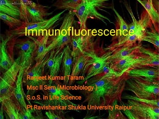
- 1. Immunofluorescence Ranjeet Kumar Taram Msc ll Sem (Microbiology ) S.o.S. in Life Science Pt Ravishankar Shukla University Raipur
- 2. Introduction Immunofluorescence is a technique allowing the visualization of a specific antigen by bindIng a specific antibody chemically conjugated with a fluorescent dye such as fluorescin isothiosynate. The most commonly used fluorescent dye are fluorescin and rhodamin. Other dye such as phycoerythrin and phycobiliproteins, an intensely colored and highly fluorescent pigment. This molecules can be conjugated to the Fc region of an antibody molecule without affecting the specificity of the antibody and visualize the immune complex. FLUORESCENCE MICROSCOPE (SoS IN LIFE SCIENCES)
- 3. Principle Fluorescence is the property of the certain molecules to absorb light at one wavelength and emit light at larger wavelength when it is illuminated by light of a different wavelength. the fluorescent can be visualized using fluorescence microscopy. The immunofluorescence technique allows for the visualization of of the presence as well as the distribution of target molecules in a sample. Each of fluorescent dye or fluorochrome below absorbs light at one wavelength and emits light at a larger wavelength.
- 4. Fluorescin, an organic dye that is most widely used labeled for immunofluorescence procedure, absorbs blue light (490nm) and emits an intense yellow green fluorescence (517nm) Rhodamin another organic dye absorbs in the yellow green range (515nm) and emits a deep red fluorescence (546nm) Phycoerythrin :-It is an efficient absorber of light and a brilliant emitter of red fluorescence. In this technique antibody molecules are tagged with a fluorescent dye. When this antibody molecules bind with antigen form immune complex and this immune complexes are easily visualized or detected by the color light emission when excited by the light of the appropriate wavelength.
- 5. Sample preparation for immunofluorescence
- 6. Types of immunofluorescence Direct immunofluorescence - In this method, only a single antibody is used that is chemically linked to a fluorochrome. The antibody recognizes the target molecules and binds to it, and form immune complex that is detected via microscopy.
- 7. Advantages of direct immunofluorescence • • direct conjugation of the antibody reduce the number of steps in the staining procedure making the process faster and reduce background signal by avoiding some is used with antibody cross reactivity direct visualisation
- 8. indirect immunofluorescence indirect immunofluorescence uses two antibodies the unlabelled primary antibody specifically binds to the target molecule and the secondary antibody which carries the flow Chrome recognise the primary antibody and bind to it.
- 9. Advantages of indirect immunofluorescence • • • indirect methods avoid the loss of antibody that is usually occurs during the conjugation indirect methods increase the sensitivity of staiming because multiple molecules of the fluorochrome reagent bind to each primary antibody molecule. Requires only one Level antibody to identify many proteins sim labelled secondary antibody can be used to Wine bind too many different proteins
- 10. Applications • • • • • to identify the number of sub population of lymphocytes notably the cd4 and cd8 T-cell population. Identifying bacterial species detecting antigen antibody complex in autoimmune disease detecting complement component in tissues To localising hormones and other cellular product stain in situ study of evolution of cells in suspension cultured cell, tissue beads.
- 11. Limitations • • • Photobleaching: The cells lose their emission property i.e intensity of fluorescence with time. increased background noises: Due to multiple attachment of Secondary antibody. Non specific fluorescence: Involves the loss of a Probe's specifity due to flurophore , from improper fixation, or from dried out specimen.
- 12. Reference Goldsby, Richard A., Thomas J. Kindt, and Barbara A. Osborne. "Kuby immunology. 4th." USA. W (2000). Im, Kyuseok, et al. "An Introduction to Performing Immunofluorescence Staining." Biobanking. Humana Press, New York, NY, 2019 299-311 Wilson, K., & Walker, J. (Eds.). (2000). Principles and techniques of practical biochemistry. Cambridge University Press.
- 13. Thank you