
Recommended
More Related Content
What's hot
What's hot (20)
Similar to Structure And function of skin .pdf
Similar to Structure And function of skin .pdf (20)
Recently uploaded
Recently uploaded (20)
Structure And function of skin .pdf
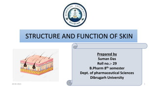
- 1. STRUCTURE AND FUNCTION OF SKIN Prepared by Suman Das Roll no.:- 29 B.Pharm 8th semester Dept. of pharmaceutical Sciences Dibrugarh University 30-05-2022 1
- 2. CONTENTS INTRODUCTION TO SKIN STRUCTURE OF SKIN FUNCTION OF SKIN BIBLIOGRAPHY 30-05-2022 2
- 3. INTRODUCTION Skin is the body’s largest and primary protective organ It covers entire external surface of the body It has a surface area about 1.5-2 m2 It contains accessory glands, hairs and nails Serves as 1st order physical barrier against the environment 30-05-2022 3
- 5. skin is made up of 3 layers Epidermis: The outermost layer of skin, provides a water barrier and contributes to skin tone Dermis: Found beneath the epidermis, contains connective tissue, hair follicles, blood vessels, lymphatic vessels and sweat glands Hypodermis: Deep to the dermis, not exactly the part of skin, it is the subcutaneous layer comprising of areolar and adipose tissue. Also contains larger vessels and nerves. 30-05-2022 5
- 6. 1. STRATUM CORNEUM Most differentiated top superficial layer, consists of 25-30 layers of flattened keratinocytes that contains mostly keratin 2. STRATUM LUCIDERM Lies below the stratum corneum. It is only present in the thick skin of fingereprints, palms and soles 3. STRATUM GRANULOSUM Middle and superficial layer of the epidermis. It is non- keratinized. 1-3 cells thick spindle shaped cells enriched with keratohyaline. 4. STRATUM SPINOSUM This layer contain 8-10 layers of cells. The cells have lots of desmosomes, which anchors the cells to each other. 5. STRATUM GERMINATIVUM It is the deepest layer of epidermis. Cells are non- nucleated. This layer contain melanocytes. EPIDERMIS It composed of stratified keratinised squomous epithelium, it contains four principle type of cells such as keratinocytes(90%), melanocytes, langerhans cells and merkel cells. It is divided into 5 distinct layers 30-05-2022 6
- 7. DERMIS Dermis is the second deepest region lying in betweeen Epidermis and Hypodermis It is formed from connective tissue containing collagen and elastin fibres The principal cells found in this layer are fibroblasts, macrophages and adipocytes Blood vessels, nerve fibre and hair follicles are embedded in this layer The superficial portion dermis called papillary layer , consist of areolar connective tissue containing elastin fibres the surface area is greatly increased by small finger like projections called dermal papilae this dermal papilae contain tactile receptors called corpuscles of touch or meissner corpuscles that are sensitive to touch The reticular region of dermis attached to the hypodermis consist of dense irregular connective tissue, collagen and elastic fibres This collagen and elastin fibres provides strength, extensibility and elasticity to the skin 30-05-2022 7
- 8. HYPODERMIS It is the subcutaneous layer which lies deep to the dermis It is not part of the skin This layer consist of areolar and adipose tissue This layer also contains nerve endinngs called lamellate(panician) corpuscles that are sensitive to pressure It serves as storage for fat Contains large blood vessels that supply the skin 30-05-2022 8
- 9. FUNCTIONS OF SKIN 2. SENSONY FUNCTION: Free nerve endings on the skin are sensitive to pain, touch, heat and cold. 1. PROTECTION: acts as a barrier preventing entry of pathogens 3. TEMPERATURE REGULATION: Regulation of body temperature at about 36.9o with a variation 0.50 and 0.750 is one of the important function 4. PRODUCTION OF VITAMIN D: Sunlight action on fatty tissue within skin causes the production of vit. D, essential for calcium absorption from the diet. 5. OTHER : Storage of fat, excretion of water, salts and very small amout of urea 30-05-2022 9
- 10. BIBLIOGRAPHY https://www.ncbi.nlm.nih.gov/books/NBK470464/ (29/04/2022; 12.10 A.M.) https://www.slideshare.net/docjikisha/structure-of-skin-124339625 (29/04/2022; 3.15 P.M.) A text book of cosmetic science; Dr. Aijaz Sheikh, Dr. Subhash Deshmane et al.; S.Vikas and Company; 2019; 1st edition; page no.:- 29-35 Cosmetic Sciences: concepts and principles; Dr. Kamala pathak, Dr. Ankur Vandya; Nirali Prakashan; 2018; 1st edition; page no. 4.1- 4.5 30-05-2022 10