SlideShare a Scribd company logo
1 of 29
INTEGUMENTARY
SYSTEM
INTEGUMENTARY SYSTEM
SKIN is the largest organ in our body. It is made up of the skin and
its accessory structures, which include the nails; hair; sebaceous, or
oil, glands; and sweat glands.
MAJOR FUNCTIONS OF
THE SKIN
3
• Protection
The skin protects the body from
mechanical trauma, pathogens, and
environmental damage.
• Excretion
The skin excretes waste and impurities
via sweat.
• Thermoregulation
The skin maintains a stable internal
body temperature through negative
feedback loops.
• Vitamin d synthesis
The skin synthesizes vitamin D when
exposed to ultraviolet radiation.
• Sensation
The skin has sensory receptors that
detect internal and external
environmental change such as heat,
cold and/or pain.
THE SKIN
4
• EPIDERMIS
• DERMIS
(Also known as the cutaneous membrane)
Main components of skin:
5
THE EPIDERMIS
the outermost and thickest layer, and it's made of
multiple layers of developing cells, which are called
keratinocytes.
Keratinocytes
 Produce Keratin
 New keratinocytes push up, older, dead
cells are shred off, forming skin flakes or
dandruff.
Melanocytes
 a protein pigment or coloring
substance, called melanin.
Langerhan cells
 Protects the skin and underlying
tissue from pathogens
 Phagocytes of the immune system
(epi – on top, dermis – skin)
THE EPIDERMIS
6
Layers of the
epidermis
 Stratum Corneum
 Stratum Lucidum
 Stratum Granulosum
 Stratum Spinosum
 Stratum Basale
THE EPIDERMIS
7
Layers of the epidermis
Stratum LuCIDUM
 Deep to the Stratum Corneum
 Found in thick skin
THE EPIDERMIS
8
Layers of the epidermis
Stratum Basale
 Contains small round cells
called basal cells
 It also contains melanocytes,
which are responsible for
producing melanin
THE EPIDERMIS
9
Layers of the epidermis
Stratum Spinosum
 Thickest stratum of the
epidermis
 Undergo mitosis
 Help to synthesize vitamin D
THE EPIDERMIS
10
Layers of the epidermis
Stratum Granulosum
 Excretes a lipid-based
substance
 Named for prominent granules
THE EPIDERMIS
11
Layers of the epidermis
Stratum Corneum
 Most superficial layer of the
epidermis
 Several layers of dead,
flattened keratinocytes
 Sheds dead keratinocytes
THE EPIDERMIS
12
Keratinocyte life cycle
 Begins in the Stratum Spinosum
 Sheds in the Stratum Corneum
 Can shed from environmental and physical stress
 Shed cells are replaced by mitosis
 Cells primarily divide at night
THE EPIDERMIS
13
THICK VS. THIN SKIN
THICK SKIN
 Consists of all five layers of the
epidermis with a thick Stratum
Corneum
 Contains sweat glands
 Found in palms of hands and
soles of feet
THE EPIDERMIS
14
THICK VS. THIN SKIN
THIn SKIN
 Consists of only four layers of
the epidermis, lacking the
Stratum Lucidum
 Equivalent to the size of a
sheet of printing paper
15
THE DERMIS
is made up of elastic connective tissue that gives
flexibility to the skin.
 Houses blood supply
 Anchors epidermis in place
Two layers:
 Papillary Layer
 Reticular Layer
16
THE DERMIS
 Most superficial layer of the dermis
 Consists of loose connective tissue
Papillary Layer
 Collagen fibers anchor dermis and
epidermis together
 Dermal Papillae:
Papillary layer
Reticular layer
Dermal papillae Papillary plexus
Cutaneous plexus
DERMIS
 Found on the surface of the papillary
layer
 Houses tiny blood vessels called
capillaries
 Houses sensory receptors called
Tactile (Meissner) corpuscles
17
THE DERMIS
Reticular Layer
 Deepest and thickest layer of the dermis
 Consists of dense irregular connective tissue
 Collagen fibers that strengthen the dermis
 Elastic fibers that allow skin to revert after stretching
 Proteoglycans that hydrate the skin
 Blood vessels and accessory structures (hair, sweat
glands, sebaceous glands)
 Sensory receptors, such as Pacinian (lamellated)
corpuscles that respond to changes in pressure and
vibration
18
THE DERMIS
SKIN MARKINGS
 Interactions between the dermis and epidermis are
shown on the skin as small lines called “skin
markings”.
 Thick collagen fibers arrange the dermal papillae into
dermal ridges.
 The epidermis will blend in with the dermal papillae
that lies underneath, creating epidermal ridges.
Epidermal ridges function to increases gripping ability
of hands and feet.
 Tiny sweat pores open along the ridges to form a thin
film called a fingerprint.
 Gaps found in between bundles of collagen will indent
to form cleavage lines, also known as tension lines.
 As the reticular layer tightly fixes to deeper structures
of the skin, this creates a deep crease called a flexure
line.
19
THE HYPODERMIS
is made of fat and connective tissue.
 helps insulate deeper tissues
 provides padding to the body and
helps the skin bind to the rest of the
body
 Subcutaneous fat allows for thermal
insulation and provides an energy
reservoir
 contains collagen, fibers, adipose tissue
(fat cells), connective tissue, larger
nerves and blood vessels
 Target of subcutaneous injections due
to it’s vascularity and therefore absorbs
drugs quickly
20
SKIN PIGMENTATION
MELANIN
Melanin ranges in colors of orange-red to black. Melanin is produced by melanocytes in
the stratum basale It is composed of two “tyrosine” amino acids that are joined by the
enzyme “tyrosinase”. More melanin is synthesized when exposed to UV radiation
(tanning). Melanin functions to:
 Protect keratinocytes from mutating due to UV exposure.
 Prevent the skin from synthesizing too much Vitamin D in response to radiation
Melanin can be unevenly distributed throughout the skin:
 Moles are produced when a high amount of melanocytes are proliferated in one
spot.
 Freckles are are produced when pigment is concentrated in one spot from a high
amount of melanin production.
 Albinism is when melanocytes fail to produce the tyrosinase enzyme. This lack of
skin pigmentation can increase the risk of DNA damage of keratinocytes.
21
SKIN PIGMENTATION
Other Pigments That Affect Skin Color
There are two other pigments that contribute to skin color, carotene and hemoglobin.
Hemoglobin:
 Gives skin a pinkish hue
 Protein found in red blood cells that binds and transports oxygen
 Turns a bright-orange red color when oxidized
Carotene:
 Gives yellow-orange pigment
 Lipid-soluble molecule
 Usually ingested in diet from yellow and orange foods
 Accumulates in stratum corneum
22
ACCESSORY STRUCTURES
 Shaft - Part of the hair that projects from the surface of the skin.
 Root - Part of the hair that is embedded in the dermis of the skin.
 Hair Papilla - Indentation at the base of the hair bulb that contains blood vessels.
 Hair Bulb - Structure at the deep end of the hair follicle.
 Hair Matrix - Structure at the base of hair bulb that produces new hairs via mitosis.
 Hair Follicle - Infolding surrounding the hair root.
 Epithelial Root Sheath - Has two parts:
*Outer component: Anchors hair follicle to the dermis.
*Inner component: Anchors tightly to the root.
 Dermal Root Sheath - Supports the hair follicle and separates it from the dermis.
 Arrector Pili Muscles - Tiny bands of smooth muscle that causes hairs to stand up
when they contract, this is known as piloerection. Piloerection occurs when we are
cold or frightened, giving the skin what we know as “goosebumps”.
Structures of the Hair
They are many structures that make up the hair, with the root and shaft
being the main two:
23
ACCESSORY STRUCTURES
Nail plate - Part of the nail that rests on top of the epidermal nail bed.
Nail bed - Deep to the nail plate, nourishes and protects the nail.
Nail body - Visible part of the nail.
Nail root - Lies under the skin.
Nail matrix - Part of the nail with living, dividing cells. Supplies oxygen to
the nail, sight of nail growth.
Proximal nail fold - Covers the edge of the root.
Eponychium - Also known as the “cuticle”, found at the base of the nail,
protects matrix from infection.
Nail folds - Overlapping of skin that borders the nail laterally and
medially.
Hyponychium - Skin that lies under the free edge of the nail.
Lunula - Crescent shaped area where keratin accumulates.
NAILS
protect our underlying tissue and enable gripping and manipulation
24
ACCESSORY STRUCTURES
Sudoriferous (sweat) glands have four types:
Eccrine sweat glands: Released through sweat pores. Produces sweat that contains
antimicrobial compounds to prevent the growth of pathogens. Also functions in
thermoregulation.
Apocrine sweat glands: Sweat released into hair pores. Only in certain parts of the
body such as armpits, areolas, and the anal area. Sweat metabolized by bacteria which
an odor.
Ceruminous Glands: Secretes a thick fluid called cerumen (earwax) into hair follicles.
Cerumen lines the ears and functions to lubricate the eardrum. Traps particles before they
the eardrum.
Mammary Glands: Produces a sweat called milk. Milk contains, proteins, lipids, sugars,
and immune cells to nourish a newborn.
GLANDS
The skin has two main types of glands, sudoriferous glands that produce sweat, and sebaceous
glands which produce sebum (oil). Both are located in the dermis of the skin.
25
PATHOLOGY OF THE SKIN
The most common type of skin pathology is a wound, a wound is any disruption of
the skin’s integrity. They may disrupt the epidermis, dermis, or deeper. There are
different kinds of wounds:
Lacerations (cuts)
Burnscancers
Skin
Wound treatment can vary amongst severity. For instance, a laceration may be treated
with sutures (stitches), where as a burn may be treated with surgical repair.
26
PATHOLOGY OF THE SKIN
First-degree burns: The most minor, also called “superficial burns”
because only the epidermis is damaged. Symptoms are erythema
(redness) and minor pain. Usually require no treatment, no blisters or
permanent damage.
Second-degree burns: Also called “partial thickness burns”. Damage is
caused to the epidermis and either some or all of the dermis. Symptoms
are pain, blistering, scarring. Usually require medical treatment.
Third-degree burns: Most damaging, also known as “full thickness
burns”. Damages the epidermis, dermis, and hypodermis. Muscle and
bone may also be damaged. Symptoms are severe scarring, lost of hair
follicles, and dehydration due to fluid loss. Swelling and infection may
also occur. Treatment may result in skin grafting.
BURNS
A burn is a skin wound caused by heat, extreme cold, chemicals,
and/or radiation. There are three classifications of burn wounds, all
based on the extent of tissue damage.
27
PATHOLOGY OF THE SKIN
Basal Cell Carcinoma: The most common type of skin cancer, a cancer of
keratinocytes in the stratum basale. Usually found in skin that is frequently
exposed to UV radiation. Forms a nodule with a central crater that develops
into an ulcer. Can be completely treated by surgical removal.
Squamous Cell Carcinoma: Second most common. A cancer of keratinocytes
in the stratum spinosum. Most commonly found on the head and neck.
Forms scaly patches that ulcerates or bleed. Can be surgically removed.
Malignant Melanoma: Most severe, cancer of melanocytes. More likely to
metastasize due to the arms of cancerous cells. Arms of melanocytes allow
cancerous cells to extend down to the blood vessels of the dermis and
cardiovascular & lymphatic system. Treatment can include surgical removal,
chemotherapy, and radiation therapy.
Skin Cancer
Cancer is a very common skin disease. It is caused due to mutations in the
DNA that cause cells to lose control over their cell cycle. This can lead to
tumors, which is a cluster of undifferentiated cells. Tumor cells can
metastasize, or travel to other parts of the body, alter the structure of a
tissue and prevent the tissue from functioning properly.
28
PATHOLOGY OF THE SKIN
 Asymmetrical shape: Both sides of the mole are uneven.
 Border irregularity: Mole has jagged edges or blurriness.
 Color: Mole is blue-black, or different colors.
 Diameter: Mole is larger than 6mm or the size of a pencil
eraser.
 Evolving: Mole is continuously changing in shape and size.
Skin Cancer
Malignant Melanoma mole can be detected early on and is
distinguishable from other skin cancers using the “ABCDE” rule:
THANK YOU!

More Related Content

Similar to Integumentary system prep.pptx

The Integumentary System Bethany, Susan
The Integumentary System Bethany, SusanThe Integumentary System Bethany, Susan
The Integumentary System Bethany, Susanwbuchberg
 
Skin structures and functions ppt Dr.Mumux
Skin structures and functions ppt Dr.MumuxSkin structures and functions ppt Dr.Mumux
Skin structures and functions ppt Dr.MumuxMumux Mirani
 
Anatomy of skin
Anatomy of skinAnatomy of skin
Anatomy of skinA Y
 
5. The Integument and Related Structures
5. The Integument and Related Structures5. The Integument and Related Structures
5. The Integument and Related StructuresSUNY Ulster
 
Anatomy and physiology of skin .pptx
Anatomy and physiology of skin .pptxAnatomy and physiology of skin .pptx
Anatomy and physiology of skin .pptxShabnamSabu1
 
The integumentary system (slide show)
The integumentary system (slide show)The integumentary system (slide show)
The integumentary system (slide show)William Banaag
 
EVA KIPTOO DENTAL PRESENTATION - Copy.pptx
EVA KIPTOO DENTAL PRESENTATION - Copy.pptxEVA KIPTOO DENTAL PRESENTATION - Copy.pptx
EVA KIPTOO DENTAL PRESENTATION - Copy.pptxboaznabiswa
 
Integumentary system
Integumentary systemIntegumentary system
Integumentary systemSoneeshah
 
Janiola,structure-skincolor.pptx
Janiola,structure-skincolor.pptxJaniola,structure-skincolor.pptx
Janiola,structure-skincolor.pptxNelJaniola
 
Ch. 4 Skin And Body Membranes
Ch. 4   Skin And Body MembranesCh. 4   Skin And Body Membranes
Ch. 4 Skin And Body MembranesWesley McCammon
 
SKIN CONDITIONS.ppt
SKIN                                 CONDITIONS.pptSKIN                                 CONDITIONS.ppt
SKIN CONDITIONS.pptAnthonyMatu1
 
INTEGUMENTARY SYSTEM BY EVA KIPTOO.pptx
INTEGUMENTARY SYSTEM BY EVA KIPTOO.pptxINTEGUMENTARY SYSTEM BY EVA KIPTOO.pptx
INTEGUMENTARY SYSTEM BY EVA KIPTOO.pptxNabiswaboazWangila
 
Integumentary System.pptx
Integumentary System.pptxIntegumentary System.pptx
Integumentary System.pptxSheetal Patil
 
Skin presentation
Skin presentationSkin presentation
Skin presentationSUJIT DAS
 

Similar to Integumentary system prep.pptx (20)

The Integumentary System Bethany, Susan
The Integumentary System Bethany, SusanThe Integumentary System Bethany, Susan
The Integumentary System Bethany, Susan
 
The Integumentary System
The Integumentary SystemThe Integumentary System
The Integumentary System
 
Skin structures and functions ppt Dr.Mumux
Skin structures and functions ppt Dr.MumuxSkin structures and functions ppt Dr.Mumux
Skin structures and functions ppt Dr.Mumux
 
Anatomy of skin
Anatomy of skinAnatomy of skin
Anatomy of skin
 
5. The Integument and Related Structures
5. The Integument and Related Structures5. The Integument and Related Structures
5. The Integument and Related Structures
 
TAM1
TAM1TAM1
TAM1
 
Anatomy and physiology of skin .pptx
Anatomy and physiology of skin .pptxAnatomy and physiology of skin .pptx
Anatomy and physiology of skin .pptx
 
Anatomy and physiologyof skin
Anatomy and physiologyof skinAnatomy and physiologyof skin
Anatomy and physiologyof skin
 
The integumentary system (slide show)
The integumentary system (slide show)The integumentary system (slide show)
The integumentary system (slide show)
 
Skin structure and development
Skin structure and developmentSkin structure and development
Skin structure and development
 
EVA KIPTOO DENTAL PRESENTATION - Copy.pptx
EVA KIPTOO DENTAL PRESENTATION - Copy.pptxEVA KIPTOO DENTAL PRESENTATION - Copy.pptx
EVA KIPTOO DENTAL PRESENTATION - Copy.pptx
 
Integumentary system
Integumentary systemIntegumentary system
Integumentary system
 
Janiola,structure-skincolor.pptx
Janiola,structure-skincolor.pptxJaniola,structure-skincolor.pptx
Janiola,structure-skincolor.pptx
 
Ch. 4 Skin And Body Membranes
Ch. 4   Skin And Body MembranesCh. 4   Skin And Body Membranes
Ch. 4 Skin And Body Membranes
 
Skin and Hair
Skin and HairSkin and Hair
Skin and Hair
 
Integument
IntegumentIntegument
Integument
 
SKIN CONDITIONS.ppt
SKIN                                 CONDITIONS.pptSKIN                                 CONDITIONS.ppt
SKIN CONDITIONS.ppt
 
INTEGUMENTARY SYSTEM BY EVA KIPTOO.pptx
INTEGUMENTARY SYSTEM BY EVA KIPTOO.pptxINTEGUMENTARY SYSTEM BY EVA KIPTOO.pptx
INTEGUMENTARY SYSTEM BY EVA KIPTOO.pptx
 
Integumentary System.pptx
Integumentary System.pptxIntegumentary System.pptx
Integumentary System.pptx
 
Skin presentation
Skin presentationSkin presentation
Skin presentation
 

Recently uploaded

Quarter 4 Peace-education.pptx Catch Up Friday
Quarter 4 Peace-education.pptx Catch Up FridayQuarter 4 Peace-education.pptx Catch Up Friday
Quarter 4 Peace-education.pptx Catch Up FridayMakMakNepo
 
HỌC TỐT TIẾNG ANH 11 THEO CHƯƠNG TRÌNH GLOBAL SUCCESS ĐÁP ÁN CHI TIẾT - CẢ NĂ...
HỌC TỐT TIẾNG ANH 11 THEO CHƯƠNG TRÌNH GLOBAL SUCCESS ĐÁP ÁN CHI TIẾT - CẢ NĂ...HỌC TỐT TIẾNG ANH 11 THEO CHƯƠNG TRÌNH GLOBAL SUCCESS ĐÁP ÁN CHI TIẾT - CẢ NĂ...
HỌC TỐT TIẾNG ANH 11 THEO CHƯƠNG TRÌNH GLOBAL SUCCESS ĐÁP ÁN CHI TIẾT - CẢ NĂ...Nguyen Thanh Tu Collection
 
Employee wellbeing at the workplace.pptx
Employee wellbeing at the workplace.pptxEmployee wellbeing at the workplace.pptx
Employee wellbeing at the workplace.pptxNirmalaLoungPoorunde1
 
Difference Between Search & Browse Methods in Odoo 17
Difference Between Search & Browse Methods in Odoo 17Difference Between Search & Browse Methods in Odoo 17
Difference Between Search & Browse Methods in Odoo 17Celine George
 
Hierarchy of management that covers different levels of management
Hierarchy of management that covers different levels of managementHierarchy of management that covers different levels of management
Hierarchy of management that covers different levels of managementmkooblal
 
ENGLISH 7_Q4_LESSON 2_ Employing a Variety of Strategies for Effective Interp...
ENGLISH 7_Q4_LESSON 2_ Employing a Variety of Strategies for Effective Interp...ENGLISH 7_Q4_LESSON 2_ Employing a Variety of Strategies for Effective Interp...
ENGLISH 7_Q4_LESSON 2_ Employing a Variety of Strategies for Effective Interp...JhezDiaz1
 
ECONOMIC CONTEXT - LONG FORM TV DRAMA - PPT
ECONOMIC CONTEXT - LONG FORM TV DRAMA - PPTECONOMIC CONTEXT - LONG FORM TV DRAMA - PPT
ECONOMIC CONTEXT - LONG FORM TV DRAMA - PPTiammrhaywood
 
Atmosphere science 7 quarter 4 .........
Atmosphere science 7 quarter 4 .........Atmosphere science 7 quarter 4 .........
Atmosphere science 7 quarter 4 .........LeaCamillePacle
 
Crayon Activity Handout For the Crayon A
Crayon Activity Handout For the Crayon ACrayon Activity Handout For the Crayon A
Crayon Activity Handout For the Crayon AUnboundStockton
 
Earth Day Presentation wow hello nice great
Earth Day Presentation wow hello nice greatEarth Day Presentation wow hello nice great
Earth Day Presentation wow hello nice greatYousafMalik24
 
Proudly South Africa powerpoint Thorisha.pptx
Proudly South Africa powerpoint Thorisha.pptxProudly South Africa powerpoint Thorisha.pptx
Proudly South Africa powerpoint Thorisha.pptxthorishapillay1
 
Keynote by Prof. Wurzer at Nordex about IP-design
Keynote by Prof. Wurzer at Nordex about IP-designKeynote by Prof. Wurzer at Nordex about IP-design
Keynote by Prof. Wurzer at Nordex about IP-designMIPLM
 
DATA STRUCTURE AND ALGORITHM for beginners
DATA STRUCTURE AND ALGORITHM for beginnersDATA STRUCTURE AND ALGORITHM for beginners
DATA STRUCTURE AND ALGORITHM for beginnersSabitha Banu
 
MULTIDISCIPLINRY NATURE OF THE ENVIRONMENTAL STUDIES.pptx
MULTIDISCIPLINRY NATURE OF THE ENVIRONMENTAL STUDIES.pptxMULTIDISCIPLINRY NATURE OF THE ENVIRONMENTAL STUDIES.pptx
MULTIDISCIPLINRY NATURE OF THE ENVIRONMENTAL STUDIES.pptxAnupkumar Sharma
 
Planning a health career 4th Quarter.pptx
Planning a health career 4th Quarter.pptxPlanning a health career 4th Quarter.pptx
Planning a health career 4th Quarter.pptxLigayaBacuel1
 
AMERICAN LANGUAGE HUB_Level2_Student'sBook_Answerkey.pdf
AMERICAN LANGUAGE HUB_Level2_Student'sBook_Answerkey.pdfAMERICAN LANGUAGE HUB_Level2_Student'sBook_Answerkey.pdf
AMERICAN LANGUAGE HUB_Level2_Student'sBook_Answerkey.pdfphamnguyenenglishnb
 
ROOT CAUSE ANALYSIS PowerPoint Presentation
ROOT CAUSE ANALYSIS PowerPoint PresentationROOT CAUSE ANALYSIS PowerPoint Presentation
ROOT CAUSE ANALYSIS PowerPoint PresentationAadityaSharma884161
 
Framing an Appropriate Research Question 6b9b26d93da94caf993c038d9efcdedb.pdf
Framing an Appropriate Research Question 6b9b26d93da94caf993c038d9efcdedb.pdfFraming an Appropriate Research Question 6b9b26d93da94caf993c038d9efcdedb.pdf
Framing an Appropriate Research Question 6b9b26d93da94caf993c038d9efcdedb.pdfUjwalaBharambe
 

Recently uploaded (20)

Quarter 4 Peace-education.pptx Catch Up Friday
Quarter 4 Peace-education.pptx Catch Up FridayQuarter 4 Peace-education.pptx Catch Up Friday
Quarter 4 Peace-education.pptx Catch Up Friday
 
HỌC TỐT TIẾNG ANH 11 THEO CHƯƠNG TRÌNH GLOBAL SUCCESS ĐÁP ÁN CHI TIẾT - CẢ NĂ...
HỌC TỐT TIẾNG ANH 11 THEO CHƯƠNG TRÌNH GLOBAL SUCCESS ĐÁP ÁN CHI TIẾT - CẢ NĂ...HỌC TỐT TIẾNG ANH 11 THEO CHƯƠNG TRÌNH GLOBAL SUCCESS ĐÁP ÁN CHI TIẾT - CẢ NĂ...
HỌC TỐT TIẾNG ANH 11 THEO CHƯƠNG TRÌNH GLOBAL SUCCESS ĐÁP ÁN CHI TIẾT - CẢ NĂ...
 
Employee wellbeing at the workplace.pptx
Employee wellbeing at the workplace.pptxEmployee wellbeing at the workplace.pptx
Employee wellbeing at the workplace.pptx
 
Rapple "Scholarly Communications and the Sustainable Development Goals"
Rapple "Scholarly Communications and the Sustainable Development Goals"Rapple "Scholarly Communications and the Sustainable Development Goals"
Rapple "Scholarly Communications and the Sustainable Development Goals"
 
Difference Between Search & Browse Methods in Odoo 17
Difference Between Search & Browse Methods in Odoo 17Difference Between Search & Browse Methods in Odoo 17
Difference Between Search & Browse Methods in Odoo 17
 
Hierarchy of management that covers different levels of management
Hierarchy of management that covers different levels of managementHierarchy of management that covers different levels of management
Hierarchy of management that covers different levels of management
 
ENGLISH 7_Q4_LESSON 2_ Employing a Variety of Strategies for Effective Interp...
ENGLISH 7_Q4_LESSON 2_ Employing a Variety of Strategies for Effective Interp...ENGLISH 7_Q4_LESSON 2_ Employing a Variety of Strategies for Effective Interp...
ENGLISH 7_Q4_LESSON 2_ Employing a Variety of Strategies for Effective Interp...
 
ECONOMIC CONTEXT - LONG FORM TV DRAMA - PPT
ECONOMIC CONTEXT - LONG FORM TV DRAMA - PPTECONOMIC CONTEXT - LONG FORM TV DRAMA - PPT
ECONOMIC CONTEXT - LONG FORM TV DRAMA - PPT
 
OS-operating systems- ch04 (Threads) ...
OS-operating systems- ch04 (Threads) ...OS-operating systems- ch04 (Threads) ...
OS-operating systems- ch04 (Threads) ...
 
Atmosphere science 7 quarter 4 .........
Atmosphere science 7 quarter 4 .........Atmosphere science 7 quarter 4 .........
Atmosphere science 7 quarter 4 .........
 
Crayon Activity Handout For the Crayon A
Crayon Activity Handout For the Crayon ACrayon Activity Handout For the Crayon A
Crayon Activity Handout For the Crayon A
 
Earth Day Presentation wow hello nice great
Earth Day Presentation wow hello nice greatEarth Day Presentation wow hello nice great
Earth Day Presentation wow hello nice great
 
Proudly South Africa powerpoint Thorisha.pptx
Proudly South Africa powerpoint Thorisha.pptxProudly South Africa powerpoint Thorisha.pptx
Proudly South Africa powerpoint Thorisha.pptx
 
Keynote by Prof. Wurzer at Nordex about IP-design
Keynote by Prof. Wurzer at Nordex about IP-designKeynote by Prof. Wurzer at Nordex about IP-design
Keynote by Prof. Wurzer at Nordex about IP-design
 
DATA STRUCTURE AND ALGORITHM for beginners
DATA STRUCTURE AND ALGORITHM for beginnersDATA STRUCTURE AND ALGORITHM for beginners
DATA STRUCTURE AND ALGORITHM for beginners
 
MULTIDISCIPLINRY NATURE OF THE ENVIRONMENTAL STUDIES.pptx
MULTIDISCIPLINRY NATURE OF THE ENVIRONMENTAL STUDIES.pptxMULTIDISCIPLINRY NATURE OF THE ENVIRONMENTAL STUDIES.pptx
MULTIDISCIPLINRY NATURE OF THE ENVIRONMENTAL STUDIES.pptx
 
Planning a health career 4th Quarter.pptx
Planning a health career 4th Quarter.pptxPlanning a health career 4th Quarter.pptx
Planning a health career 4th Quarter.pptx
 
AMERICAN LANGUAGE HUB_Level2_Student'sBook_Answerkey.pdf
AMERICAN LANGUAGE HUB_Level2_Student'sBook_Answerkey.pdfAMERICAN LANGUAGE HUB_Level2_Student'sBook_Answerkey.pdf
AMERICAN LANGUAGE HUB_Level2_Student'sBook_Answerkey.pdf
 
ROOT CAUSE ANALYSIS PowerPoint Presentation
ROOT CAUSE ANALYSIS PowerPoint PresentationROOT CAUSE ANALYSIS PowerPoint Presentation
ROOT CAUSE ANALYSIS PowerPoint Presentation
 
Framing an Appropriate Research Question 6b9b26d93da94caf993c038d9efcdedb.pdf
Framing an Appropriate Research Question 6b9b26d93da94caf993c038d9efcdedb.pdfFraming an Appropriate Research Question 6b9b26d93da94caf993c038d9efcdedb.pdf
Framing an Appropriate Research Question 6b9b26d93da94caf993c038d9efcdedb.pdf
 

Integumentary system prep.pptx

  • 2. INTEGUMENTARY SYSTEM SKIN is the largest organ in our body. It is made up of the skin and its accessory structures, which include the nails; hair; sebaceous, or oil, glands; and sweat glands.
  • 3. MAJOR FUNCTIONS OF THE SKIN 3 • Protection The skin protects the body from mechanical trauma, pathogens, and environmental damage. • Excretion The skin excretes waste and impurities via sweat. • Thermoregulation The skin maintains a stable internal body temperature through negative feedback loops. • Vitamin d synthesis The skin synthesizes vitamin D when exposed to ultraviolet radiation. • Sensation The skin has sensory receptors that detect internal and external environmental change such as heat, cold and/or pain.
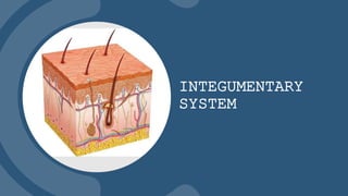
  • 4. THE SKIN 4 • EPIDERMIS • DERMIS (Also known as the cutaneous membrane) Main components of skin:
  • 5. 5 THE EPIDERMIS the outermost and thickest layer, and it's made of multiple layers of developing cells, which are called keratinocytes. Keratinocytes  Produce Keratin  New keratinocytes push up, older, dead cells are shred off, forming skin flakes or dandruff. Melanocytes  a protein pigment or coloring substance, called melanin. Langerhan cells  Protects the skin and underlying tissue from pathogens  Phagocytes of the immune system (epi – on top, dermis – skin)
  • 6. THE EPIDERMIS 6 Layers of the epidermis  Stratum Corneum  Stratum Lucidum  Stratum Granulosum  Stratum Spinosum  Stratum Basale
  • 7. THE EPIDERMIS 7 Layers of the epidermis Stratum LuCIDUM  Deep to the Stratum Corneum  Found in thick skin  Found in thick skin
  • 8. THE EPIDERMIS 8 Layers of the epidermis Stratum Basale  Contains small round cells called basal cells  It also contains melanocytes, which are responsible for producing melanin
  • 9. THE EPIDERMIS 9 Layers of the epidermis Stratum Spinosum  Thickest stratum of the epidermis  Undergo mitosis  Help to synthesize vitamin D
  • 10. THE EPIDERMIS 10 Layers of the epidermis Stratum Granulosum  Excretes a lipid-based substance  Named for prominent granules
  • 11. THE EPIDERMIS 11 Layers of the epidermis Stratum Corneum  Most superficial layer of the epidermis  Several layers of dead, flattened keratinocytes  Sheds dead keratinocytes
  • 12. THE EPIDERMIS 12 Keratinocyte life cycle  Begins in the Stratum Spinosum  Sheds in the Stratum Corneum  Can shed from environmental and physical stress  Shed cells are replaced by mitosis  Cells primarily divide at night
  • 13. THE EPIDERMIS 13 THICK VS. THIN SKIN THICK SKIN  Consists of all five layers of the epidermis with a thick Stratum Corneum  Contains sweat glands  Found in palms of hands and soles of feet
  • 14. THE EPIDERMIS 14 THICK VS. THIN SKIN THIn SKIN  Consists of only four layers of the epidermis, lacking the Stratum Lucidum  Equivalent to the size of a sheet of printing paper
  • 15. 15 THE DERMIS is made up of elastic connective tissue that gives flexibility to the skin.  Houses blood supply  Anchors epidermis in place Two layers:  Papillary Layer  Reticular Layer
  • 16. 16 THE DERMIS  Most superficial layer of the dermis  Consists of loose connective tissue Papillary Layer  Collagen fibers anchor dermis and epidermis together  Dermal Papillae: Papillary layer Reticular layer Dermal papillae Papillary plexus Cutaneous plexus DERMIS  Found on the surface of the papillary layer  Houses tiny blood vessels called capillaries  Houses sensory receptors called Tactile (Meissner) corpuscles
  • 17. 17 THE DERMIS Reticular Layer  Deepest and thickest layer of the dermis  Consists of dense irregular connective tissue  Collagen fibers that strengthen the dermis  Elastic fibers that allow skin to revert after stretching  Proteoglycans that hydrate the skin  Blood vessels and accessory structures (hair, sweat glands, sebaceous glands)  Sensory receptors, such as Pacinian (lamellated) corpuscles that respond to changes in pressure and vibration
  • 18. 18 THE DERMIS SKIN MARKINGS  Interactions between the dermis and epidermis are shown on the skin as small lines called “skin markings”.  Thick collagen fibers arrange the dermal papillae into dermal ridges.  The epidermis will blend in with the dermal papillae that lies underneath, creating epidermal ridges. Epidermal ridges function to increases gripping ability of hands and feet.  Tiny sweat pores open along the ridges to form a thin film called a fingerprint.  Gaps found in between bundles of collagen will indent to form cleavage lines, also known as tension lines.  As the reticular layer tightly fixes to deeper structures of the skin, this creates a deep crease called a flexure line.
  • 19. 19 THE HYPODERMIS is made of fat and connective tissue.  helps insulate deeper tissues  provides padding to the body and helps the skin bind to the rest of the body  Subcutaneous fat allows for thermal insulation and provides an energy reservoir  contains collagen, fibers, adipose tissue (fat cells), connective tissue, larger nerves and blood vessels  Target of subcutaneous injections due to it’s vascularity and therefore absorbs drugs quickly
  • 20. 20 SKIN PIGMENTATION MELANIN Melanin ranges in colors of orange-red to black. Melanin is produced by melanocytes in the stratum basale It is composed of two “tyrosine” amino acids that are joined by the enzyme “tyrosinase”. More melanin is synthesized when exposed to UV radiation (tanning). Melanin functions to:  Protect keratinocytes from mutating due to UV exposure.  Prevent the skin from synthesizing too much Vitamin D in response to radiation Melanin can be unevenly distributed throughout the skin:  Moles are produced when a high amount of melanocytes are proliferated in one spot.  Freckles are are produced when pigment is concentrated in one spot from a high amount of melanin production.  Albinism is when melanocytes fail to produce the tyrosinase enzyme. This lack of skin pigmentation can increase the risk of DNA damage of keratinocytes.
  • 21. 21 SKIN PIGMENTATION Other Pigments That Affect Skin Color There are two other pigments that contribute to skin color, carotene and hemoglobin. Hemoglobin:  Gives skin a pinkish hue  Protein found in red blood cells that binds and transports oxygen  Turns a bright-orange red color when oxidized Carotene:  Gives yellow-orange pigment  Lipid-soluble molecule  Usually ingested in diet from yellow and orange foods  Accumulates in stratum corneum
  • 22. 22 ACCESSORY STRUCTURES  Shaft - Part of the hair that projects from the surface of the skin.  Root - Part of the hair that is embedded in the dermis of the skin.  Hair Papilla - Indentation at the base of the hair bulb that contains blood vessels.  Hair Bulb - Structure at the deep end of the hair follicle.  Hair Matrix - Structure at the base of hair bulb that produces new hairs via mitosis.  Hair Follicle - Infolding surrounding the hair root.  Epithelial Root Sheath - Has two parts: *Outer component: Anchors hair follicle to the dermis. *Inner component: Anchors tightly to the root.  Dermal Root Sheath - Supports the hair follicle and separates it from the dermis.  Arrector Pili Muscles - Tiny bands of smooth muscle that causes hairs to stand up when they contract, this is known as piloerection. Piloerection occurs when we are cold or frightened, giving the skin what we know as “goosebumps”. Structures of the Hair They are many structures that make up the hair, with the root and shaft being the main two:
  • 23. 23 ACCESSORY STRUCTURES Nail plate - Part of the nail that rests on top of the epidermal nail bed. Nail bed - Deep to the nail plate, nourishes and protects the nail. Nail body - Visible part of the nail. Nail root - Lies under the skin. Nail matrix - Part of the nail with living, dividing cells. Supplies oxygen to the nail, sight of nail growth. Proximal nail fold - Covers the edge of the root. Eponychium - Also known as the “cuticle”, found at the base of the nail, protects matrix from infection. Nail folds - Overlapping of skin that borders the nail laterally and medially. Hyponychium - Skin that lies under the free edge of the nail. Lunula - Crescent shaped area where keratin accumulates. NAILS protect our underlying tissue and enable gripping and manipulation
  • 24. 24 ACCESSORY STRUCTURES Sudoriferous (sweat) glands have four types: Eccrine sweat glands: Released through sweat pores. Produces sweat that contains antimicrobial compounds to prevent the growth of pathogens. Also functions in thermoregulation. Apocrine sweat glands: Sweat released into hair pores. Only in certain parts of the body such as armpits, areolas, and the anal area. Sweat metabolized by bacteria which an odor. Ceruminous Glands: Secretes a thick fluid called cerumen (earwax) into hair follicles. Cerumen lines the ears and functions to lubricate the eardrum. Traps particles before they the eardrum. Mammary Glands: Produces a sweat called milk. Milk contains, proteins, lipids, sugars, and immune cells to nourish a newborn. GLANDS The skin has two main types of glands, sudoriferous glands that produce sweat, and sebaceous glands which produce sebum (oil). Both are located in the dermis of the skin.
  • 25. 25 PATHOLOGY OF THE SKIN The most common type of skin pathology is a wound, a wound is any disruption of the skin’s integrity. They may disrupt the epidermis, dermis, or deeper. There are different kinds of wounds: Lacerations (cuts) Burnscancers Skin Wound treatment can vary amongst severity. For instance, a laceration may be treated with sutures (stitches), where as a burn may be treated with surgical repair.
  • 26. 26 PATHOLOGY OF THE SKIN First-degree burns: The most minor, also called “superficial burns” because only the epidermis is damaged. Symptoms are erythema (redness) and minor pain. Usually require no treatment, no blisters or permanent damage. Second-degree burns: Also called “partial thickness burns”. Damage is caused to the epidermis and either some or all of the dermis. Symptoms are pain, blistering, scarring. Usually require medical treatment. Third-degree burns: Most damaging, also known as “full thickness burns”. Damages the epidermis, dermis, and hypodermis. Muscle and bone may also be damaged. Symptoms are severe scarring, lost of hair follicles, and dehydration due to fluid loss. Swelling and infection may also occur. Treatment may result in skin grafting. BURNS A burn is a skin wound caused by heat, extreme cold, chemicals, and/or radiation. There are three classifications of burn wounds, all based on the extent of tissue damage.
  • 27. 27 PATHOLOGY OF THE SKIN Basal Cell Carcinoma: The most common type of skin cancer, a cancer of keratinocytes in the stratum basale. Usually found in skin that is frequently exposed to UV radiation. Forms a nodule with a central crater that develops into an ulcer. Can be completely treated by surgical removal. Squamous Cell Carcinoma: Second most common. A cancer of keratinocytes in the stratum spinosum. Most commonly found on the head and neck. Forms scaly patches that ulcerates or bleed. Can be surgically removed. Malignant Melanoma: Most severe, cancer of melanocytes. More likely to metastasize due to the arms of cancerous cells. Arms of melanocytes allow cancerous cells to extend down to the blood vessels of the dermis and cardiovascular & lymphatic system. Treatment can include surgical removal, chemotherapy, and radiation therapy. Skin Cancer Cancer is a very common skin disease. It is caused due to mutations in the DNA that cause cells to lose control over their cell cycle. This can lead to tumors, which is a cluster of undifferentiated cells. Tumor cells can metastasize, or travel to other parts of the body, alter the structure of a tissue and prevent the tissue from functioning properly.
  • 28. 28 PATHOLOGY OF THE SKIN  Asymmetrical shape: Both sides of the mole are uneven.  Border irregularity: Mole has jagged edges or blurriness.  Color: Mole is blue-black, or different colors.  Diameter: Mole is larger than 6mm or the size of a pencil eraser.  Evolving: Mole is continuously changing in shape and size. Skin Cancer Malignant Melanoma mole can be detected early on and is distinguishable from other skin cancers using the “ABCDE” rule: