^In Pietermaritzburg Hager Werken Embalming +27789155305 Compound Powder in ...
Embryogenesis of the_human_respiratory_system
1. CBU INTERNATIONAL CONFERENCE ON INNOVATIONS IN SCIENCE AND EDUCATION
MARCH 21-23, 2018, PRAGUE, CZECH REPUBLIC WWW.CBUNI.CZ, WWW.JOURNALS.CZ
1
EMBRYOGENESIS OF THE HUMAN RESPIRATORY SYSTEM
Olexandr Tsyhykalo1
, Аlla Khodorovska2
, Iryna Popova3
Abstract: One of the topical issues of morphology is studying general regulations of development and structural formation
dynamics of respiratory system. The aim of the study was to determine peculiarities of the embryogenesis of respiratory system
organs during prenatal development in humans. Research was conducted on 22 series of histological specimens of human
embryos which were 4,5-8,0 mm of parieto-cocygeal lengths (PLC), and by using complex morphological methods of study
(morphometry, histological assessment, three-dimensional reconstruction). It was established that the source of human lungs
primordium is a traheopulmonary primordium, which at the end of 4th
week of human prenatal development is represented by
an odd bud-shaped entity which departs with an acute angle from the ventral wall of the foregut and is located in front of
foregut. The beginning of the 5th
week of human prenatal development is considered to be a critical period, which holds
intensive processes of organogenesis of the respiratory system and is a possible time for the occurrence of some congenital
defects or anomalies and structural variants. Sources of pulmonary vessels are islands of intraorgan hematopoiesis and
extraorgan main vessels, communication between which occurs during the end of 4th
and at the start of 5th
weeks of human
prenatal development.
UDC Classification Number: 611
Key words: embryogenesis, respiratory system, morphogenesis, human prenatal development
Introduction
The direction of morphology research is the study of the patterns of development and dynamics of the
formation and structural organization of the human respiratory system during prenatal development. The
clarification of the regularities of organogenesis of the respiratory system will allow a better
understanding of the etiopathogenesis of birth defects and variants of structural variants, as was said by
Akhtemiychuk et al. (2014). Dem'yanenko (2012) suggests that the data on the peculiarities of
organogenesis of the upper respiratory tract and lungs is depleted, which contributes to development of
new improved methods of prevention, diagnosis and treatment of congenital and acquired pathology in
pulmonology and thoracic surgery. The analysis of scientific sources indicates the fragmentation and
contradiction of data on pulmonary system organogenesis, and the formation of a histological structure
of the respiratory organs, as is seen in publications of Makar et al. (2009), Pavlov and Essev (2014),
Hasyuk et al. (2011), and others. Comprehensive studies using the latest methods of processing
histological data will allow a better approach to solving current medical and social problem - reducing
morbidity and mortality from respiratory pathology.
Objective.
The research was conducted on 22 specimen series of human embryos with 4,5-8,0 mm of parieto-
coccygeal length (PCL) by using complex morphological research methods (histological methodic,
morphometry, graphic and three-dimensional computer reconstruction and statistical analysis) to
determine developmental peculiarities of respiratory organs during the prenatal period of human
ontogenesis.
Materials and methods
Materials were received from the Chernivtsi regional municipal medical institution “Pathologists
office”. Research was also conducted on a series of histological sections from the museum's collection
of the Department of Histology, Cytology and Embryology and the Department of Human Anatomy
named after Turkevich of the Higher State Educational Establishment of Ukraine “Bukovinian State
Medical University”. Studies have been conducted in compliance with the main provisions of The
Declaration of Helsinki (DoH), World Medical Association's (WMA), on the ethical principles of
conducting scientific-medical research with participation of a human (1964-2000), and in compliance
with the Ministry of Health of Ukraine №6 on February 13, 2006. The work is a fragment of planned
scientific research work of the Department of Histology, Cytology and Embryology of Higher Medical
1
Department of Histology, Cytology and Embryology, Bukovinian State Medical University, Chernivtsi,
Ukraine, tsyhykalo.olexandr@bsmu.edu.ua
2
Department of Histology, Cytology and Embryology, Bukovinian State Medical University, Chernivtsi,
Ukraine, khodorovska.alla@bsmu.edu.ua
3
Department of Histology, Cytology and Embryology, Bukovinian State Medical University, Chernivtsi,
Ukraine, popova_i@bsmu.edu.ua
2. CBU INTERNATIONAL CONFERENCE ON INNOVATIONS IN SCIENCE AND EDUCATION
MARCH 21-23, 2018, PRAGUE, CZECH REPUBLIC WWW.CBUNI.CZ, WWW.JOURNALS.CZ
2
Institution of Ukraine "Bukovinian State Medical University" – “Patterns of morphogenesis and
structural and functional features of tissues and organs in human ontogenesis” (state registration number
0116U002938).
Results and discussion
Traheopulmonary primordium was found it embryos of 4,5 mm of PCL (at the end of the 4th
week of
prenatal development) and is represented by bud–shaped odd formation which goes from the ventral
wall of the foregut with an acute angle and is located in front of it, as is shown in Figure 1. The
primordium of the respiratory system has an irregular shape with a narrowed bulb–shaped upper pole
(132 μm embryo of 5,0 mm of PCL and 220 μm - in 6,0 mm PCL embryo) and an expanded lower pole
(380 μm in embryos of 5,0 mm of PCL and 760 μm in objects 6,0 mm of PCL), which is the part form
which lungs are later formed. The longitudinal length of the embryos during this period was 484 and
880 μm, respectively.
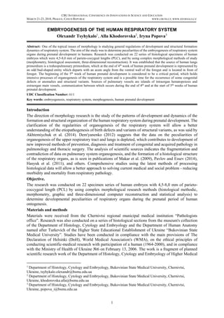
Figure 2: Three-dimensional computer reconstruction of a human embryo of 4,5 mm of PCL. Anterior
frontal projection. A – Lineaments of a heart, blood vessel, oral sinus and anterior gut. B – depicts
only lineaments of the oral sinus and anterior gut. Magnification 12,5х
. In Figure 2: 1 – anterior gut;
2 – lung bud; 3 – primordium of esophagus; 4 – dorsal aorta; 5 – cardinal veins; 6 – primordium of
an eye ball; 7 – head; 8 – lineaments of oral sinus cavity; 9 – primordium of pharynx; 10 – heart.
Source:, Tsyhykalo, Khodorovska, Popova (2017)
Figure 1: Microphotograph of the sagital section of a human embryo of 4,5 mm of PCL stained with
hematoxylin and eosin. Magnification: lens 8х
, eyeglass 7х
. In this Figure: 1 – rudiment of trachea; 2
- rudiment of the left main bronchus; 3 - hepatic diverticulum; 4 - heart; 5 - head; 6 - germ of mandible;
7 - aorta; 8 - the germ of the spinal column; 9 - mesonephros; 10 - primary colon.
Source:, Tsyhykalo, Khodorovska, Popova (2017)
3. CBU INTERNATIONAL CONFERENCE ON INNOVATIONS IN SCIENCE AND EDUCATION
MARCH 21-23, 2018, PRAGUE, CZECH REPUBLIC WWW.CBUNI.CZ, WWW.JOURNALS.CZ
3
The bronchopulmonary primordium at the end of the 4th
week of is surrounded by splanhno-pleurae and
has a simple histological structure: its major mass is the mesenchyma, whose cells are relatively
compact. From the inside of the mesenchyma, an epithelial tube is determined, and it is connected with
the lumen of the foregut. The distal end of this tube is divided into two channels, which end blindly and
have almost the same diameter – the primordiums of the main bronchi, as shown in Figure 2. The length
of the trachea primordium is 264 μm (in embryo of 5,0 mm of PCL) and 396 μm (embryo of 6,0 mm of
PCL); the right bronchus has a length of 180 μm in an embryo of 5,0 mm of PCL and 276 μm in an
embryo of 6,0 mm of PCL, left – 206 μm and 292 μm, respectively.
In embryos of 5,0 mm and 6,0 mm of PCL, diameter of trachea lumen is 88 μm and 92 μm, and the
diameter of the main bronchi is 60 μm and 68 μm, respectively. The walls of the trachea and the main
bronchi have almost the same histological structure, represented by a high multi-row epithelium, the
nuclei of which are oval and 4,0-6,0 mcm in diameter, and in histological specimens of 10 micrometers
they form 3-4, and sometimes even 5 rows. The nuclei are located eccentrically in the cytoplasm - close
to the apical poles of the cell, and the protoplasmic part is concentrated near the basal membrane. The
boundaries between the cells of the epithelium (stained with haemotoxylin and eosin) are not clear. It
should be noted that in different parts of the mesenchymal rudiment of the organ, there occurs an
accumulation of formed blood elements – islets of intraorganic hematopoiesis.
We found the development of lungs originates from two embryonic rudiments - endodermal and
mesenchymal. It should be noticed that from the endodermal rudiment paired epithelial tubes are formed,
and from the mesenchymal – odd tubes. Single mesenchymal tube surrounds the continuous layer of
trachea and main bronchi and passes without a clear border into the mesenchyme of intestinal tube. We
believe that researchers who described the early stages of the development of pair lung rudiments, refer
only to its endodermal part.
In embryos of 7,0-7,5 mm of PCL the lungs primordium forms two lateral protrusions, directed along
dorsolateral rudiment of esophagus. This feature should be considered as the initial stage of forming
lungs as a pair organ. The above mentioned structures are shown in Figure 3, both of them are elongated
and their direction coincides with the longitudinal axis of the embryo body. The top pole of lungs’
primordium is located behind the heart; middle and lower parts, and behind a massive (at this stage)
liver. At that time a prominent asymmetry in sizes of right and left lungs rudiments become noticeable.
The longitudinal size of right lung is 550 μm, transverse – 374 μm; of the left lung – 500 μm and 330
μm, respectively. The length of right main bronchus primordiums is 300 μm, left – 346 μm and the
diameter of lumen in both structures is less than 100 μm. The thickness of the mesenchyma layer, which
surrounds main bronchi, ranges from 110 μm (median semicircle) to 220 μm (lateral semicircle).
Figure 3: Three-dimensional computer model reconstruction of the upper part of the human embryo
5,1 mm of PCL. The right posterior-lateral projection. In Figure 2: 1 - the rudiment of the brain; 2 -
eye; 3 - mouth bay; 4 - bronchopulmonary germ; 5 - notohord; 6 - the heart; 7 - the germ of the right
upper extremity; 8 - the germ of the spinal cord; 9 - dorsal aorta; 10 – precardial vein.
Source:, Tsyhykalo, Khodorovska Popova (2017)
4. CBU INTERNATIONAL CONFERENCE ON INNOVATIONS IN SCIENCE AND EDUCATION
MARCH 21-23, 2018, PRAGUE, CZECH REPUBLIC WWW.CBUNI.CZ, WWW.JOURNALS.CZ
4
Already at that time (7,0-7,5 mm of PCL ) there is a noticeable asymmetry in the size of the rudiments
of the right and left lungs. The longitudinal size of the right lung is 550 μm, the transverse 374 μm; in
the left lung - 500 μm and 330 μm, respectively. The length of the right main bronchus primordium is
300 μm, the left one is 346 μm; the diameter of the lumen of both structures does not exceed 100 μm.
The thickness of mesenchyma layer, which surrounds the main bronchi, ranges from 110 μm (medial
semicircle) to 220 μm (lateral semicircle).
The rudiments of the main bronchi are lined with a high multi-row epithelium, whose thickness reaches
28 mcm. It is located on a well-defined basal membrane. The nuclei of the epitheliocytes are oval, with
a diameter of 4-6 mcm, at histological sections (10μm thick), located in 3 or 4 rows. The nuclei of the
epitheliocytes occupy predominantly an apical position. The boundaries of cells are not clearly
expressed.
In embryos of 8,0 mm of PCL in the lungs’ primordiums lateral protrusions are clearly determined. The
longitudinal size of right lung is 600,0 μm, transverse size – 440 μm; the left lung size - 550 μm and
430,0 μm respectively (as is shown in Figure 4).
Figure 4: The graphical reconstruction of series of histological sections of the human embryo of 8,0
mm of PCL. Right lateral projection. Magnification 30х
. In this Figure: 1 - aorta; 2 - celiac trunk; 3 -
superior mesenteric artery; 4 - liver; 5 - stomach; 6 - duodenum; 7 - right lung; 8 - premordiums of
the vertebrae; 9 - heart; 10 - mandible.
Source:, Tsyhykalo, Khodorovska, Popova (2017)
Inside the mesenchymal rudiment of the lungs is clearly observed the primordium of the trachea and the
main bronchi. The primordium of left main bronchus is a blind tube length of 350 μm and the diameter
is 110 μm. The primordium of right main bronchus is at a distance of 90 μm from the bifurcation of the
trachea and is dichotomously divided into blind branches of almost the same diameter – 110 and 112
μm. The lower branch in directed as a continuation of the main bronchus trunk, and the upper branch is
directed almost horizontally, deviating in lateral direction. Its length is 132 μm. All bronchial branches
in their blind ends form small expansions.
Because the bronchial lumen at this stage of development is relatively large, the primordium of lungs
(bronchopulmonary area lateral protrusion rudiments) on the frontal histological sections is bag-shaped
with a wall thickness of 66-78 μm. The wall of trachea and bronchi through the entire length has the
same structure and does not differ from that in embryo of length 7,0-7,5 mm of PCL.
The fact that in the absence of external signs of lung rudiment division into the lobes, the bronchial tree
is already beginning to branch out, indicating that the endodermal lining of the lungs slightly outstrips
in its mesenchymal development, and therefore plays a leading role in the formation of the lungs. In
addition, it should be noted that starting from the 5th
week of prenatal development, there is already
asymmetry not only in the size of the right and left lungs, but also in the branching of the bronchi.
5. CBU INTERNATIONAL CONFERENCE ON INNOVATIONS IN SCIENCE AND EDUCATION
MARCH 21-23, 2018, PRAGUE, CZECH REPUBLIC WWW.CBUNI.CZ, WWW.JOURNALS.CZ
5
Conclusions
The source of human lung primordium is the traheopulmonary primordium, which at the end of the 4th
week of prenatal development is represented by an odd bud-shaped entity which departs with an acute
angle from the ventral wall of the foregut and is located in front of the foregut. The beginning of the 5th
week of human prenatal development is considered to be a critical period, which holds intensive
processes of organogenesis of the respiratory system and is a possible time for the occurrence of some
congenital defects and structural variants. Sources of pulmonary vessels are islands of intraorgan
hematopoiesis and extraorgan main vessels, communication between which occurs during end of the 4th
and the start of the 5th
weeks of prenatal development. We consider it expedient to find out the
preconditions of congenital anatomy of organs of the respiratory system in human by using the latest
methods of morphological research.
References
Akhtemiychuk Yu.T. Slobodyan O.M., Lavrov L.P. (2014) Prenatalʹnyy rozvytok orhaniv i struktur orhanizmu. [Prenatal
development of organs and structures of the organism.] Experimental and Clinical Medicine, 3(64), 18-21.
Dem'yanenko I.O. (2012) Morfolohichni ta histokhimichni osoblyvosti rannʹoho histohenezu trakheyi i lehenʹ v umovakh
ektopichnoyi implantatsiyi. [Morphological and histochemical features of early histogenesis of trachea and lungs under ectopic
implantation conditions.] Ukrainian morphological almanac, 10(4), 40-42.
Hasyuk Yu.A., Zachepylo S.V., Khaver O.A. (2011) Embrionalʹnyy histohenez epitelialʹnykh tkanyn hortani. [Embryonic
histogenesis of the epithelial tissues of the larynx]. Svit medytsyny ta biolohiyi [World of Medicine and Biology], 3, 148-152.
Holovatsʹkyy A.S. (2001) Embriotopohrafichni osoblyvosti lehenevykh ven, arteriy ta bronkhiv u zarodkovomu periodi
prenatalʹnoho ontohehenezu lyudyny [Embryoto-peculiarities of pulmonary veins, arteries and bronchi in the embryonic period
of human prenatal ontogenesis]. Scientific newsletter of Uzhgorod University, series "Medicine", 13, 27-30.
Makar B.H., Popelyuk O-M. V., Yakovetsʹ K.I. (2009) Suchasni pohlyady na morfohenez i topohrafo-anatomichni
vzayemovidnoshennya hortani v rannʹomu ontohenezi lyudyny. [Modern views on morphogenesis and topographic-anatomical
interactions of the larynx in early ontogeny of man (review of literature).] Bukovinian Medical Bulletin, 13(2), 100–103.
Michai Szpinda, Marcin Daroszewski, Alina Wosniak, Anna Szpinda, et al. (2012) Celestyna Mila-Kierzenkowska. Tracheal
dimensions in human fetuses: an anatomical, digital and statistical study. Surg Radiol Anat., 34, 317–323.
Pavlov A.V., Essev L.I. (2014) Gistofiziologiya epiteliya trakhei u krys v postnatal'nom ontogeneze. [Histophysiology of the
epithelium of the trachea in rats in postnatal ontogenesis.] Morphology, 146 (6), 80-86.
Proskurnya S.A. (2013) Osoblyvosti elastychnoho karkasu krovonosnykh sudyn lehenʹ v normi. [The features of the elastic
framework of the blood vessels of the lungs in norm.] Bulletin of Biology and Medicine. 1(1), 196-198.
Zeng, Xin, et al. (1998) VEGF enhances pulmonary vasculogenesis and disrupts lung morphogenesis in vivo. Developmental
dynamics, 211(3), 215-27.