Volumetric-Based Analysis of In-Vivo and Ex-Vivo Quantita-tive MR Diffusion P...
BPPV inner ear work HP
1. Morphological evaluation of the common crus of
the semicircular canals of patients with BPPV
Paolo Gargiulo (1,2), Agnes Czenek (2), Hannes Petersen (3,4)
1) Heilbrigðis og upplýsingatæknideild, Landspítalinn
2) Heilbrigðisverkfræðisvið, tækni og verkfræðideild, Háskólinn í Reykjavík
3) Háls-, nef- og eyrnadeild Landspitalans, Skurðsvið.
4) Líffærafræði, Læknadeild Háskóla islands.
Introduction
BPPV is the most common cause of vertigo. Although there
are system causes the presentation is unilateral. Therefore it
is interesting to investigate if there are morphological
changes causing this lateralization. For the calcium crystals
of the utriculus to enter the posterior semicircular canal,
they have to traverse the common crus. Differences between
right and left might explain the unilaterally of the disease.
The study aim is to search morphological differences
between healthy and diseased inner ears developing
measurements tools which allow to calculate the total length
and the diameter of the common crus as well as it’s
angulation using CT imaging and image processing
techniques.
Methods
4 voluntary patients suffering from BPPV are enrolled into
the study. Special CT scanning protocol is employed to scan
the region of interest between temporal and zygomatic bone
with image matrix of 768 × 768 pixels, slice thickness of 0.670
mm, tube voltage of 140 KV, and pixel size of 0.247 mm,
resulting in a total of about 250–300 CT slices. The images
are then imported into MIMICS (platform for medical
image processing) for segmentation and computation.
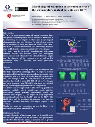
The inner ears are segmented in the following partitions:
cochlea, vestibule, lateral semicircular canal, anterior
semicircular canal, posterior semicircular canal and
common crus (Figure 1). The lengths of every segmented
element is calculated on the healthy and on the BPPV side
(Table 1) as well the lengths on the common crus: Anterior
vestibualr, posterior vestibular and length (Figure 2 and
Table 2).
Finally the inner ear angulations as seen in Figure 3 is
calculated in the same way (Table 3)
Preliminary Results
Accurate 3D model of the human inner ear is possible with
the advanced protocol used in this study. The preliminary
results show that the size (mm3
) of common crus is greater
and coresponds to the side of BPPV in all patients.
.
Figure 1: segmentation of human inner ear
Figure 2: Common crus lenghts
Figure 3: Schematic representation for the
angulations measurements
Patients
Anterior
semicircular
canal
(BPPV)
Anterior
semicircular
canal
(Healthy)
Posterior
semicircular
canal (BPPV)
Posterior
semicircular
canal
(Healthy)
Lateral
semicircular
canal
(BPPV)
Lateral
semicircul
ar canal
(Healthy)
Comon
crus
(BPPV)
Comon
crus
(Healthy)
1 7,71 10,20 9,93 9,98 6,59 9,40 5,41 4,97
2 20,81 12,74 26,50 15,69 23,38 12,37 9,48 6,50
3 12,86 14,68 19,08 15,99 12,75 14,81 4,96 4,95
4 9,96 8,34 15,82 13,10 14,20 8,41 4,88 3,97
Patients
Anterior
vestibular
(BPPV)
Anterior
vestibular
(Healthy)
Posterior
vestibular
(BPPV)
Posterior
vestibular
(Healthy)
Lenght
(BPPV)
Lenght
(Healthy)
1 2,20 1,86 2,27 1,78 2,25 2,28
2 2,04 2,30 2,40 2,34 2,78 2,84
3 2,07 1,84 1,52 1,78 2,15 2,01
4 2,46 2,19 1,97 2,34 2,32 2,53
BPPV Healthy
Patient 1 133,18 129,71
Patient 2 120,79 126,23
Patient 3 115,96 118,32
Patient 4 115,86 116,11
Table 1: lenght measurments of semicircular canals and common
crus for healthy and BPPV inner ears
Table 2: lenght measurments of common crus for healthy and BPPV
inner ears
Table 3: Angulations measurements
of common crus for healthy and
BPPV inner ears