2. Our synthesis on the mastoid (or rather the entire
middle ear cleft) is presented in sections 2-4, where we
discuss three main questions:
(1) Does the developmental morphology of the middle
ear cleft condition the physiology of the mastoid?
(2) Does functional morphology of the middle ear cleft
influence the physiology of the mastoid?
(3) How does the balance of pressure variations in the
pneumatic spaces of the temporal bone constitutes a
basic requirement for healthy functioning?
Most of the information presented here reflects already
established knowledge, which surgeons are already or
should be aware of.
After addressing these topics on the mastoid
morphology and physiology, an overview of the
operation procedures using the mastoid as surgical
approach for the tympanic cavity is given in section 5
with their implication, risks and/or merits, which is of
interest and controversy to practicing ear surgeons
since the 1950’s [1,2]
.
With this review, we hope to fill the missing gaps in the
surgeon’s knowledge of the developmental and
functional aspects of the mastoid, and hope to educate
and help them in making a justified choice of surgical
method.
Middle Ear Cleft Developmental Morphology
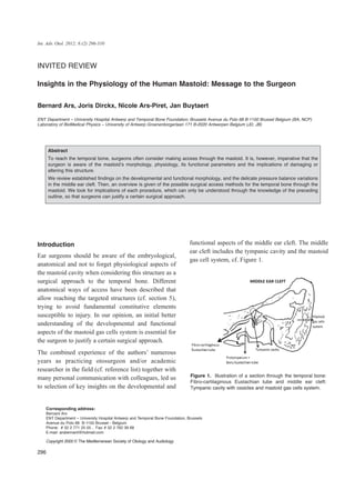
Because the mastoid gas cells system is an integral part
of the middle ear cleft, cf. Figure 1, its development
directly depends on the development of the whole
middle ear cleft [3-6]
.
The tympanic cavity is endodermal. It originates at about
four weeks of fetal life from the first pharyngeal pouch,
which grows laterally and expands rapidly to pre-form
two fundamental structures: the distal part forms the
tubotympanic recess, which will become the primitive
tympanic cavity, and the proximal part constricts to form
the fibrocartilaginous Eustachian tube [7]
.
During the first twelve weeks of fetal life, the
cartilaginous cells, precursors of the ossicles, are
embedded in loose mesenchyme, which limits the
expansion of the future tympanic cavity. This is
invaded by the epithelium of the future
fibrocartilaginous Eustachian tube. It divides into four
sacci [8]
, which expand in four different directions,
lining the tympanic cavity with epithelium and
enveloping the ossicles:
- The saccus anticus extending in a cranio-frontal
direction will form the anterior pouch of von Trôltsch;
- The saccus medius forms the attic, extending upward
and usually breaking into three smaller sacculi;
- The saccus superior extending posteriorly and
laterally between the malleus and the long crus of the
incus, forming the posterior pouch of von Trôltsch;
- The saccus posterior extending along the
hypotympanum, to form the round window niche,
sinus tympani and oval window niche.
The antrum, a lateral extension of the epitympanum,
starts to form at about twenty-two weeks. The mastoid
gas cells develop as an outgrowth of the antrum.
Epithelial buds from the tympanic cavity and antrum
extend to adjacent areas of the temporal bone, after
osteoclastic resorption of bone or differentiation of
bone marrow into loose mesenchyme. Thus, mastoid
buds from the antrum penetrate the temporal bone,
giving rise to the mastoid gas cells. The degree of
pneumatization of the mastoid varies greatly in normal
temporal bones. The age at which the gas cells develop
is subject to huge individual variation.
Expansion of the tympanic cavity is virtually complete
by about thirty-three weeks of fetal life. The
epitympanum follows approximately four weeks later.
Because of the fusion of the periosteal layers of the
otic capsule and the tympanic process of the squamous
bone, the mastoid process begins to develop at about
twenty-nine weeks. Formation of the antrum starts
when the epitympanum becomes angled posteriorly
and is well developed by the thirty-fifth week. As early
as this, cavitation extends into the mastoid.
Pneumatization of the tympanic cavity or tympanum is
followed by that of the associated gas spaces [9]
. At 34
weeks, pneumatization of the mastoid may only just
have started, and this progresses during infancy and
childhood. As the mastoid grows, the antrum shrinks in
size relatively, and assumes a more medial position.
When the antrum is completely formed and in place,
the ventro-lateral wall continues to grow until puberty
or beyond, giving rise to the mastoid bone.
297
Insights in the Physiology of the Human Mastoid: Message to the Surgeon
3. In contrast to the labyrinth and tympanic cavity, the
postnatal growth of the mastoid can be seen in its
length, width and depth [10,11]
. It is governed by two
principal forces: the first is external, caused by the
traction of the muscles of the neck and mainly of the
sterno-cleido-mastoidian muscle. The second is
internal and consists in the progression of the buds of
expansion of the tubo-tympanic epithelium following
the resorption of the embryonic mesenchyme.
The postnatal development of the mastoid is neither
uniform nor symmetrical, but it is obvious. The
evolution of general morphology shows that after an
accelerated growth during the first two years of the
life, the development continues, relatively more
progressive, in the child and the teenager. The internal
configuration is even more prone to variations. Its
development leads to the creation of a more or less
high number of gas cells that vary greatly in size and
which are grouped around a larger cell of constant
anatomical localization, i.e. the antrum.
Number, size and volume of the mastoidian cells are
individual characteristics. The external size of the
mastoid never bodes the width of the contained gas cells
system; so, very small mastoids can perfectly be well-
pneumatized mastoids [8,9]
. In fact, growth seems to be
controlled by many intricate factors such as: heredity,
environment, nutrition, gas exchanges and frequency of
infections [12-14]
. It is obvious that the infections during
childhood, just like the other developmental incidents,
carry a real inhibiting effect on the pneumatization.
However, as soon as the end of the infection, or as soon
as the lifting of the brake, pneumatization may start
again. However, it is really quite difficult to specify if
infections are the only ones able to act as a causal agent,
stimulating the activity of the osteoblasts or, on the
contrary, if this infection does not result from ‘local
morphological predisposing factors’.
Middle Ear Cleft Functional Morphology
The tympanic cavity is constricted in its superior third
by the inter-attico-tympanic diaphragm, which is a bony
membranous barrier perforated by two small permanent
openings: the anterior tympanic isthmus, which is
situated between the tensor tympani tendon and the
stapes; and the posterior tympanic isthmus, which is
between the double posterior ligament of the incus and
the bony posterior tympanic wall.
This anatomic barrier divides the middle ear cleft into
two separate compartments: an anterio-inferior one,
principally devoted to the mucociliary clearance
function, and a postero-superior one, more devoted to
the gas exchange function. The barrier forms a
diaphragm that is composed of two complementary
types of structures: mucosal folds and bones with
muscular and ligament structures: the head and neck of
the malleus, the body and short process of the incus, the
tensor tympani muscle, the anterior and lateral mallear,
and the double posterior incudal ligaments. With this
concept of partition, we are better able to understand the
mechanisms involved in the pathogenesis of otitis
media [6,15]
. Clinical as well as surgical management will
thereby be enhanced.
The antero-inferior compartment of the middle ear cleft,
which is situated under the diaphragm, includes the pro,
meso- and hypo-tympanum and is covered by secretory
or non-secretory ciliated cells that enable mucociliary
clearance. It consists of a less rigid chamber because of
the presence of the eardrum. Because of the
fibrocartilaginous Eustachian tube, it opens into an
intermittently ventilated gas pocket. It communicates
with the postero-superior compartment by both the
anterior and posterior tympanic isthmi. It is often the
site of secondary bacterial infections from the
rhinopharynx. An inflammatory process involving the
mucosa of the antero-inferior middle ear cleft
compartment leads to dysfunction in mucociliary
clearance and to the accumulation of mucus.
The postero-superior compartment of the middle ear
cleft, which is situated above the diaphragm, includes
the epi- and retrotympanum, aditus ad antrum, antrum,
and mastoid gas cells system. It is covered by a richly
vascularized cuboidal epithelium that is devoted
primarily to gas exchange. It consists of a rigid
chamber and an open non-ventilated gas pocket that
communicates with the antero-inferior compartment
via both openings. It may be the site of viral
haematogenous infections. Inflammation of the
mucosa of the postero-superior middle ear cleft
compartment impairs the gas exchange, which in turn
leads to the development of a ‘gas deficit’ in the middle
298
The Journal of International Advanced Otology
4. 299
Insights in the Physiology of the Human Mastoid: Message to the Surgeon
ear cleft, cf. section 3.2.2. The inter-attico-tympanic
diaphragm conditions the topography of the tympanic
membrane retraction pockets. When only one opening
is blocked, the postero-superior quadrant of the pars
tensa is attracted in the direction of the retrotympanum.
When both openings are completely blocked, the pars
flaccida is drawn in toward the epitympanum. In the
same way, the diaphragm also influences the invasion of
the middle ear cleft by a cholesteatoma [16-21]
.
Pressure Balance of the Temporal Bone
Pneumatic Spaces
On the quantitative point of view, gas spaces of the
middle ear cleft embody the most constitutive part of
the pneumatic spaces of the temporal bone. The
middle ear cleft consists of both the mastoid gas cells
system and the tympanic cavity. The latter gathers the
tympanum and four annexes: the epi- and hypo-
tympanum, retro- and pro-tympanum that corresponds
to the bony Eustachian tube. In fact, the middle ear
cleft consists of a set of interconnected gas cells lined
with the same respiratory mucosa. Because the gas
exchange is actually performed through the mucosa of
the cells, the total surface area of mucosa will
influence the rate of gas exchange [22,23]
.
What is then the role of the mastoid? The physiology
of the mastoid can only be understood in the process of
balance of pressure variations in the middle ear cleft.
The middle ear cleft is essentially a non-collapsible
poorly ventilated gas pocket through which sound
wave energy is transported to reach the inner ear.
Every amount of gas must be contained in a three-
dimensional container or volume V. The pressure of a
gas P is created by the molecules of gas striking the
walls of the container. It is a force per unit of area. The
physical characteristics of a gas are linked together by
the ideal gas law: P.V = n.R.T, in which n is the mass
expressed in molecule gram; R: the universal gas
constant; T: the absolute temperature. The middle ear
cleft is submitted to physiological variations in volume
and pressure. It has the capacity to maintain a steady
state in volume and pressure, which is achieved by
different regulatory mechanisms, which neutralize or
minimize the pressure and volume variations. This is
performed by adjusting, either, the quantity of gas, its
diffusion, and/or the volume of the middle ear cleft[24]
.
Gas exchanges
The gas content of the middle ear cleft is constituted
and maintained thanks to gas exchanges between the
middle ear cleft and the neighboring structures,
including the environment across the tympanic
membrane, the inner ear via the round window
membrane, the blood compartment via the mucosa and
the rhino-pharynx through the fibrocartilaginous
Eustachian tube [25-32]
.
The gases that are present in the middle ear cleft are
identical to those found in the blood and in the
atmosphere: Oxygen (O2), Carbon dioxide (CO2),
Nitrogen (N2), Argon (Ar) and Water vapor (H2O).
Their relative amounts may be expressed by their
partial pressure P(x). Nitrogen is neither produced nor
consumed at the level of the middle ear cleft. The
P(O2) in the middle ear cleft is slightly lower and the
P(CO2) is slightly higher than in venous blood. This
means that there exist a modest consumption of O2 and
production of CO2 in the middle ear cleft. Oxygen and
nitrogen are absorbed from the middle ear cleft via the
mucosa into the blood compartment. Carbon dioxide
and water vapor are diffused from the blood
compartment, via the mucosa, into the middle ear cleft.
The steady exchange of gas through the mucosa
depends on the functional properties of the cells of the
mucosa, the specific diffusion rate of the gas, which is
a constant value, characteristic for a particular gas, and
the behavior of the vascular system. The primary
purpose of the mucosa is to facilitate gas exchange
between the middle ear cleft and the blood
compartment through a constitutive tissular barrier.
To vibrate in an optimal manner, the tympano-
ossicular system must remain in balance. This means
that the intra-middle ear cleft pressure must be
equivalent to the atmospheric pressure (760 mmHg).
In ambient air, this equivalent value is obtained by
calculating the sum of the partial pressures of the four
main constitutive gases in the air: oxygen (158
mmHg), carbon dioxide (0.3 mmHg), nitrogen (596
mmHg), and water vapor (5.7 mmHg) [33]
.
However, in the middle ear cleft, the composition of
gas varies for two main reasons. The middle ear cleft
is a closed cavity that is connected with the nasal
5. 300
The Journal of International Advanced Otology
cavities by the fibrocartilaginous Eustachian tube. The
gas in the rhinopharynx, which enters the middle ear
cleft via the tube, consists of exhaled gas that contains
less oxygen and more carbon dioxide than ambient air.
This gas composition also varies because gas diffusion
occurs between the middle ear cleft and the arterial and
venous blood, via the mucosa. In blood, the partial
pressure of these gases differs. There is more oxygen
in the arterial blood (93 mmHg) and more carbon
dioxide in the venous blood (44 mmHg) because the
gas exchange occurs at the level of the pulmonary air
cells and the tissues throughout the body, respectively.
Thus, the composition of gas in the middle ear cleft
differs from that in ambient air. The gradient ‘out’
from middle ear cleft to the capillaries is 57 mmHg for
oxygen, and the gradient ‘in’ from the capillaries to the
middle ear cleft is 39 mmHg for carbon dioxide. This
should result in a negative pressure in the middle ear
cleft. However, in spite of the difference in gas
composition, the pressure in the middle ear cleft
approximates the atmospheric pressure to enable
optimal sound transmission.
The reason for this balance is threefold:
- Because the sum of the partial pressures of oxygen
and carbon dioxide is lower in the middle ear cleft (90
mmHg) than in ambient air (150 mmHg), nitrogen
exerts the higher partial pressure in the middle ear cleft
(623 mm ) because of its very slow diffusion towards
the capillaries.
- The composition of the exhaled gas that enters the
middle ear cleft via the tube contains less oxygen and
more carbon dioxide than ambient air does. This
reduces the passive diffusion of these gases through
the mucosa.
- The blood flow through the middle ear cleft mucosa
is probably low. This limits the importance of gas
diffusion and enables the oxygen and carbon dioxide
partial pressures to nearly equalize, because each has a
high diffusion rate. The same mechanism applies to
water vapor of which the diffusion rate is also very
high. Transmucosal gas exchange results in gas
absorption, cf. Figure 2.
Figure 2. Partial pressures (mm Hg) of the physiological gases for the middle ear cleft and their neighboring structures.
Values are taken Hergils et al [27] and from Felding et al [69].
6. 301
Insights in the Physiology of the Human Mastoid: Message to the Surgeon
Regulating systems
The steady state in volume and pressure into the
middle ear cleft is preserved by the way of two types
of regulating mechanisms: on the one hand, the in situ
adaptation systems and, on the other hand, both central
and peripheral nervous regulating system with retro-
control between the middle ear cleft and the muscles of
the fibrocartilaginous Eustachian tube.
The in-situ adaptation systems
The in situ adaptation systems consist mainly in the
compliance of the tympanic membrane lamina propria,
the fibrocartilaginous Eustachian tube function and the
behavior of the vascular system of the middle ear cleft
mucosa, especially that of the mastoid.
The compliance of the tympanic membrane lamina
propria
The compliance of the tympanic membrane lamina
propria is used in the case of sudden pressure changes
(altitude, flight, diving, explosion, etc.) This occurs
largely due to the viscoelastic properties of the lamina
propria and to the flexibility of the incudo-mallear
joint, which acts as a static pressure receptor for the
tympanic membrane, ensuring three-dimensional
movement into the malleus. Deformation of the
eardrum causes a volume change of the semi-rigid
middle ear cleft and can therefore compensate partially
for fast pressure changes. The eardrum is an active
pressure buffer, but also a passive pressure victim [34-38]
.
The fibrocartilaginous Eustachian tube function
The fibrocartilaginous Eustachian tube is closed most
of the time, and it therefore certainly does not function
as a ventilation hole, which keeps pressure inside the
middle ear cleft equal to the outside. Only at large
pressure differences, over 2kPa, the fibrocartilaginous
Eustachian tube will open spontaneously.
Changes in position seem to affect the function of the
fibrocartilaginous Eustachian tube. The mean volume
of gas passing through the tube in upright position is
reduced by one third when the body is elevated at 20
degrees to horizontal position. It is reduced by two
thirds in horizontal position. This is the result of the
increase in the venous tissular pressure around the
fibrocartilaginous Eustachian tube [39]
.
The vascular system in combination with the mastoid
gas cell system
The behavior of the vascular system plays an
important regulatory role in the physiological balance
of pressure variations in the middle ear cleft.
Variations in the middle ear cleft blood flow,
associated with variations in the permeability of the
vessels, allow ample adaptation to normal gas pressure
fluctuations [40,41]
. The normal extensive variations
observed in middle ear cleft pressure over a 24-hours
period, which appear regularly, are related to the
vascular adaptations required by body position and
sleep [42]
. The mastoid gas cell system constitutes the
most important volume of the middle ear cleft and
therefore represents the major part of the middle ear
cleft mucosal area available for vascular gas
exchange[43]
.
The amount of gas present in the mastoid is important
in regulating middle ear cleft pressure for two reasons.
First, the physical, anatomical, properties of mastoid
volume affect compliance: the greater the volume, the
more compliant the system. Second reason: the surface
area affects mucosal gas exchanges. Both are
important in the regulation of middle ear cleft
pressure. The gas contained in the mastoid gas cells
system acts as a physiological passive pressure buffer.
The size of the mastoid gas cells system varies
between individuals to a great extent. However,
whatever the cause, be it genetic determination of
growth or environmental factors, there is unanimous
agreement that the small mastoid gas cell system has a
pathological association. The smaller the system, the
faster are the deviation from normal pressures.
However, nature is a matter of balance! It is not a
question of determining the volume of the mastoid in
order to predict its clinical potential. In normal
conditions and provided with healthy mucosa, a small
mastoid, in an adult, is normal in so far as it constitutes
the complete outcome of normal and standard
development, i.e. the individual simply possesses the
morphological particularity of being ‘small’. A small
7. mastoid in an adult is normal, in that it is sufficient for
the physiological balance of pressure variations in
middle ear cleft. This small mastoid is no more likely
to develop pathology than another is. However, when
the mucosa undergoes inflammatory process, a small
mastoid in an adult, e.g. as outcome of disturbed or
failed development, is certainly at a disadvantage
when it is exposed to excessive variations in pressure.
This mastoid is ‘too small’ compared to the rest of the
middle ear cleft. Fluctuations in middle ear cleft
pressure in the presence of a small mastoid gas cells
system will result in greater forces applied to the
tympanic membrane, when compared with the same
pressure changes in a larger mastoid gas cells system.
If the expansion reservoir is too small, it cannot play
its role adequately.
In normal conditions and healthy mucosa, a slight
negative pressure is present in the middle ear cleft
compared to the outside air, created by exchanges of
gas between the cleft and the blood compartment. Gas
exchanges through the mucosa of the middle ear cleft
are induced by the gradient of partial pressure of gases
between the cleft and the capillaries of the sub-
mucosal connective tissue. Given the capacity of gas
diffusion in the middle ear cleft, partial pressures of
those gases aim at a balance on both sides of the
mucosa barrier of the middle ear cleft. The progressive
diffusion of the gases is capable of modifying the gas
composition in the middle ear cleft as well as in the
circulating blood. The gas, which penetrates into the
middle ear cleft from the rhinopharynx, is not outside
air. Its composition is rather close to exhaled air.
Quantitatively, the main gas that enters the middle ear
cleft is of course nitrogen. Nitrogen diffuses much
more slowly in the blood than the other gases (35 times
more slowly than CO2 and 1.8 times less than the O2)
[44]
, cf. Figure 3.
Considering this nitrogen in steady state, there is a
difference in partial pressure between the middle ear
cleft and the blood flow around the middle ear cleft.
This steady state corresponds more or less to the total
amount of pressure difference (54 to 56 mmHg)
between middle ear cleft and blood. In steady state,
oxygen, carbon dioxide and water vapor have nearly
the same partial pressures in the middle ear cleft as
well as in the blood compartment. Consequently, an
302
The Journal of International Advanced Otology
Figure 3. Gas exchanges between the middle ear cleft and the blood compartment through healthy (a) and inflamed mucosa (b).
8. increase in blood flow into the middle ear cleft will be
of little effect on exchanges of these gases, except
perhaps during the night, when carbon dioxide
increases and oxygen becomes reduced in blood flow.
On the other hand, nitrogen maintains always a higher
partial pressure into the middle ear cleft as compared
with the blood. Therefore, there is a clear and
continuous elimination of nitrogen from the middle ear
cleft in direction of the blood compartment. This leads
to the negative pressure bias in the middle ear cleft. In
normal conditions, this elimination is regularly
compensated by the gas contribution originating from
the rhinopharynx. A variation in blood flow could
consequently explain variations in nitrogen absorption.
Inflammatory processes increase both the number and
diameter of the blood vessels, thus increasing
vascularization of the mucosa. In the middle ear cleft,
the ratio ventilation / perfusion is respectively
4.75x10⁻³ and 3.98x10⁻³ in normal and inflammatory
conditions. This means that the middle ear cleft is 210
times more perfused than ventilated under normal
conditions; and 251 times more perfused than
ventilated under inflammatory conditions. The result
of this increased blood perfusion is a higher diffusion
of nitrogen from the middle ear cleft in direction of the
blood. This explains the apparent contradiction
between the increase in thickness and barrier of the
mucosa and the increase of the gas elimination in
inflammatory conditions [45]
. The normally slightly
balanced middle ear cleft negative pressure thus
increases. The higher nitrogen absorption would
normally be sufficiently compensated through the
fibrocartilaginous Eustachian tube, but could now
generate pathological negative pressures into the
middle ear cleft compared with the atmospheric
pressure. This condition carries a “gas deficit” which
could constitute a mutual physio-pathologic support to
the development of chronic otitis media.
The central and peripheral nervous regulating system
In the middle ear cleft, the steady state in volume and
pressure close to the atmospheric pressure is
maintained not only by the way of the in-situ
adaptation systems, but also by the way of a peripheral
and central nervous regulating system with retro-
control between the middle ear cleft and the muscles of
the fibrocartilaginous Eustachian tube.
The mucosa of the middle ear cleft manages the quality
of the gas contained into the middle ear cleft,
especially its composition, mainly by means of chemo-
sensitive sensors that act on the neuro-capillary
system.
The fibrocartilaginous Eustachian tube manages the
quantity (volume) of the gas contained into the middle
ear cleft by means of baro-sensitive sensors that effect
on the neuro-muscular system.
Sensors located in the middle ear cleft
Chemo-sensors
Within the middle ear cleft, the innervation of the
mucosa originates from the autonomic nervous system,
which gives and receives information’s at the same
time from sympathic and parasympathic networks [46,47]
.
Adrenergic nerve endings have been displayed; most
of them being in the vicinity of vessels. They probably
control their tonus. The peptidergic system non-
adrenergic and non-cholinergic would also play a role.
The presence of peptidic mediators such as P
substance, vasoactive intestinal peptide (VIP) or
calcitonin-gene related peptide (CGRP) has been
demonstrated. It has been established that these
mediators give rise to vasodilatation as well as an
increase in the vascular permeability.
At the level of the middle ear cleft, chemo-sensors
situated within the glomic tissue of the mucosa give
answers to variations in the gas composition and give
impulses to reflexes of gas transfer from the rhino-
pharynx in the direction of the middle ear cleft.
Baro-sensors
The presence of baro-sensors has been reported at the
level of the middle ear cleft as well as at the level of
the tympanic membrane.
At the level of the tympanic membrane, stretching
sensitive sensors have been described. They are
corpuscles situated into the lamina propria. Their
303
Insights in the Physiology of the Human Mastoid: Message to the Surgeon
9. sensitivity should fit the tympanic membrane
movements. Moreover, they might play a role in the
detection of pressure variations in the middle ear
cleft[48-52]
.
The tensor tympani muscle does not appear to be
involved in active dilatation of the fibrocartilaginous
Eustachian tube. However, there may be stretch
sensors in the tympanic membrane that could be
related to modulation of the middle ear cleft pressure
through the tensor tympani muscle, thereby affecting
the tensor veli palatine muscle opening of the
fibrocartilaginous Eustachian tube [53]
.
Sensors located at the pharyngeal orifice of the
fibrocartilaginous Eustachian tube
Opening of the fibrocartilaginous Eustachian tube
occurs when pressure imbalance takes place on both
sides of the tympanic membrane [54]
.
On the one hand, baro-sensors are situated in the
vicinity of the pharyngeal orifice of the
fibrocartilaginous Eustachian tube [55]
, and on the other
hand, within the middle ear cleft and most likely in the
tympanic membrane. In this way, they suggest a neural
connection between the sensory sensors in the
tympanic membrane and the tubal muscles.
The opening of the fibrocartilaginous Eustachian tube
would not be limited to a movement associated with or
consecutive to yawning or swallowing, but could be
controlled by a system whose retro-control could be
initiated in the tympanic membrane.
Neural connection between sensors
The presence of a feedback system acting from the
middle ear cleft to the tubal muscles as well as afferent
nerves from the sensors located in the middle ear cleft
to the salivary nucleus of the brain stem were
described. This nucleus receives also the efferent
fibers of both the sensitive and sensorial sensibilities
from the V and IX. This nucleus also gives efferent
fibers towards the swallowing muscles as well as
fibers, which regulate the vasomotor tonus of the
arteriole and pre-capillary sphincters from the active
tissular areas, mainly the mastoidian ones. Therefore,
this is consistent with the description of a complex
system with a regulation loop with central nervous
control [56-59]
.
Discussion
We summarize the above in section 5.1. In the
following section 5.2, we consider most
mastoidectomy procedures. This means surgery to
remove hollow, gas-filled mastoid cells, either because
of infection (removal of pus, necrotic bone,
cholesteatoma, granulations therein) or to reach the
tympanic cavity or inner ear (for corrective surgery,
prosthesis or implants).
The implications of each operation procedure
mentioned can only be fully understood by the
knowledge of the previously accounted sections.
How the system operates
The gas exchange that governs the balance of middle
ear cleft pressure variations is performed mainly into
the mucosa of the middle ear cleft and through the
fibrocartilaginous Eustachian tube.
In the middle ear cleft, the steady state in volume and
pressure is maintained near the atmospheric pressure,
on the one hand, by the working of local mechanisms
of adaptability and on the other hand, by both central
and peripheral nervous systems of regulation with
retro-control between the middle ear cleft and the
muscles of the fibrocartilaginous Eustachian tube.
The middle ear cleft is a poorly ventilated, but a highly
perfused organ. Gas exchanges depend on variations in
middle ear cleft blood flow. In a similar fashion to
alveoli in the lung, the direction of gas exchange is
predicted on the differences in partial pressure of the
component gases in the middle ear cleft and in the
blood compartment.
When the mucosa is healthy, there is a balance of gas
exchanges because oxygen and nitrogen are absorbed
by the mucosa on the same rate at which carbon
dioxide is expelled. For a given mucosal blood flow,
nitrogen is the exchange rate limiting factor of the
middle ear gas loss, because it diffuses slower than
carbon dioxide and oxygen which equilibrate
relatively quickly with venous blood.
304
The Journal of International Advanced Otology
10. Inflammatory processes increase blood flow. The more
inflamed, and thus vascularized, the mucosa, the
greater is the rate of gas absorption, cf. Figure 3.
In case of inflammatory process in the mastoid gas cell
system, what is the most accurate surgical procedure to
restore the physiology? Because of the unhealthy status
of the mastoid, opening is essential to permit drainage
of the inflammatory secretion, removal of
inflammatory granulated mucosa and creation of an
enlarged gas reservoir. It is important for a surgeon to
be aware of the physiological function of the mastoid
gas cells, and the consequences of an operation upon it.
How to operate the system
When surgical approaches to the middle ear cleft are
compared and considered, the decision of course
depends on the specific pathological condition (the
site, the extent of disease, the presence or absence of
mucosal inflammation and Eustachian-tube
dysfunction) and the availability of surgical and
healthcare accommodations. The operation must thus
be tailored to the patient and situation [60]
. An overview
of the different surgical options with some insights in
their merits, risks or consequences will now be listed.
Two main mastoidectomy categories exist – canal wall
up and canal wall down – with many subcategories.
Mastoidectomy and access to the tympanic cavity can
be initiated either by retroauricular (surgical incision
behind the ear) or endaural approach (through the
external ear canal), and each approach can continue
through the bone by transmeatal or transcortical route[61]
.
Canal wall up
This procedure, where the postero-superior bony
partition between the external ear canal and mastoid is
kept intact, is commonly referred to as canal wall up
(CWU). Subclassifications for this procedure are
simple mastoidectomy, complete mastoidectomy,
closed (cavity) mastoidectomy, cortical
mastoidectomy, combined approach tympanoplasty
and classic intact canal wall mastoidectomy. The two
main subcategories are discussed.
Simple mastoidectomy
Simple mastoidectomy involves the removal of the gas
cells of the mastoid without disturbing the contents of
the tympanic cavity. The operation is performed
retroauricular or endaural, and the surgeon opens the
mastoid bone and removes the inflamed gas cells,
without opening of the antrum or the attic
(epitympanum).
Complete mastoidectomy
Some authors describe complete mastoidectomy as
simple mastoidectomy with a facial recess approach.
Removal of all of the mastoid gas cells along the
tegmen, sigmoid sinus, presigmoid dural plate and
posterior wall of the external auditory canal is then
necessary; however, the external auditory canal wall
still remains up. In this case, opening of the aditus ad
antrum allows access to the epitympanum, and the
incus and malleus may be removed for greater access.
Implications
These CWU methods have less post-operative care.
Furthermore, after a canal wall up procedure, the
mastoid can retain some of its native cuboidal
nitrogen-absorbing epithelium.
Recurrence or persistence of the pathology is known to
be common [61]
. In case of an inflammatory process,
again both the number and the diameter of the blood
vessels increases, thereby increasing the diffusion of
gas. Middle ear cleft negative pressure increases and is
no longer fully compensated, and can thus become
pathological again [15,62]
.
Canal wall down
When the postero-superior bony partition between the
external auditory canal and mastoid is not preserved,
one speaks of canal wall down (CWD). Synonyms or
subclassifications for this procedure are open (cavity)
mastoidectomy, atticotomy, classical radical operation,
Bondy’s operation, atticoantrotomy and retrograde
mastoidectomy. We distinguish two categories:
Radical mastoidectomy
The procedure that converts the entire mastoid cavity,
the external ear canal and the tympanic cavity with the
epitympanum into one open(-air) cavity, is called
radical mastoidectomy. The malleus, incus and chorda
tympani are removed for better access. The innermost
305
Insights in the Physiology of the Human Mastoid: Message to the Surgeon
11. ossicle (the stapes) is left behind to later offset the
hearing loss. The method may sometimes be called
open-mastoido-epitympanectomy (OME) [63]
.
Tympanic membrane and ossicles are not
reconstructed, thus exteriorizing the middle ear cleft.
The pro-tympanum can be obliterated with soft tissue
to reduce the risk of a chronic otorrhea.
Modified radical mastoidectomy
This approach can be described as a radical
mastoidectomy maintaining the tympanic cavity.
Either some ossicles are left in place; or ossicular
reconstruction is used to repair the bones
(ossiculoplasty). Furthermore, the original eardrum is
preserved or rebuilt through tympanic membrane
grafting (tympanoplasty), with possible relocation
deeper into the middle ear, to separate the mucosal
lined middle ear space from the mastoid cavity and ear
canal. The method may sometimes be called open-
mastoido-epitympanectomy with tympanoplasty
(OMET) [63]
.
Implications
The canal wall down technique enables the easy and
full removal of the nitrogen-absorbing mucosa of the
mastoid, and thus offers the best chance of full
elimination of residual disease. The new epithelial
lining of the mastoid bowl is a stratified keratinizing
epithelium. Gas diffusion is no long occurring.
With radical mastoidectomy, the eardrum and middle
ear structures may be completely removed, thus
causing loss of hearing, and patients require life-long
otologic care of their open middle ear cleft. Recurrent
infection of this middle ear is possible.
Nowadays, modified radical mastoidectomy is
therefore preferred by most surgeons over radical
mastoidectomy [63]
, as it creates a more closed, dry and
self-cleansing middle ear.
Canal wall reconstruction
First a CWD is performed, giving increased
intraoperative exposure allowing full removal of the
nitrogen absorbing mastoid epithelium, before the
actual reconstruction of the postero-superior canal
wall. The method is thus sometimes referred to as
open-closed mastoidectomy.
Canal wall reconstruction is often combined with total
or partial obliteration of the mastoid, using either local
flaps (muscle, periosteum, fascia) or free grafts (bone
chips, cartilage, fat, hydroxyapatite, bone pâté) [1,64]
.
Implications
The mastoid obliteration techniques reduce the
transmucosal gas absorption in the middle ear cleft. In
addition, the tympanum is isolated from the attic and
mastoid. This design prevents recurrent postoperative
retraction pockets if the fibro-cartilaginous Eustachian
tube does not function properly [65]
. Thus, the
recurrence rates of pathologies that lead to a ‘gas
deficit’ in the middle ear cleft are reduced.
The main setback of this surgical technique is the
suppression of the buffer effect of this great gaseous
area.
Even if regular controls are performed by accurate
computed tomography imaging (M.R.I.) of the
obliterated mastoid cavity, long-term complications of
such a technique, including intracranial cholesteatoma
and/or abscess, may occur.
Thoughts on antro-attico-mastoidectomy
The surface area of the mastoid cavity is greatly
decreased after most mastoidectomy procedures as the
fine maze of gas cells is reduced or removed. Because
the passive pressure buffer function is partially
governed by the mucosal surface area, it may not be
desirable to remove all gas cells when the mastoid
contains healthy cells lined with thin normal mucosa.
Taking this and our previous overview into account,
these authors prefer CWU, where possible, but
consider simple mastoidectomy to be insufficient.
Optimally, an antro-attico-mastoidectomy, taking care
to preserve the scutum (sharp bony spur formed by the
lateral wall of the tympanic cavity and the superior
wall of the external auditory canal) and the posterior
bony auditory canal wall must be performed with the
aim of restoring the function of gas exchange to the
postero-superior part of the middle ear cleft. Antro-
attico-mastoidectomy is a CWU procedure by
retroauricular approach, which is then superiorly and
anteriorly enlarged, obtaining access to the attic and
306
The Journal of International Advanced Otology
12. antrum. In this situation, the opening of the antero-
superior compartment allows specific cleaning with
restoration of the gas exchanges. If necessary, this
could be combined with Jansen’s posterior (attico)
tympanotomy [66]
for more inferior and posterior
access.
So, in our opinion, the posterior wall of the external
auditory bony canal must be preserved or at least
reconstructed, overcoming the inconveniences derived
from the open mastoid cavity, making superior hearing
outcomes more likely [67]
, and as to constitute an
impenetrable barrier giving the mastoid gas cell system
the means of performing its function[68]
.
Conclusion: Message to the Surgeon
The mastoid gas cells system has a physiological
passive pressure buffer role in the balance of pressure
variations in the middle ear cleft, for two main reasons:
(1) The physical anatomical properties of the middle
ear cleft volume affect compliance: the greater the
volume, the more compliant the system;
(2) The surface area of the mastoid gas cells system
greatly affects the mucosal gas exchange.
It is our understanding that most otosurgeons too often
only think about the mechanical approach of middle
ear cleft surgery, while they should be aware of all the
functional parameters of the mastoid and middle ear
cleft, so they can justify a certain surgical approach
and its consequences. The operated middle ear cleft is
often conceived as permeated by gaseous flows.
However, one should always consider the physiologic
aspect of an operation involving the mastoid. The
operated middle ear cleft should, if circumstances
allow, result in a cavity still covered by a mucosa
through which gas diffusion occurs so that each gas –
nitrogen N2, oxygen O2, carbon dioxide CO2 and water
vapor H2O – has its own specific diffusion rate
influencing the healthy balance of pressure variations
in the middle ear cleft.
Acknowledgments
Financial support was given by the Research
Foundation – Flanders (FWO-Vlaanderen).
References
1. Estrem SA, Highfill G (1999) Hydroxyapatite canal
wall reconstruction/mastoid obliteration. Otolaryngol
Head Neck Surg 120(3):345-349
2. Haberman RS (2004) Middle ear and mastoid
surgery. Thieme
3. Bast Th, Anson S (1947) The Temporal Bone and
the Ear. Springfield, IL: CC Thomas Publications
4. Merchant SN, Nadol JB Jr. (2010) Schucknect's
Pathology of the Ear. PMPH-USA
5. Proctor B (1964) The development of the midd1e
ear spaces and their surgical significance. J Laryngo1
Otol 78(7):630-649
6. Ars B (2008) Chronic Otitis Media. Pathogenesis-
Oriented Therapeutic Management. Kugler
Publications, Amsterdam, The Netherlands
7. Ars B, Ars-Piret N (2003) Developmental
morphology of the middle ear cleft. In: Ars B (ed)
Fibrocartilaginous Eustachian tube – Middle ear cleft.
Kugler Publications, Amsterdam, The Netherlands, pp
33-42
8. Proctor B (1989) Surgical anatomy of the Ear and
Temporal Bone. Thieme, New York
9. Allam AF (1969) Pneumatization of the temporal
bone. Ann Otol Rhinol Laryngol 78(1):49-64
10. Eby Th, Nadol J (1986) Postnatal growth of the
human temporal bone: implications for cochlear
implants in children. Ann Otol Rhinol Laryngol
95:356-364
11. Ars B. (2008) Pathogenesis of Chronic Otitis
Media – Local morphological predisposing factors. In:
Chronic Otitis Media. Pathogenesis-Oriented
Therapeutic Management, ed. B. Ars, Kugler
Publications, Amsterdam, The Netherlands, pp 13-25.
12. Diamant M. (1940) Otitis and pneumatisation of
the mastoid bone. Acta Oto-Laryngologica XLIS: 1-
144.
13. Friedman I. (1956) Mastoid pneumatisation. J
Laryngol 69: 27 and 588.
14. Tumarkin A. (1957) Importance of mastoid air cell
system. J Laryngol 65: 257-259.
15. Ars B (2007) Morphofunctional partition of the
middle ear cleft. J. Advanced Otology. (3):31-39.
307
Insights in the Physiology of the Human Mastoid: Message to the Surgeon
13. 16. Ars B, Ars-Piret N. (1997) Morphofunctional
partition of the middle ear cleft. Acta Oto-Rhino-
Laryngol Belg 51: 181-184.
17. Ars B, Ars-Piret N. (2008) Morphofunctional
partition of the middle ear cleft. In: Chronic Otitis
Media. Pathogenesis-Oriented Therapeutic
Management, ed. B. Ars, Kugler Publications,
Amsterdam, The Netherlands, pp 3-11.
18. Ars B, Wuyts F, Van de Heyning P. (1997)
Histomorphometric study of the normal middle ear
mucosa. Preliminary results supporting the gas
exchange function in the postero-superior part of the
middle ear cleft. Acta Otolaryngol (Stockh) 117: 704-
707.
19. Matanda R, Van de Heyning P, Bogers J, Ars B.
(2006) Behaviour of middle ear cleft mucosa during
inflammation: Histomorphometric study. Acta
OtoLaryngol 126: 905-909.
20. Lim D. (1979) Normal and pathological mucosa of
the middle ear and eustachian tube. Clin. Otolaryngol
4: 213-234.
21. Sadé J. (1966) Middle ear mucosa. Arch
Otolaryngol 84: 137-164.
22. Ars B. (2003) Balance of Pressure Variation in the
Middle Ear Cleft. In: Ars B (ed.) Fibrocartilaginous
eustachian tube – Middle ear cleft. Kugler
Publications, Amsterdam, The Netherlands, pp. 57-66.
23. Ars B, Ars-Piret N. (1994) Middle ear pressure
balance under normal conditions. Specific role of the
middle ear structures. Acta Otorhinolaryngol Belg 48:
339-342.
24. Piiper J. (1965) Physiological equilibria of gas
cavities in the body. In: Fenn W, Rahn H (eds.)
Handbook of physiology, Section 3: Respiration.
Washington, DC: American Physiological Society pp.
1205-1218.
25. Elner A. (1976) Normal gas exchange in the
human middle ear. Ann Otol Rhinol Laryngol 85: 161-
164.
26. Hergils L, Magnuson B. (1997) Middle ear gas
composition in pathologic conditions: Mass
spectrometry in otitis media with effusion and
atelectasis Ann Otol Rhinol Laryngol 106: 743-745.
27. Hergils L, Magnuson B. (1998) Nasal gas
composition in humans and its implications on middle
ear pressure. Acta Otolaryngol (Stockh) 118: 697-700.
28. Ingestedt S, Ivarsson A, Jonson B. (1967)
Mechanics of the human middle ear. Pressure
regulation in aviation and diving. A non-traumatic
method. Acta Otolaryngol 228S: 221-258
29. Luntz M, Sadé J. (1993) Dynamic measurement of
gas composition in the middle ear. I. Technique. Acta
Otolaryngol 113: 349-352.
30. Sadé J, Ar A. (1997) Middle ear and auditory tube:
middle ear clearance, gas exchange, and pressure
regulation. Otolaryngol Head Neck Surg 116: 499-
524.
31. Sadé J, Luntz M. (1993) Dynamic measurement of
gas composition in the middle ear. Steady state values.
Acta Otolaryngol 113: 353-357.
32. Tideholm B, Jönsson S, Carlborg B, Welinder R,
Grenner J. (1996) Continuous 24-hour measurement of
middle ear pressure. Acta Otolaryngol (Stockh) 116:
581-588.
33. Doyle J. (2000) Middle ear pressure regulation.
In: The function and mechanics of normal, diseased
and reconstructed middle ears, pp 3-21, edt by J.
Rosowski and S. Merchant, Kugler Publications, The
Hague, The Netherlands.
34. Ars B, Decraemer W, Ars-Piret N. (1989) The
lamina propria and cholesteatoma. Clin Otolaryngol
14: 471-475.
35. Ars B, Dirckx J, Decraemer W, Ars-Piret N.
(1989) Faulty aeration of the middle ear cavities and
subsequent behavior of the tympanic membrane.
Conference on the Eustachian tube and middle ear
diseases, Geneva, Switzerland, October 20-29,
1989.Amsterdam/Berkeley/Milano: Kugler & Ghedini
Publications pp. 365-371.
36. Dirckx J, Decraemer W (1990) Human tympanic
membrane deformation under static pressure. Hear Res
51: 93-106.
37. Dirckx J, Decraemer W, Larsson Ch, von Unge M.
(1998) Volume displacement of the Mongolian gerbil
pars flaccida as a function of pressure. Hear Res 118:
35-46.
38. Sadé J. (2001) Hyperectasis: the hyperinflated
tympanic membrane: the middle ear as an actively
controlled system. Otol Neurotol 22: 133-139.
39. Miura M, Takahashi H, Sugimaru T, Honjo I.
(1996) Influence of surface condition of mucosa of
308
The Journal of International Advanced Otology
14. Eustachian tube on tubal compliance. Acta
Otolaryngol 116: 840-844.
40. Alper C, Tabari R, Seroky J, Doyle W. (1995)
Effects of dopamine, dobutamine and phentolamine on
middle ear pressure and blood flow in cynomolgus
monkeys. Acta Otolaryngol 115: 55-60.
41. Luntz M, Levi D, Sadé J, (1995) Herman M.
Relationship between the gas composition of the
middle ear and the venous blood at steady state.
Laryngoscope 105: 510-512.
42. Dirckx JJJ, Gaihede M, Jacobsen H, Buytaert
JAN, Aernouts JEF (2008) Pressure fluctuations in the
normal and intact middle ear and its relation to speed
of transmucosal gas exchange. Ars B (ed) Chronic
Otitis Media. Pathogenesis-Oriented Therapeutic
Management. Kugler Publications, Amsterdam, The
Netherlands, pp 155-170
43. Doyle J. (2007) The mastoid as a functional rate-
limiter of middle ear pressure change. Int J Ped
Otorhinolaryngol 71: 393-402.
44. Kania R. (2006) Modélisation expérimentale et
mathématique des échanges gazeux transmuqueux de
l’oreille moyenne en conditions normales et
inflammatoires. Thèse de Doctorat de l’Université de
Paris 6. pp. 155.
45. Ar A, Herman Ph, Lecain E, Wassef M, Thran Ba
Huy P, Kania R. (2007) Middle ear gas loss in
inflammatory conditions: The role of mucosa
thickness and blood flow. Resp Physiol Neurobiol 155:
167-176.
46. Ito J, Oyagi S, Honso I. (1993) Autonomic
innervations in the middle ear and pharynx. Acta
Otolaryngol (Stockh) 506S: 90-93.
47. Widemar L, Hellstrom S, Schultzberg M, Stenfors
L. (1985) Autonomic innervation of the tympanic
membrane. An immunocytochemical and
histofluorescence study. Acta Otolaryngol 100: 58-65.
48. Lim D, Jackson D, Bennett J. (1975) Human
middle ear corpuscles. A light and electron
microscopic study. Laryngoscope 85: 1725-1737.
49. Nagai T. (1995) Innervation of the tympanic
membrane. Acta Oto-Rhino-Laryngologica Belg 49:
117-120.
50. Nagai T, Tono T. (1989) Encapsulated nerve
corpuscles in the human tympanic membrane. Arch
Otorhinolaryngol 246: 169-172.
51. Rockley T, Hawke W. (1992) The middle ear as a
baro-receptor. Acta otolaryngol 112: 816-823
52. Wilson J. (1907) The nerves and nerve endings in
the membrane tympani. J Comp Neurol Psychol 17:
459-468.
53. Gannon P, Eden A. (1987) A specialized
innervation of the tensor tympani muscle in Macaca
fascicularis. Brain Res 404: 257-262.
54. Poe D. (2003) Opening and closure of the
fibrocartilaginous eustachian tube, In: Ars B (ed.)
Fibrocartilaginous eustachian tube – Middle ear cleft.
The Hague, The Netherlands: Kugler Publications pp.
49-56.
55. Gumidi G. (1981) Nasopharyngeal mecano-
receptors and their role in autoregulation of
endolymphatic pressure. ORL 60: 43-56.
56. Eden A, Gannon P. (1987) Neural control of
middle ear aeration. Arch Otolaryngol Head Neck
Surg 113: 133-137.
57. Eden A, Laitman J, Gannon P. (1990) Mechanisms
of middle ear aeration: Anatomic and physiologic
evidence in primates. Laryngoscope 100: 67-75.
58. Estève D. (2003) Tubomanometry and pathology.
In: Ars B (ed.) Fibrocartilaginous Eustachian tube –
Middle ear cleft. The Hague, The Netherlands: Kugler
Publications pp. 159-175.
59. Gaihede M. (2005) Determination of Middle Ear
Pressure in Diseased Ears. In: Dirckx J, Sadé J. Middle
ear pressure regulation: Basic research and clinical
observation. Otol Neurotol 26: 300-309.
60. Syms MJ, Luxford WM (2003) Management of
cholesteatoma: status of the canal wall. Laryngoscope
113(3):443-448
61. Tos M (1995) Manual of middle ear surgery: Vol.2
Mastoid surgery and reconstructive procedures.
Thieme
62. Ars B, Ars-Piret N (1993) Tympano-ossicular
allograft tympanoplasty. In: Ars B and Ars-Piret N
(eds) A manual of techniques. Kugler Publications,
Amsterdam, The Netherlands, pp 1-55
63. Fisch U, May J, Linder T (2008) Tympanoplasty,
mastoidectomy, and stapes surgery. Thieme
64. Mehta RP, Harris JP (2006) Mastoid obliteration.
Otolaryngol Clin North Am 39(6):1129-1142
309
Insights in the Physiology of the Human Mastoid: Message to the Surgeon
15. 65. Gantz BJ, Wilkinson EP, Hansen MR (2005)
Canal Wall Reconstruction Tympanomastoidectomy
with Mastoid Obliteration. Laryngoscope 115:1734-
1740
66. Geza JJ (1967) The posterior route to the middle
ear: Posterior tympanotomy. The Laryngoscope
77(3):306-316
67. Gehrking E (2010) Osteoplastic atticoantrotomy
with autologous bone chips and a bony attic strut in
cholesteatoma surgery. Eur Arch Otorhinolaryngol
267:1055-1066
68. Sade J, (2005) The effective and rational treatment
of secretory otitis media (Invited review), (1): 36-43
69. Felding J, Rasmussen J, Lildholdt T. (1987) Gas
composition of the normal and ventilated middle ear
cavity. Scand J Clin Lab Invest 47 (suppl 186): 31-41.
310
The Journal of International Advanced Otology

![Our synthesis on the mastoid (or rather the entire
middle ear cleft) is presented in sections 2-4, where we
discuss three main questions:
(1) Does the developmental morphology of the middle
ear cleft condition the physiology of the mastoid?
(2) Does functional morphology of the middle ear cleft
influence the physiology of the mastoid?
(3) How does the balance of pressure variations in the
pneumatic spaces of the temporal bone constitutes a
basic requirement for healthy functioning?
Most of the information presented here reflects already
established knowledge, which surgeons are already or
should be aware of.
After addressing these topics on the mastoid
morphology and physiology, an overview of the
operation procedures using the mastoid as surgical
approach for the tympanic cavity is given in section 5
with their implication, risks and/or merits, which is of
interest and controversy to practicing ear surgeons
since the 1950’s [1,2]
.
With this review, we hope to fill the missing gaps in the
surgeon’s knowledge of the developmental and
functional aspects of the mastoid, and hope to educate
and help them in making a justified choice of surgical
method.
Middle Ear Cleft Developmental Morphology
Because the mastoid gas cells system is an integral part
of the middle ear cleft, cf. Figure 1, its development
directly depends on the development of the whole
middle ear cleft [3-6]
.
The tympanic cavity is endodermal. It originates at about
four weeks of fetal life from the first pharyngeal pouch,
which grows laterally and expands rapidly to pre-form
two fundamental structures: the distal part forms the
tubotympanic recess, which will become the primitive
tympanic cavity, and the proximal part constricts to form
the fibrocartilaginous Eustachian tube [7]
.
During the first twelve weeks of fetal life, the
cartilaginous cells, precursors of the ossicles, are
embedded in loose mesenchyme, which limits the
expansion of the future tympanic cavity. This is
invaded by the epithelium of the future
fibrocartilaginous Eustachian tube. It divides into four
sacci [8]
, which expand in four different directions,
lining the tympanic cavity with epithelium and
enveloping the ossicles:
- The saccus anticus extending in a cranio-frontal
direction will form the anterior pouch of von Trôltsch;
- The saccus medius forms the attic, extending upward
and usually breaking into three smaller sacculi;
- The saccus superior extending posteriorly and
laterally between the malleus and the long crus of the
incus, forming the posterior pouch of von Trôltsch;
- The saccus posterior extending along the
hypotympanum, to form the round window niche,
sinus tympani and oval window niche.
The antrum, a lateral extension of the epitympanum,
starts to form at about twenty-two weeks. The mastoid
gas cells develop as an outgrowth of the antrum.
Epithelial buds from the tympanic cavity and antrum
extend to adjacent areas of the temporal bone, after
osteoclastic resorption of bone or differentiation of
bone marrow into loose mesenchyme. Thus, mastoid
buds from the antrum penetrate the temporal bone,
giving rise to the mastoid gas cells. The degree of
pneumatization of the mastoid varies greatly in normal
temporal bones. The age at which the gas cells develop
is subject to huge individual variation.
Expansion of the tympanic cavity is virtually complete
by about thirty-three weeks of fetal life. The
epitympanum follows approximately four weeks later.
Because of the fusion of the periosteal layers of the
otic capsule and the tympanic process of the squamous
bone, the mastoid process begins to develop at about
twenty-nine weeks. Formation of the antrum starts
when the epitympanum becomes angled posteriorly
and is well developed by the thirty-fifth week. As early
as this, cavitation extends into the mastoid.
Pneumatization of the tympanic cavity or tympanum is
followed by that of the associated gas spaces [9]
. At 34
weeks, pneumatization of the mastoid may only just
have started, and this progresses during infancy and
childhood. As the mastoid grows, the antrum shrinks in
size relatively, and assumes a more medial position.
When the antrum is completely formed and in place,
the ventro-lateral wall continues to grow until puberty
or beyond, giving rise to the mastoid bone.
297
Insights in the Physiology of the Human Mastoid: Message to the Surgeon](data:image/gif;base64,R0lGODlhAQABAIAAAAAAAP///yH5BAEAAAAALAAAAAABAAEAAAIBRAA7)