2. INTRODUCTION
Ocular toxoplasmosis is a recurrent retinochoroiditis caused by Toxoplasma
gondii obligate intracellular parasite
Most common posterior uveitis in immunocompetent individuals.
2222
3. HISTORY
1st observed in rodents by Nicolle and Manceaux in 1908
Nicolle and Manceaux named the parasite Toxoplasma gondii: Toxoplasma
from the Greek word toxon, meaning arc, to describe the small crescentic
shape of the tachyzoites
Splendore identified the schizogenous form of reproduction and the
formation of true cysts
The word gondii is the name of a North African desert rodent which is
related to the organism that T.gondii was originally found in.(Freij et.al)
3
5. EPIDEMIOLOGY
Infects one third of world’s population.
Infects at least 500 million persons worldwide
At least 50% of the adult population in the United States has the chronic
symptomless form of the disease.
Prospective study in Sierra Leone identified toxoplasmosis as the most common
cause of uveitis.
7
6. In Nepal, over 50% of those coming to hospital, not only for uveitis, but for
other disorders such as malignancies and obstetric problems, had antibodies
to toxoplasma, with over 5% being IgM positive, indicative of a recent
infection
8
7. Geographic Distribution
Toxoplasmosis is found worldwide.
Common in warm, humid climates and at lower altitudes.
Higher prevalence in tropical areas
Lower prevalence is found in arid regions, in cold climates, and at high
altitudes
9
8. Toxoplasma gondii
10
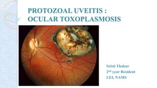
- Obligate intracellular protozoan parasite
- Phylum Apicomplexa.
- Found in the host's tissues and body fluids, such as saliva, milk, semen, urine, and
peritoneal fluid
.
11. Life cycle
Two distinct phases:
Asexual phase - which occurs in all hosts
Sexual phase - only in the intestinal epithelium of the definitive host.
Definitive host - Felines, especially domestic cats - sustain both sexual and
asexual reproduction
Intermediate host group - Humans and many other animals - cattle, pigs, sheep,
and poultry - support asexual reproduction
14
13. PRINCIPAL MODES OF TRANSMISSION:
Ingestion of undercooked, infected meat containing tissue cysts
Ingestion of contaminated water, fruit, or vegetables with oocysts
Inadvertent contact with cat feaces, cat litter, or soil containing oocysts
Transplacental transmission with primary infection during Pregnancy
Blood transfusion
Organ transplantation
17
15. definitive host infected- ingesting meat containing tissue cysts/tachyzoites from
intermediated hosts or by ingesting sporulated oocysts present in the soil and shed in
the feces of feline hosts..
asexual cycle starts - susceptible host ingests mature oocysts, tissue cysts contain
bradyzoites, or tachyzoites present in body secretions and raw meat. Tachyzoites reach
the stomach-envade enterocytes - destroyed by gastric acid, but. tachyzoites are very
active and may penetrate the oral mucosa. Digestive enzymes break down the walls of
both oocysts and tissue cysts, releasing sporozoites and bradyzoites. These enter cells
ofthe intestinal tract and transform into rapidly multiplying tachyzoites that rupture the
cells, releasing free tachyzoites. Extracellular tachyzoites or tachyzoites within
leukocytes are transported throughout the body via the lymphatic system and
bloodstream, and they can invade any organ or tissue.initial infection - acute phase of
the disease. Once infected, the host produces specific antibodies, which bind to the
extracellular tachyzoites, initiating immune-mediated eradication of the free parasite.
humoral immunity is ineffective against intracellular parasites and the cellular immune
response is called upon to attack the parasitized cells, reducing intracellular
multiplication and causing the tachyzoites to encyst. When the immune system has 19
16. Incubation Period
10 to 23 days after ingesting contaminated meat,
5 to 20 days after exposure to infected cats.
29
17. Manifests as a focal retinochoroiditis with necrotizing granulomatous
inflammation of retina with reactive granulomatous involvement of choroid,
vitreous, anterior uvea.
Mononuclear infiltrates – surround retinal blood vessels
Disruption/Migration of RPE.
After resolution retinochoroidal Scar is seen.
PATHOLOGYAND PATHOGENESIS
30
18. Pathogenesis depends upon a delicate balance between host immunity and
parasite virulence.
Adaptive immune response is mediated by CD4+ T lymphocytes and
macrophages.
Th-1 helper reaction leads to pro-inflammatory cytokines : IL-12, Interferon-
Ý and TNF-alpha
31
20. 33
The number of bradyzoite
increases
Mechanical streching and
release of bradyzoite
Conversion in to
tachyzoites
Invades contagious cells-
retinitis/chorioretinitis
23. Systemic
Acquired disease
Nonimmunocompromised
◦ lymphadenopathy in 90% of patients
◦ fever, malaise, and sore throat
◦ More severe disease can occur, affecting muscle, skin, brain, heart, and
kidney, as well as other organs.
◦ Death - rarely
Immunocompromised patient - fulminant central nervous system (CNS) -
rapidly leads to death.
37
26. Toxoplasmosis during pregnancy
Toxoplasmosis is a part of TORCH syndrome.
Only pregnant women with primary active infection leads to congenital
Toxoplasmosis
Development of active immunity once, protects subsequent pregnancies.
Vertical transmission - third trimester
First trimester - spontaneous abortion or birth of an infant with severe
disease.
40
27. Rate of Transmission
Develop infection at least 6-
9 months before pregnancy –
Pt immune – rare
transmission
within 2–3 months before
conception - 1% or below
risk of transmission but a
high risk of miscarriage
41
28. Retinochoroiditis – 70% - 90 %
Bilateral
Predilection for the posterior pole and macula
Congenital toxoplasmosis
42
30. Some - born with clinical signs of active infection
Neurologic involvement or generalized disease
Central nervous system involvement
◦ Encephalomyelitis
◦ Paralysis,
◦ Meningismus,
◦ Seizures,
◦ Respiratory Disturbances
◦ Hydrocephalus Or Microcephalus,
◦ Intracranial Calcifications,
◦ Failure To Thrive.
44
34. Ocular
Consequence of reactivation of congenitally acquired infection
Peripheral retinochoroidal scars - 82% patients
Strong predilection for the posterior pole, particularly the macular region
(based on comparison of the total retinal area) – 76 %
recurrent disease, and two thirds of patients present with relapses
49
35. SYMPTOMS:
Unilateral acute or subacute onset of floaters
Blurring
Photophobia.
Loss of vision
50
CLINICAL FEATURES
36. SIGNS
Anterior segment:
Granulomatous or nongranulomatous inflammatory reaction.
‘Spill-over’ anterior uveitis :Granulomatous inflammation with mutton fat KP’s,
posterior synechiae, fibrin deposition, and Koeppe and Busacca nodules.
Corneal edema - due to endothelial dysfunction.
51
37. Posterior segment
Vitritis - severe
Retino-choroiditis with vitritis—usually intensely
yellowish white or grey focal lesions, overlying
vitreous inflammatory haze—may give “headlight
in fog” appearance
52
39. Recurrent lesions - at the borders of old
toxoplasma retinochoroidal scars - so-called
satellite lesions
Usually single but can be multiple
Newly acquired ocular toxoplasmosis -
unilateral, solitary, active lesions without
evidence of previous retinochoroidal
scarring
54
40. Ophthalmoscopically - a yellowish-white
or gray exudate with ill-defined borders
caused by surrounding retinal edema
Size of the lesion - 1/10 of a disc
diameter to two quadrants of the retina. .
55
41. Subretinal neovascularization
◦ neovascularization regressed with resolution of the
inflammation. 57
Retinal ischemia
associated with
severe retinal
vasculitis
Inflammatory
reactions alone
Neovascularization of the
retina
42. Vascular involvement
Either in the vicinity of the active lesion or in the distant retina
Typically - diffuse or segmental vasculitis
Involves primarily the veins, but arterial involvement is not uncommon.
Complications
◦ Retinal hemorrhage
◦ Vascular obstruction
◦ Vascular shunting
◦ Neovascularization.
58
44. Optic nerve
Optic neuritis or papillitis associated with edema.
Direct extension of cerebral infection through the sheath of the optic nerve.
Patient with toxoplasma papillitis may present without evidence of a focus of
retinitis
60
45. Atypical Forms
PUNCTATE OUTER RETINAL
TOXOPLASMOSIS
small multifocal gray-white lesions that
develop in the deep layers of the retina and
retinal pigment epithelilun
Acute lesions resolve, leaving behind fine,
granular, white scars, but they frequently
recur.
significant optic nerve involvement and
atrophy.
61
46. NEURORETINITIS
active lesions localized to the juxtapapillary
region, aggressively involving the retina and
optic nerve
initially presents as severe papillitis with disc
hemorrhages, venous engorgement, and
overlying vitritis .
NEURITIS
Papillitis
62
47. MULTIPLE PSEUDORETINITIS
simultaneous presence of retinal lesions, which appear to be active.
PERIPHERAL LESIONS
simulating the snow-banking of pars planitis
63
48. ANTERIOR UVEITIS
granulomatous iridocyclitis without evidence of retinal toxoplasmosis - develop
in both immunocompetent and immunocompromised patients
FUCHS' HETEROCHROMIC IRIDOCYCLITIS
Toxoplasmosis rate higher - ranges between 8% and 65%
UNILATERAL PIGMENTARY RETINOPATHY
sequela of chronic recurrent ocular toxoplasmosis.
64
49. Healing of the retinitis associated with decrease in retinal edema and
flattening of the lesion with evidence of scar formation surrounded by
variable amounts of pigments
Punched out scar with
underlying sclera
resulting from extensive
retinal and choroidal
necrosis surrounded by
pigment proliferation
a conglomerate or
proliferated
retinal pigment
cells
Small and
appear as a
pigment clump
in the retina
65
51. Toxoplasma in:
Immunocompetent Immunocompromised
Isolated lesion
Often unilateral
Multifocal lesion
Bilateral
White fluffy focus of necrotizing retinitis
seen with associated retinal edema,
vasculitis and vitritis
Less vitiritis and lesions may simulate the
appearance of viral retinitis, such as acute
retinal necrosis or CMV retinitis
Secondary non-granulomatous inflammation
of the adjacent choroid and sclera
Symptoms are:
Necrosis from multiplication of the
parasite can cause multiple abscesses in
nervous tissue, with the symptoms of a
mass lesion.
Chorioretinitis, myocarditis and
pneumonitis
Severe Fulminant CNS disease(
toxoplasmic encephalitis
69
◦ Lymphadenopathy in 90% patients
◦ Fever, malaise and sore throat
53. Diagnosis of toxoplasmosis
◦ Serological tests,
◦ Polymerase chain reaction (PCR),
◦ Histological demonstration of the parasite and/or its antigens (i.e.
immunoperoxidase stain),
◦ Or isolation of the organism
54. Serological tests
Sabin-feldman dye test
Complement fixation (CF) test
Hemagglutination test
Immunofluorescence antibody test (IFAT)
Enzyme-linked immunosorbent assay (ELISA)
Immunoblotting (IB)
Immunosorbent agglutination assay (ISAGA)
74
55. Interpretation of serological tests
Newborns- IgM + : confirms congenital infection
Measurement of IgAAb titers may also be useful in a diagnosis of congenital
toxoplasmosis in a fetus or newborn
During this period, IgM production is often weak and presence of IgG Ab
may indicate passive transfer of maternal Ab in utero.
IgA antibodies usually disappear by 7 months.
75
56. Ocular fluid antibody assessment
Goldmann–witmer coefficient
Intraocular production of specific anti-toxoplasma Ab may be computed using
goldmann– witmer coefficient
Based on the correlation between titers of specific antibodies to t. Gondii in
aqueous humor or serum versus the globulin titers in the same fluids
77
57. When the coefficient is less than 2 in an immunocompetent patient - no
active ocular toxoplasmosis.
If the ratio is between 2 and 4, - active ocular disease,
Ratio greater than 4 - diagnostic of active ocular toxoplasmosis.
78
58. Polymerase Chain Reaction (PCR)
Used to detect T. gondii DNA in body fluids and tissues.
Used to diagnose congenital, ocular, cerebral and disseminated
toxoplasmosis.
PCR performed on amniotic fluid has revolutionized the diagnosis of fetal T.
gondii infection
PCR has allowed detection of T. gondii DNA in brain tissue, cerebrospinal
fluid (CSF), vitreous and aqueous fluid, bronchoalveolar lavage (BAL) fluid,
urine, amniotic fluid and peripheral blood
79
59. Imaging.
OCT : At the site of the active lesion, retinal layers are hyperreflective, with
irregular hyperreflective formations and some degree of posterior optical
shadowing
80
Red arrows indicate disorganization of the
inner/middle highly reflective layers (HRLs)
adjacent the active lesion site;
Yellow arrow shows the disorganization of
retinal layers (“smudge effect”)
Green arrows indicate hyperreflective
signals in the overlying vitreous
60. B-scan ultrasonic imaging: exclude RD in presence of severe vitritis
FAF: monitoring of inflammatory activity
CT brain: in immunocompromised pt, as these patients will have
concomitant CNS involvement
81
61. CT scan will show Ring Enhancing Lesions with darker areas of surounding
edema that are typical of toxoplasmosis.
82
65. AIM
ocular toxoplasmosis is a self limiting disease which recover over 4- 8 week.
But it has no cure till date as no drug are found to be effective against tissue
cyst
To reduce
1. the risk of permanent visual loss
2. recurrent retinochoroiditis
3. the severity and duration of acute symptoms.
89
66. Criteria for treatment
Lesion within the temporal arcade;
Lesion abutting the optic nerve or threatening a large retinal vessel
Lesion that has induced a large degree of hemorrhage
Lesion that has induced enough of a vitreal inflammatory response that the vision
has dropped below 20/40 in a previously 20/20 eye, or at least has sustained a
two-line drop from the visual acuity before the acute infection
91
67. Congenital Toxoplasma retinochoroiditis in the first year of life
A newborn diagnosed with congenital toxoplasmosis, regardless of the
presence of ocular lesions
Any lesion in an immunocompromised host
Relative indication - case of multiple recurrences that develop marked vitreal
condensation.
92
69. Pyrimethamine
Mode of action (MOA)
- Interrupts the metabolic cycle of parasite
- Inhibiting the dihydrofolate-reductase enzyme
- Preventing the conversion of folic acid to folinic acid, which is essential in both
DNA and RNA synthesis
Adverse effects
- Dose-related bone marrow suppression (10%)
- Leukopenia
- Thrombocytopenia
- Megaloblastic anemia
- Simulating folinic acid deficiency 94
70. Loading dose Maintenance dose
Adults 75-100 mg 25-50mg/day for 30-60
days
Children 4mg/kg 1mg/kg/day divided in 2
doses
Newborns 1mg/kg/day divided in 2
doses
Newborns should be treated daily for the first 6 months and then 3 times/wk for
their first year of life
95
71. Complete blood cell counts – weekly
Stopped if the platelet count falls below 100,000/ml or the leukocyte count
falls below 4000 cells
Combined with folinic acid : 5 mg 3 times a week
Retard thrombocytopenia, leukopenia and folate defciency
Contraindicated in the first trimester of pregnancy - teratogenicity.
96
72. Sulfonamides
MOA:
Structural analogues and competitive antagonists of paraminobenzoic acid
(PABA)
Prevent normal utilization of PABA for the synthesis of folic acid by the
parasites
Adverse effects :
◦ Crystalluria, hematuria, and renal damage
◦ Acute hemolytic anemia
◦ Agranulocytosis
◦ Hypersensitivity reactions - photosensitivity to a severe stevensjohnson
97
73. Contraindicated
◦ Glucose 6-phosphate Dehydrogenase Deficiency
◦ Third Trimester Of Gestation
Doses:
◦ Adults: 2 g loading dose followed by 1 g every 6 hr for 30-60 days
◦ Children: 100 mg/kg/day divided every 6 hr
◦ Newborns - daily for their first year of life.
Dosage: 100 mg/kg/day divided into 2 doses.
98
74. Clindamycin
MOA- inhibits ribosomal protein synthesis
Adverse effects : Pseudomembranous colitis, skin rashes, diarrhea
Dose:
◦ Adult: 300 mg every 6 hours for 30-40 days
◦ Children: 16-20 mg/kg/day divided every 6 hr
Intravitreal therapy with 1 mg of clindamycin and 0.4 mg of dexamethasone as a
local treatment option
Intravitreal injection of clindamycin and dexamethasone might be an acceptable
alternative to the classic triple-drug treatment in ocular toxoplasmosis.
99
75. Co-trimoxazole
MOA :
- sulfamethoxazole inhibits the incorporation of PABA in the synthesis of folic acid
- trimethoprim prevents reduction from dihydrofolate to tetrahydrofolate.
Dose : 160/800 mg (one tablet) every 12 hrs for 30-40 days
(trimethoprim 160 mg /sulfamethoxazole 800 mg)
combination with prednisolone
lower-cost
.
100
76. Azithromycin
MOA:
- inhibits ribosomal protein synthesis.
- effective against the encysted forms of the parasite (the bradyzoites) in vitro
500-1000mg/day for 3 wk
Reduce rate of recurrence of retinochoroiditis
Used in combination with pyrimethamine, folinic acid and Prednisolone is a
newer regimen
101
77. Atovaquone
MOA: interferes mitochondrial electrical transport chain.
Potent action against tachyzoites
Theoretically attacks encysted bradyzoites but does not seem to prevent
recurrence in vivo
750 mg every 6 hr for 4-6 wks
No serious adverse effects
102
78. Spiramycin :
Macrolide antibiotic and antiparasitic : protein synthesis inhibitor
reduces rate of tachyzoite transmission to fetus
Teratogenicity: (-) DOC: in pregnancy
Newborns with congenital toxoplasmosis are commonly treated with
pyrimethamine and sulfonamides (plus folinic acid) for 1 year
Pregnancy: 500 mg every 6 hr for 3 wk; regimen may be repeated after 21 days.
Adults: 500-750 mg every 6 hr for 30-40 days
Children: 100 mg/kg/day divided every 6 hr
103
79. Always should be combined with specific anti-toxoplasma agent—
pyrimethamine + sulfadiazine ‘classic’ therapy or ‘triple’ therapy and sometimes
supplementation with clindamycin
Steroids—after 24 to 48hours of antimicrobial therapy and stopped before
discontinuation of anti parasitic therapy
Used with adjuvant anti microbial therapy
Done with caution in immunocompromised patients
104
Prednisolone (1 mg/kg)
80. Treatment :updates
Triple drug therapy:
PYRIMETHAMINE, SULFADIAZINE & PREDINISOLONE
- greater reduction in the size of the retinal lesion compared with patients
receiving other treatment regimens or no treatment.
Quadruple therapy:
PYRIMETHAMINE, SULFADIAZINE, PREDNISOLONE &
CLINDAMYCIN
107
81. • Bactrim(2 tabs bid), sulfamethoxazole and trimethoprim is as effective as
pyrimethamine /sulfadiazine for lesions outside fovea.
Patients had resolution of active retinochoroiditis associated with improved
vision.
Trimethoprim-sulfamethoxazole was a safe and effective substitute for
sulfadiazine and pyrimethamine in treating ocular toxoplasmosis
108
82. Pregnancy
Treatment of recurrent ocular toxoplasmosis during : chosen carefully and only
started if clearly necessary
Teratogenic drugs>>used with caution
Drug Of Choice: Spiramycin
Intravitreal therapy for reactivated disease, or systemic treatment with
azithromycin, clindamycin and possibly prednisolone may be appropriate.
Specific treatment to prevent transmission to the fetus is not generally given
except in newly acquired infection
109
83. Treatment failure
Immune-mediated disease should be considered if active retinitis persists for more
than 4 months on appropriate antibiotics
Evidence of immune sensitization to retinal antigens supports the use of
corticosteroid acutely to minimize exposure to and stimulation by retinal antigens.
110
85. Laser photocoagulation
For extramacular chronically exudative lesions in individuals nonresponsive to
or not tolerating systemic therapy.
Pars Plana Vitrectomy
• For removal of persistent vitreous opacity or to relieve vitreoretinal traction that
may lead to retinal detachment
• Also removes antigenic proteins with inflammatory cells from vitreous.
112
86. MAIN FACTORS INFLUENCING TREATMENT ON ACTIVE
TOXOPLASMIC RETINOCHOROIDITIS
Immune status of individual
Location and size of active lesion
Presence of macular and/or optic disc edema
Degree of vitritis and of decreased vision
Clinical course
Special situations (newborns, pregnant women, drug allergy)
Adverse effects of antiparasitic drugs and corticosteroids
113
87. COURSE AND PROGNOSIS
Toxoplasmic retinochoroiditis: recurrent disease
~ 2/3rd of patients develop reactivations later in life
more common in congenital > postnatally acquired toxoplasmosis
Occur especially in first year after previous episode.
Some patients, however, sustain long-lasting disease remission
114
88. Prognosis depends on
Immune status and age
of patient
Size and location of
lesions
Poor prognosis:
Local complications such as
Persistent vitreous opacities
Macular edema
Epiretinal membranes
Extensive retinochoroidal
scarring
Choroidal neovascularization
Optic atrophy
Retinal detachment
115
89. Prevention
Meat should be cooked to 60°C (140°F) for at least 15 minutes or frozen to
temperatures below - 20°C for at least 24 hours to destroy the cysts.
Any contact with cat feces should be avoided.
Hands should be washed after touching uncooked meat and after contact with
cats or soil that could be contaminated with cat feces.
Consumption of raw eggs and nonpasteurized milk, particularly goat's milk,
should be avoided
116
90. Fruits and vegetables should be adequately washed before ingestion
Daily cleaning of cat litter box removes the oocysts before they become
infectious, because they need 1 to 3 days after excretion to undergo
sporulation.
Blood transfusions and organ transplants from seropositive donors should be
avoided if the recipient is seronegative
117
91. CONCLUSION
Toxoplasmosis is a recurrent and progressively destructive ocular and
systemic disease, with potentially blinding and even fatal consequences.
Formulation of primary prevention strategies
Once chronic infection has been established and the tissue form has
encysted, there is no effective treatment to eradicate the organism.
Host immune system plays a vital role in modulating the course of disease.
Tissue cysts lie dormant, - reactivate when immune surveillance falters.
118
92. Bibliography
Diagnosis and Treatment of Uveitis : C. Stephen Foster and Albert T. Vitale
Uveitis – Fundamentals and clinical practice : Nussenblact and Whitcup
Myron and Yanoff – 5th edition
Kanski Clinical Opthalmology – 8th Edition
American association of Opthalmology - Intraocular Inflammation and
Uveitis – 2016-17
Ryan – Retina -6th edition
119
Toxoplasmosis can cause severe, life-threatening disease, especially in newborns and immunosuppressed patients, but the majority of T. gondii infections in immunocompetent patients remain asymptomatic
1908 T. gondii was first found in the brain of the North African rodent the gondi, by Nicolle and Manceaux5 and then by Splendore6 in a rabbit in Brazil.
Acquired toxoplasmosis with ocular manifestations was not described until 1940, when Pinkerton and Weinman noted retinal lesions in a young adult with generalized disease.
KanskiOcular toxoplasmosis is a common cause of posterior uveitis (30-50%)1
In Nepal, 50% of those coming to hospital for uveitis and other disorders like malignancies and obstetric problems had antibodies to toxoplasma, 5.7% being IgM positive2…1.Infectious causes of posterior uveitis, review article by Efrem D. Mandelcorn published in Canadian Journal of Ophthalmology, Feb 2013
2.Rai SK, Upadhyay MP, Shrestha HG: Toxoplasma infection in selected patients in Kathmandu, Nepal. Nepal Med Coll J 2003;5(2):89-91
Toxoplasmosis is the result of infection by Toxoplasma gondii.. cosmopolitan’ parasite, being found all over the world.. The Apicomplexa are a large phylum of parasitic alveolates. Most of them possess a unique form of organelle that comprises a type of plastid called an apicoplast, and an apical complex structure
• Oocysts or soil form(containing sporozoites), which are shed in the feces.
• Tachyzoites or infectious form, rapidly multiplying organisms found in the tissues.
• Bradyzoites, slowly multiplying organisms found in the tissues.
• Tissue cysts or latent form: walled structures, often found in the muscles and central nervous system (CNS), containing dormant T. gondii bradyzoites
Major strains.. Atypical strains as well as mixed infections have being identified in many parts of the world and seem to be common in Brazil.
most studied is SAG 1 or p30. This major surface antigen has a molecular mass between 27 and 30 kDa. It is useful in the serologic diagnosis of infection12 and may play a role in the parasite’s ability to invade a cell.. A second antigen that has been characterized is SAG 2 or p22. This cell surface antigen (molecular mass 22 kDa) can participate in antibody-dependent, complement-mediated lysis of the tachyzoite.. F3G3 antigen. This 58-kDa antigen is cytoplasmic and not expressed on the cell surface. Passive transfer of antibody that reacts to this antigen has been successful in protecting animals from a lethal challenge by the Toxoplasma organism.
The asexual cycle starts - susceptible host ingests mature oocysts, tissue cysts contain bradyzoites, or tachyzoites are present in body secretions and raw meat. Tachyzoites that reach the stomach-envade enterocytes - destroyed by gastric acid, but. tachyzoites are very active and may penetrate the oral mucosa. Digestive enzymes break down the walls of both oocysts and tissue cysts, releasing sporozoites and bradyzoites. These organisms then enter cells ofthe intestinal tract and transform into rapidly multiplying tachyzoites that rupture the cells, releasing free tachyzoites. Extracellular tachyzoites or tachyzoites within leukocytes are transported throughout the body via the lymphatic system and bloodstream, and they can invade any organ or tissue. This initial infection characterizes the acute phase of the disease. Once infected, the host produces specific antibodies, which bind to the extracellular tachyzoites, initiating immune-mediated eradication of the free parasite. However, humoral immunity is ineffective against intracellular parasites and the cellular immune response is called upon to attack the parasitized cells, reducing intracellular multiplication and causing the tachyzoites to encystY When the immune system has eliminated the tachyzoites, symptoms disappear, and the chronic phase ensues. The sexual cycle takes place exclusively in the feline intestine, and it is unclear why this phase occurs only in members of the cat family. Cats may initially become infected by eating contaminated meat containing tissue cysts or by ingesting sporulated oocysts. In the cat's intestine, the tachyzoites invade the ~pithelial cells and start to multiply by schizogony. During this process, gametocytes are formed and fertilized to produce oocysts. The time interval between the infection and the appearance of oocysts in the feces depends on the form of the organism ingested and varies from 3 to 24 days. Excretion continues for up to 20 days, with shedding of as many as 12 million oocysts in a single day. In general, once a cat has cleared the initial infection it will not shed oocysts again. However, if the cat becomes infected with Isospora felis, recurrent oocyst shedding may occur.34
sexual cycle takes place exclusively in the feline intestine.- initially become infected by eating contaminated meat containing tissue cysts or by ingesting sporulated oocysts. In the cat's intestine, the tachyzoites invade the ~pithelial cells and start to multiply by schizogony. During this process, gametocytes are formed and fertilized to produce oocysts. The time interval between the infection and the appearance of oocysts in the feces depends on the form of the organism ingested and varies from 3 to 24 days. Excretion continues for up to 20 days, with shedding of as many as 12 million oocysts in a single day. In general, once a cat has cleared the initial infection it will not shed oocysts again. However, if the cat becomes infected with Isospora felis, recurrent oocyst shedding may occur.34
These enter cells ofthe intestinal tract - transform into -tachyzoites that rupture the cells, - free tachyzoites. Extracellular tachyzoites or tachyzoites within leukocytes are transported throughout the body via the lymphatic system and bloodstream, - invade any organ or tissue.initial infection - acute phase of the disease. Once infected, the host produces specific antibodies, - bind to the extracellular tachyzoites, initiating immune-mediated eradication of the free parasite. humoral immunity is ineffective against intracellular parasites and the cellular immune response is called upon to attack the parasitized cells, reducing intracellular multiplication and causing the tachyzoites to encyst. When the immune system has eliminated the tachyzoites, symptoms disappear, - chronic phase ensues
Oocysts are produced only in the feline intestinal cells
Within 1 to 21 days after shedding, the oocysts undergo sporulation and become mature, infective oocystS.33 These mature forms contain two sporocysts, each ofwhich contains four sporozoites. The ingestion of mature oocysts can cause infection in either an intermediate or the definitive host. Sporulation does not occur below 4°C or above 37°C, thus explaining the lower incidence of toxoplasmosis in areas with extreme temperatures.
Invade all mammalian cells except nonnucleated erythrocytes and are found extracellulary as well as intracellularly in various organs…
, leading to cell lysis, direct tissue damage, and subsequently to a strong, potentially destructive immune response
Endodyogeny-A form of asexual reproduction, favoured by parasites such as Toxoplasma gondii, in which two daughter cells are produced inside a mother cell, which is then consumed by the offspring prior to their separation.
, which eventually becomes part of the cyst's capsule.. The wall of a mature cyst is composed of a cOmbination of both host and parasitic components, so the bradyzoites are protected from the host's immune system. The cysts are very resistant and can remain dormant in "i' the host for years without tissue damageY
Mononuclear inflammatory infiltrates surrounding retinal blood vessels .. The inflammatory process can extend to underlying sclera. A retinochoroidal scar is left (chorioretinal adhesion) after resolution of inflammation, with variable proliferation of RPE. Intact T. gondii cysts without reactive inflammation may be found in histologically normal retina
Classically, the initial lesion starts in the superfiCIal retina. As the retinitis progresses, involvement of the full-thickness retina, adjacent choroid, vitreous, and even sclera may occur.
Rpe hyperplasia..
Slowly, the borders of the lesion become more defined, the exudates andvitritis diminish, and the lesion shows an elevated central area with a whitish-gray to brown discoloration. after a variable time period, pigmentation occurs, particularly in.the margins of the lesion. The time required for a retu10chorOldal lesion to heal varies, depending on the size of the lesion, the treatment delivered, the immunologic condition of the host, and the strain of T. gondii…
Rupture of such cysts releases bradyzoites that convert to tachyzoites, establishing active parasite proliferation locally, with subsequent cell lysis and release of cytotoxic mediators and eliciting a vigorous necrotizing granulomatous response that may also lead to further tissue damage.30,31 Hypersensitivity to retinal antigens may also play a role in maintaining intraocular inflammation during recurrences.32
7.12.1 Histopathology of Toxoplasmic retinochoroiditis, Showing Extensive Necrosis of the Neurosensory retina and retinal Pigment Epithelium (rPE). the choroid displays reactive diffuse granulomatous inflammation (hematoxylin-eosin stain, original magnification 400×). toxoplasmic cysts are seen within the necrotic retina, staining with Gomori’s methenamine silver (Gms, inset). (courtesy n. rao, University of southern california.)
severity of congenital toxoplasmosis is inversely related to the time of gestational exposure, …when the fetus may be exposed to maternal blood. Fortunately, third-trimester infection usually results in a subclinical form of the disease…Most auth~rities agree that transplacental transmission rates are lower if the mother receives treatment during pregnancy.. present as a subclinical or chronic infection. The newborn mayor may not have retinochoroidal scars , intracranial calcifications (Fig. 33-6), or other sequelae of intrauterine infection. The identification of subclinical infection is ilnportant, because early treatment improves the prognosis.68 Some infants with congenital toxoplasmosis are born with clinical signs of active infection.
The first trimester - 15% chance but Severity of disease in neonate is more
Second trimester - 25% risk
Third trimester - 65% chance but Severity of disease in neonate is less usually asymptomatic
occurring in less than 10% of newborns with congenital toxoplasmosis.12,19 The leading clinical manifestation, however, is retinochoroiditis, present in up to 80% of newborns at birth
They may present at birth with ….but the neurologic is more frequent.
This neonatal form is severe and patients frequently develop ocular ane}. neurologic sequelae even with treatment
Typical wagon wheel scar..
occasionally,the infant is normal at birth and develops actIve dIsease In the first few months of life. This form is more common in premature infants and results in severe disease, but it may also occur in full-term infants, in whom it is less severe
70% of immunocompetent patients who acquire toxoplasmosis are completely symptom free..Cervical nodes are involved more frequently, followed by suboccipital, supraclavicular, axillary, inguinal, and mediastinal nodes. Involved lymph nodes are llsually bilateral, discrete, nontender, and nonsuppurative and they vary in firmness.
Reasopn unclear but some authorties says - parasites first invade the eye through fhe posterior ciliary art~ries or the optic nerve. Invasion of the eye by way of the optic nerve may give rise to juxtapapillary Toxoplasma retino~ho~oiditis. Some other thoughts as to the macular predIlectIOn for Toxoplasma - ea:1ier vascul~rization of the posterior pole than the penphery dunng development and the fact that the fetal vasculature contains end arterioles. In addition, there may be entrapment of free parasites, or parasites within macrophages, in the terminal capillaries of the fovea…classic teaching recurrence is the result of release of T. gondii from cysts. Cysts may rupture and release live organisms that actively invade the retina, or cysts may simplyrelease antigens that stimulate an inflammatory retinochoroiditis. Alternatively, an autoimmune response may develop to retinal antigens such as the retinal S-antigen, which results in retinochoroiditis..may be the .result of reinfection. ImmunIty from a primary Toxoplasma infection is not sufficient to prevent reinfection with a new strain of T. gondii.
‘Spill-over’ anterior uveitis : common
A single inflammatory focus of fluffy white retinitis or retinochoroiditis associated with a pigmented scar (‘satellite lesion’) is typical.
Lesions tend to involve posterior pole.
IOP: elevated
This process is believed to develop as a result of a hypersensitivity reaction to Toxoplasma antigen, because live T. gondii has never been dem~nstratedin the anterior segment of an immunocompetent patient. ..
mimic's Fuchs uveitis syndromeRetina/choroid lesion
Fluffly white/brown
Vitritis more
Fluffy/domeshaped, well demarcated
Vitreous involvement may occur as a localized or diffuse exudate, inflammatory cells, pigment, or hemorrhage. Vitreous opacities tend to be slowly reabsorbed and may persist for years after complete resolution of the retinal lesion. When there is severe and prolonged vitreous involvement, vitreous contraction, posterior vitreous detachment, or even retinal detachment may occur.
Denovo, relatively uncommon; more frequently in immunocompromised patients
Recurrent lesions are usually single and typically develop at the mergins of retinochoriodal scar called satellite lesions.
Classically, the initial lesion starts in the superfiCIal retina. As the retinitis progresses, involvement of the full-thickness retina, adjacent choroid, vitreous, and even sclera may occur. Slowly, the borders of the lesion become more defined, the exudates andvitritis diminish, and the lesion shows an elevated central area with a whitish-gray to brown discoloration. after a variable time period, pigmentation occurs, particularly in.the margins of the lesion. The time required for a retu10chorOldal lesion to heal varies, depending on the size of the lesion, the treatment delivered, the immunologic condition of the host, and the strain of T. gondii
produced by antigen-antibody complex deposition in the vessel wall, as well as localized mononuclear cell infiltrates (Fig. 33-14).
Fundus Appearance of Toxoplasmic retinochoroiditis, With an Active Exudative Lesion in the Absence of retinochoroidal Scars (Isolated Focal Lesion). inset shows surrounding retinal edema and periarteriolar exudates (Kyrieleis arteriolitis
(the presence of exudates or periarterial plaques not associated with leakage or vascular obstruction)
first and second decades of life
congenital or acquired
bilateral in a third of the cases
Because the process is localized to the outer retinal layers, there is little or no overlying vitritis. Thus, even without foveal lesions, these patients may suffer significant visual loss as a result of optic neuropathy. , and some patients present with classic Toxoplasma retinochoroiditis in one eye and the punctate form in the fellow eyeP..erhaps this fonn is an immune phenomenon related to exposure of retinal antigens..patients with ocular toxoplasmosis develop both cellular and humoral immune responses to retinal antigens. autoimmune sensitization
previously known as Jensen's choroiditis-neuroretinitis
Soon after, a juxtapapillary retinochoroiditis and macular star develop (Fig. 33-20). ToxojJlas17la neuroretinitis is an ophthalmic emergency and requires prompt treatment
However, close observation reveals just a single active Toxoplasma lesion accompanied by noncontiguous areas of retinal .edema. Once the true active lesion heals, the pseudolesions completely disappear without scarring
anterior uveitis is either a hypersensitivity reaction to Toxoplasma antigen or a Toxoplasma infection in the anterior segment. However, the parasite has never been demonstrated in the anterior segment of immunocompetent patients. 109 Several mechanisms have been proposed to explain the association between FHI and Toxoplasma retinochoroidallesions
One hypothesis suggests that primary retinochoroidal inflammation results in production of antibodies that cross-react with anterior segment antigens, causing a low-grade anterior uveitis (i.e., FHI).109 Others posit that there is no statistically significant association between FHI and ocular toxoplasmosis.l
Healing spontaneously 6-8weeks, vitreous opacity take longer to clear
Inflammatory focus replaced by well defined border with central retinochoroidal atrophy and peripheral retinal pigment epithelial hyperplasia
A healed Toxoplasma scar typically has well-defined borders with central retinochoroidal atrophy and peripheral pigment epithelial hyperplasia. In the atrophic central area, either choroidal vessels or bare sclera may be observed. Healing Toxoplasma .lesions may be complicated by proliferative vitreoretinopathy, retinal gliosis, vascular shunts, and choroidal neovascular membranes
Traction bands are also frequent, and they usually link an old scar to the optic disc (Franceschetti's syndrome) or to a neighboring scar
Punctate outer retinal toxoplasmosis—atypical manifestation featuring clusters of small (25-75 micrometre), grey white lesion involving outer retina and RPE. Vitritis is minimal but associated with exudative retinal detachment
Other diseases resembalance in immunocompromised toxo= syphilitic retinitisfungal chorioretinitis and primary ocular lymphoma
glaucoma -mechanical obstruction of the trabecular meshwork with fibrin, inflammatory cells, or inflammatory debris
Cataracts - severe vitreous inflammation or the use of local and systemic corticosteroids. Posterior subcapsular cataract is typical
Vitreous hemorrhage and tractional or rhegmatogenous retinal detachment - proliferative vitreoretinopathy and contraction ofvitreous bands. Proliferative vitreoretinopathy and tractional bands - macular dragging.
epiretinal membranes - macular pucker and cystoid macular edema.
Cystoid macular edema -chronic inflammation.
macular cyst - along with tangential traction on the retinal internal limiting membrane and the posterior hyaloid, - macular hole.
Retinal hemorrhages - retinal vein occlusion around or within active lesions.BRVO and BRAO- vessel crosses an acute Toxoplasma lesion, but venous occlusions are more common. Arteriovenous shunts in the retina and chorioretinal vascular anastomosis - complications of vascular obstruction
. Disruption ofBruch's membrane - necrotizing retinochoroiditis - choroidal neovascular membranes, - adjacent to the retinal scar or at a distant location with feeder vessels originating from the scar. Optic nerve atrophy -primary involveof the optic nerve, peripapillary'lesions, or lesions localized in the papillomacular bundle. punctate outer retinal toxoplasmosis - frequent optic nerve atrophy..
direct demonstration of the organism in tissues or body fluids, by in vitro culture, by inoculation and culture in mouse peritoneum by polymerase chain reac-.. Direct demonstration of the parasite is easiest during the acute phase, when the trophozoites can be found in body fluids such as blood, cerebrospinal fluid, urine, and breast milk.. parasite in this phase can be identified microscopically after Giemsa staining.. chronic phase, tissue cysts may be occasionally identified in biopsy samples by staining with hematoxylin and eosin or silver
neutralization test in which the patient's serum is incubated with complement and live Toxoplasrtya organisms, and a dye is employed to quantify the bound antibody. early detection of the infection and has high sensitivity and specificity in both acute and chronic phases, but it is no longer used because it requires maintenance of live virulent parasites in the laboratory,
CF test has a good sensitivity only when the level of circulating antibodies is high.
. good sensitivity and specificity in the acute and chronic phases
Ifat-early elevations in serum antibodies, and allows quantification ofIgM and IgG levels
Elisa-good sensitivity and specificity.. IgM of the test serUlll adheres to..plates precoated with anti-IgM antiserum.. detection of IgM antibodies for many months after the acute phase
ISAGA-fimmunocapture that allows the simultaneous detection of IgA and IgM anti-Toxoplasma antibodies. It has good sensitivity and specificity, allowing early diagnosis ofcongenital toxoplasmosis
Immunoblot (a type ofwestern blot) has proven to be of equal or superior sensitivity when compared with the preceding tests, and it allows an earlier diagnosis of congenital toxoplasmosis
. As IgG is passively transmitted to the fetus, its detection does not have diagnostic value. Slowly, maternal IgG decreases in the infant's circulation, and it completely disappears within 18 months.,follow up titre imp.. Serum titers that remain constant or increase in value after 1 week of life are diagnostic of fetal infection. IgM and IgA-imp
recently acquired infection will produce elevated titers of IgM, IgA, and IgE…chronic cases low titer§ of high-affinity antiToxoplasma IgG are present.. Because most of the cases - evaluated in the chronic phase, a low IgG titer is expected. However,low IgG titers may also be a sign of recent infection. To differentiate between these two possibilities, serologic testing should be repeated at a tillle interval of 2 to 4 weeks. A rising titer is indicative of recent infection.
Titer ofantibody in aqueous humor* X Concentration ofserum globulins / Titer ofantibody in serum* Concentration ofaqueous humor globulins
Step 1: Denaturation by Heat:Step 2: Annealing Primer to Target Sequence:Step 3: Extension:Step 4: End of the First PGR Cycle. Its principle is based on the use of DNA polymerase which is an in vitro replication of specific DNA sequences.. he DNA polymerase is the key enzyme that links individual nucleotides together to form the PCR product.
Diffuse unilateral subacute neuroretinitis (DUSN)
but parasite activity and multiplication can be reduced and decrease size of retinochoroidal scar…Spontaneous resolution(+): every case not treated
immunocompetent person the disease is ultimately selflimited..Here one might be concerned that the continuation of this process might lead to retinal detachment…. Although these treatment criteria are broad, some authorities believe that all active lesions should be treated. One reason for this recommendation is that active lesions, even those far from the macula, may be associated with decreased visual acuity because of macular edema, macular traction, severe vitritis, or retinal detachment. In addition, active lesions produce tachyzoites that may spread to distant retinal areas and encyst. Treatment of any active lesion reduces the number of tachyzoites and (theoretically) the chances of reactivation in crucial retinal locations.
Pyrimethamine treatment has been shown to minimize the size of the retinochoroidal scar that forms with resolution of the….. AIDS: avoid pyrimethamine or used at a lower dosage possible pre-existing BM suppression and the antagonistic effect of zidovudine when drugs are combined.
Caution in hepatic or renal failure
leucovorin
Usualy given with pyrimethamine
Adequate hydration with oral fluids to maintain a urine output of at least 1500 mllday should avoid the problem
mixture of sulfadiazine, sulfamerazine, and sulfamethazine
because of the potential for hemolytic anemia
because they dislodge the fetal bilirubin from serum albumin, causing kernicterus.
Advantages of intravitreal treatment include increased patient convenience, improved systemic side effect profile, greater drug availability, and fewer follow-up visits and hematological evaluations. Soheilian et al,2011
Potential for hemolytic anemia
Clindamydn Clindamycin inhibits ribosomal protein synthesis
Atovaquone-Interferes mitochondrial electrical transport chain.. potent action against tachyzoites, including those of very virulent strains, and it has been shown to reduce the number of cerebral tissue cysts after acute or chronic infection in the hamster mode1.
Spiramycin- drug of choice during pregnancy. It achieves a high concentration in the placenta and has no reported teratogenic effects. Spiramycin may reduce the incidence of congenital transmission
Topical steroid and mydriatic may be given for anterior uveitis.
Kanski
When antimicrobial therapy is given it kills the parasite leading to release of toxins leading to inflammation when steroids are necessary
Can reduce the risk of complications like cystoid macular edema persistent vitritis and perivascular inflammation
Periocular or intravitreal injections of depot contraindicated –scleral necrosis
Increase inflamaton vitrtisand pthisis bulbi..loss ofv ision
inhibits the incorporation of PABA in the synthesis of folic acid, whereas trimethoprim prevents reduction from dihydrofolate to tetrahydrofolate. This combination is significantly less active than the combination ofpyrimethamine and sulfadiazine but may still be effective in the treatment oftoxoplasmosis.
Rothova et al
Opremcak et al
we treat for at least 30 to 60 days in an immunocompetent patient. A positive response to treatment is defined as a sharpening of the borders of the retinochoroidal lesions and improvement of vitreous haze. When therapy is complicated by adverse effects or proves to be ineffective after 4 months, a change in therapy is recommended.
spiramycin in combination with pyrimethamine or sulfadiazine may be administered for a 3-week period. If the response is not adequate, the regimen can be repeated after 21 days.. Infants - combination of pyrimethamine, sulfadiazine, and folinic acid. A new approach to congenital toxoplasmosis is PCR of the amniotic fluid to establish the diagnosis and initiation of treatment in utero for infected fetuses.
Despite adequate treatment, some patients continue to have chronic active retinitis.. result of a particularly virulent strain of T. gondii, or it may be be
cause of a localized immune or even an autoimmune phenomenon. Many studies have demonstrated both a cellular and a humoral immune response to retinal antigens in the setting ofocular toxoplasmosis
Occasionally, patients with ocular toxoplasmosis respond to treatment including corticosteroid, but when the corticosteroid is withdrawn, active retinitis recurs despite continuous antibiotic administration. Although the reason for this recurrence has not been determined, three different mechanisms. First and most likely, this "reactivation" phenomenon may simply demonstrate immune reactivity to persistent T. gondii antigens remaining in the tissues. Second, it may represent a form oflocalized autoimmunity. Third, the diagnosis of toxoplasmosis may be erroneous.
Considered for recurrences in pregnancy, cases of drug intolerance, lesions associated with CNVM, cases with resistant medical therapy
Destroys cysts and tachyzoites and inhibits the spread of infection
Complications= retinal and vitreous hemorrhage, Epiretinal membrane and CNVM formation
Ppv.. For removal of persistent vitreous opacity or to relieve vitreoretinal traction that may lead to retinal detachment
Also removes antigenic proteins with inflammatory cells from vitreous.. immunoactivating factors, and inflammatory cells from the vitreous
This duty should be performed only by a nonpregnant individual. .