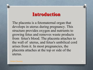
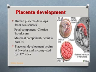
THE PLACENTA
BY
SRIDEVI DEVARAJ
Sridevi Devaraj 1
IntroductionIntroduction
The placenta is a fetomaternal organ that
develops in uterus during pregnancy. This
structure provides oxygen and nutrients to
growing fetus and removes waste products
from fetus's blood. The placenta attaches to
the wall of uterus, and fetus's umbilical cord
arises from it. In most pregnancies, the
placenta attaches at the top or side of the
uterus.
Sridevi Devaraj 2
Further Development of
Chorionic Villi
Early in the 3rd
week,
mesenchyme growth into
the primary villi forming a
core of mesenchymal
tissue. Thus the Secondary
Chorionic Villi are formed
over the entire surface of
the chorionic sac.
Some mesenchymal cells in
the secondary villi
differentiate into capillaries
and blood cells forming the
Tertiary Chorionic Villi.
The capillaries in the villi
fuse to form arteriocapillary
networks.
Sridevi Devaraj 3
Sridevi Devaraj 4
The previous formed
arteriocapillary
networks become
connected with the
embryonic heart
through vessels which
are formed in the
mesenchyme of the
chorion and
connecting stalk.
By the end of the 3rd
week, embryonic blood
begins to flow through
the capillaries in the
chorionic villi.
Sridevi Devaraj 5
Oxygen & nutrients in
the maternal blood in the
intervillous space diffuse
through the walls of the
villi and enter the
embryo’s blood.
Carbon dioxide & waste
products diffuse from
blood in the fetal
capillaries through the
wall of the chorionic villi
into the maternal blood.
DECIDUADECIDUA
Sridevi Devaraj 6
This is the endometrium of
the gravid (pregnant)
uterus.
It has four parts:
1. Decidua basalis: it
forms the maternal
part of the placenta
2. Decidua capsularis:
it covers the
conceptus
3. Decidua parietalis:
the rest of the
endometrium
4. Decidua reflexa:
Junction between
capsularis &
parietalis
PlacentaPlacenta
Human placenta is
O Discoid in shape
O Haemochorial
O Deciduate
Sridevi Devaraj 7
Placenta developmentPlacenta development
O Human placenta develops
from two sources
Fetal component- Chorion
frondosum
Maternal component- decidua
basalis
O Placental development begins
at 6 weeks and is completed
by 12th
week
Sridevi Devaraj 8
Sridevi Devaraj 9
O Until the beginning
of the 8th
week, the
entire chorionic sac
is covered with villi.
O After that, as the sac
grows, only the part
that is associated
with Decidua basalis
retain its villi.
O Villi of Decidua
capsularis
compressed by the
developing sac.
Sridevi Devaraj 10
O Thus, two types of
chorion are formed:
O Chorion
frondosum
(villous chorion)
O Chorion laeve –
bare (smooth)
chorion
O About 18 weeks
old, it covers 15-
30% of the decidua
and weights about
1 6 of fetus
ContCont..
O The villous chorion ( increase
in number, enlarge and
branch) will form the fetal
part of the placenta.
O The decidua basalis will form
the maternal part of the
placenta.
O The placenta will grow
rapidly.
O By the end of the 4th
month,
the decidua basalis is almost
entirely replaced by the fetal
part of the placenta
Sridevi Devaraj 11
Placenta at Term
O Is circular disc
O Diameter- 15-20 cm
O Weight-500gm
O Thickness-2.5 cm
O Spongy to feel
O Occupies 30% of the uterine wall
O Two surfaces- Maternal and fetal
O 4/5th
of the placenta is of fetal origin and 1/5
is of maternal origin
Sridevi Devaraj 12
Fetal surface
Sridevi Devaraj 13
O Covered by smooth and
glistening amnion
overlying the chorion
O Umbilical cord is attached
near to its centre
O Branches of the umbilical
vessels are visible beneath
the amnion as they radiate
from the insertion of the
cord.
Maternal surface
Sridevi Devaraj 14
O Rough and spongy
O Maternal blood gives
it dull red colour
O Remanants of the
decidua basalis gives
it shaggy appearance
O Divided into 15-20
cotyledons by the
septa
O Cotyledons –about 15 to
20 slightly bulging villous
areas. Their surface is
covered by shreds of
decidua basalis from the
uterine wall.
O After birth, the placenta is
always inspected for
missing cotyledons.
Cotyledons remaining
attached to the uterine
wall after birth may cause
severe bleeding.
Sridevi Devaraj 15
Margins
Margin of the placenta are
formed by fused chorionic and
the basal plate
Attachment
Placenta is attached to
the upper part of the uterine
body encouraging to the fundus
either at the posterior or
anterior wall
Sridevi Devaraj 16
Separation
OAfter delivery ,placenta separates with the
line of separation being through decidua
spongiosum (intermediate spongy layer of
the decidua basalis)
Sridevi Devaraj 17
Structure of the
placenta
Sridevi Devaraj 18
O Placenta consist of two
plates(layers)
 Chorionic plate
 The basal plate
chorionic plate
O It is lined by the amniotic
membrane on the fetal side
O The umbilical cord is
attached to it.
O Forms the roof of the
placenta
ContCont..
Sridevi Devaraj 19
O From outside inwards consists of
Syncytiotrophoblast
Cytotrophoblast
Extra embryonic mesoderm with
branches of umbilical vessels
Sridevi Devaraj 20
The basal plate
OIt consist of the structures From
outside inwards
Compact and spongy layer of
decidua basalis
Layer of Nitabuch
Cytotrophoblastic shell
Syncytiotrophoblast
Basal plate is perforated by the
spiral arteries allowing entry of
maternal blood into intervillous
space
Sridevi Devaraj 21
O Layer of Nitabuch -
Is a fibrinous layer formed at the junction
of cytotrophoblastic shell with decidua due to
fibrinoid degeneration of syncytiotrophoblast
O It prevents excessive penetration of the decidua
by the trophoblast
O Nitabuch membrane is absent in placenta
accreta and other morbidly adherent placentas
Placental barrier or membrane
Sridevi Devaraj 22
Maternal and fetal blood
are separated by
placental membrane or
barrier(0.025mm thick)
It consist of
O Endothelial lining of
fetal vessels
O Connective tissue
stroma of villus
O Basement membrane
O Cytotrophoblast
O Syncytiotrophoblast
Placental CirculationPlacental Circulation
Sridevi Devaraj 23
Placental CirculationPlacental Circulation
Utero placental circulationUtero placental circulation
O It is concerned with the circulation of the
maternal blood through the intervillous
space.
O A mature placenta has a volume of about
500 ml of blood.
O 350 ml of being occupied in the villi
system
O 150 ml lying in the intervillous space
Sridevi Devaraj 24
O Intervillous blood flow at
term is estimated to be 500-
600 ml per minute
O Blood in the inter villous
space Is replaced
completely replaced about
3-4 times per minute.
O The pressure within the
intervillous space
- During uterine
relaxation is about 10-15
mm of hg
-During uterine
contraction is about 30-50
mm of hgSridevi Devaraj 25
Arterial circulationArterial circulation
O 120-200 spiral arteries open
in to the inter villous space
O Within 12 weeks of
pregnancy the
cytotrophoblast invade the
spiral arteries up to intra
decidual portion
O Within 12-16 weeks
secondary invasion of
trophobast extend up to
radial arteries within the
myometrium.
Sridevi Devaraj 26
O Thus spiral arteries
are converted to large
bore utero placental
arteries
O Trophoblast cells that
do not take part in
villous structure
extravillous
trophoblast
these are two tyes
-endovascular
extravillous
trophoblast
-Interstitial
extravillous
trophoblast
Sridevi Devaraj 27
Venous drainageVenous drainage
O the venous blood of the inter villous space drains
through the uterine veins which pierce the basal plate
Sridevi Devaraj 28
Feto-placental circulationFeto-placental circulation
O Two umbilical arteries carries
the impure blood from the fetus
O They enter the chorionic plate
underneath the amnion
O The arteries break up in to the
small branches which enters
the small chorionic villi
O Each in turn divide in to
primary ,secondary and
tertiary vessels of the
corresponding villi.
O The blood flows in to the
corresponding venous channel
through the terminal capillary
network.Sridevi Devaraj 29
O Maternal and fetal blood flow
side by side but in opposite
direction
O This blood flow facilitate the
material exchange between
the mother and fetus
O The villous capillary pressure
is 20-40 mm of hg
O The fetal blood flow through
placenta is 400 ml /min
O This is mainly facilitated by
pumping action of heart
Sridevi Devaraj 30
Placental Function
Sridevi Devaraj 31
Transfer of gases ,nutrients and waste
products , namely
Respiratory function
Nutritive function
Excretory function
Endocrine and enzymatic function
Barrier function
Immunological function
Mechanism involved in the transfer of
substances
O Simple diffusion-O2 and CO2
O Facilitated diffusion ( carrier mediated ) –
glucose ,vitamins
O Active transfer ( against concentration
gradient )-ions
O Endocytosis- invagination of cell membrane to
form intracellular vesicle
O Exocytosis-Release of substances in the
vesicles to extracellular space
eg IgG immunoglobulin
Sridevi Devaraj 32
Respiratory function
O Although fetal respiratory movement
occurs by 11 week, no active
exchange of gases takes place
O Intake of oxygen and output of carbon
dioxide take place by simple diffusion
across the fetal membrane
O O2 delivery to the fetus is at the rate
of 8 ml/kg which is achieved by cord
blood flow of 160-320ml/min
Sridevi Devaraj 33
Excretory functionExcretory function
OWaste products from the fetus
such as urea, uric acid,
cretinine are excreted to the
maternal blood by simple
diffusion.
Sridevi Devaraj 34
Nutritive function
Fetus obtains its nutrients from the maternal blood
O Glucose- transferred to the fetus by facilitated
diffusion
O Lipids for fetal growth and development has dual
origin. They are transferred across the fetal
membrane or synthesized in the fetus
O Amino acids are transferred by active transport
O Water and electrolytes- Na, K ,Cl cross by simple
diffusion, Ca , P, and Fe cross by active transport
O Water soluble vitamins are transferred by active
transport but the fat soluble vitamins are
transferred slowlySridevi Devaraj 35
Barrier Function
O Placental membrane is thought to be a protective
barrier for the fetus against harmful agents in the
maternal blood
O Substances with large molecular weight or size like
insulin or heparin are transferred minimally
O Only IgG ( not IgA or Ig M )antibodies and antigens
can cross the placental barrier
O Most drugs can cross the placental barrier and some
can be teratogenic
O Various viruses, bacteria, protozoa can cross the
placenta and affect the fetus in uteroSridevi Devaraj 36
Immunological function
O Inspite of foreign paternally
inherited antigens in the fetus and
placenta, there is no graft rejection
due to immunological protection
provided by the placenta
Sridevi Devaraj 37
Endocrine and Enzymatic
function
O Placenta secretes various hormones – Protein
hormones like HCG, human placental
lactogen,pregnancy specific beta 1
glycoprotein,,pregnancy associated plasma
protein, steroidal hormones like estrogen and
progestrone
O Enzymes secreted are diamine oxidase-which
activates the circulatory pressor amines,
oxytocinase which neutralizes oxytocin,
phospholipase A2 which synthesizes
arachidonic acidSridevi Devaraj 38
Sridevi Devaraj 39
Sridevi Devaraj 40
OThank you

Placenta

  • 1.
  • 2.
    IntroductionIntroduction The placenta isa fetomaternal organ that develops in uterus during pregnancy. This structure provides oxygen and nutrients to growing fetus and removes waste products from fetus's blood. The placenta attaches to the wall of uterus, and fetus's umbilical cord arises from it. In most pregnancies, the placenta attaches at the top or side of the uterus. Sridevi Devaraj 2
  • 3.
    Further Development of ChorionicVilli Early in the 3rd week, mesenchyme growth into the primary villi forming a core of mesenchymal tissue. Thus the Secondary Chorionic Villi are formed over the entire surface of the chorionic sac. Some mesenchymal cells in the secondary villi differentiate into capillaries and blood cells forming the Tertiary Chorionic Villi. The capillaries in the villi fuse to form arteriocapillary networks. Sridevi Devaraj 3
  • 4.
    Sridevi Devaraj 4 Theprevious formed arteriocapillary networks become connected with the embryonic heart through vessels which are formed in the mesenchyme of the chorion and connecting stalk. By the end of the 3rd week, embryonic blood begins to flow through the capillaries in the chorionic villi.
  • 5.
    Sridevi Devaraj 5 Oxygen& nutrients in the maternal blood in the intervillous space diffuse through the walls of the villi and enter the embryo’s blood. Carbon dioxide & waste products diffuse from blood in the fetal capillaries through the wall of the chorionic villi into the maternal blood.
  • 6.
    DECIDUADECIDUA Sridevi Devaraj 6 Thisis the endometrium of the gravid (pregnant) uterus. It has four parts: 1. Decidua basalis: it forms the maternal part of the placenta 2. Decidua capsularis: it covers the conceptus 3. Decidua parietalis: the rest of the endometrium 4. Decidua reflexa: Junction between capsularis & parietalis
  • 7.
    PlacentaPlacenta Human placenta is ODiscoid in shape O Haemochorial O Deciduate Sridevi Devaraj 7
  • 8.
    Placenta developmentPlacenta development OHuman placenta develops from two sources Fetal component- Chorion frondosum Maternal component- decidua basalis O Placental development begins at 6 weeks and is completed by 12th week Sridevi Devaraj 8
  • 9.
    Sridevi Devaraj 9 OUntil the beginning of the 8th week, the entire chorionic sac is covered with villi. O After that, as the sac grows, only the part that is associated with Decidua basalis retain its villi. O Villi of Decidua capsularis compressed by the developing sac.
  • 10.
    Sridevi Devaraj 10 OThus, two types of chorion are formed: O Chorion frondosum (villous chorion) O Chorion laeve – bare (smooth) chorion O About 18 weeks old, it covers 15- 30% of the decidua and weights about 1 6 of fetus
  • 11.
    ContCont.. O The villouschorion ( increase in number, enlarge and branch) will form the fetal part of the placenta. O The decidua basalis will form the maternal part of the placenta. O The placenta will grow rapidly. O By the end of the 4th month, the decidua basalis is almost entirely replaced by the fetal part of the placenta Sridevi Devaraj 11
  • 12.
    Placenta at Term OIs circular disc O Diameter- 15-20 cm O Weight-500gm O Thickness-2.5 cm O Spongy to feel O Occupies 30% of the uterine wall O Two surfaces- Maternal and fetal O 4/5th of the placenta is of fetal origin and 1/5 is of maternal origin Sridevi Devaraj 12
  • 13.
    Fetal surface Sridevi Devaraj13 O Covered by smooth and glistening amnion overlying the chorion O Umbilical cord is attached near to its centre O Branches of the umbilical vessels are visible beneath the amnion as they radiate from the insertion of the cord.
  • 14.
    Maternal surface Sridevi Devaraj14 O Rough and spongy O Maternal blood gives it dull red colour O Remanants of the decidua basalis gives it shaggy appearance O Divided into 15-20 cotyledons by the septa
  • 15.
    O Cotyledons –about15 to 20 slightly bulging villous areas. Their surface is covered by shreds of decidua basalis from the uterine wall. O After birth, the placenta is always inspected for missing cotyledons. Cotyledons remaining attached to the uterine wall after birth may cause severe bleeding. Sridevi Devaraj 15
  • 16.
    Margins Margin of theplacenta are formed by fused chorionic and the basal plate Attachment Placenta is attached to the upper part of the uterine body encouraging to the fundus either at the posterior or anterior wall Sridevi Devaraj 16
  • 17.
    Separation OAfter delivery ,placentaseparates with the line of separation being through decidua spongiosum (intermediate spongy layer of the decidua basalis) Sridevi Devaraj 17
  • 18.
    Structure of the placenta SrideviDevaraj 18 O Placenta consist of two plates(layers)  Chorionic plate  The basal plate chorionic plate O It is lined by the amniotic membrane on the fetal side O The umbilical cord is attached to it. O Forms the roof of the placenta
  • 19.
    ContCont.. Sridevi Devaraj 19 OFrom outside inwards consists of Syncytiotrophoblast Cytotrophoblast Extra embryonic mesoderm with branches of umbilical vessels
  • 20.
    Sridevi Devaraj 20 Thebasal plate OIt consist of the structures From outside inwards Compact and spongy layer of decidua basalis Layer of Nitabuch Cytotrophoblastic shell Syncytiotrophoblast Basal plate is perforated by the spiral arteries allowing entry of maternal blood into intervillous space
  • 21.
    Sridevi Devaraj 21 OLayer of Nitabuch - Is a fibrinous layer formed at the junction of cytotrophoblastic shell with decidua due to fibrinoid degeneration of syncytiotrophoblast O It prevents excessive penetration of the decidua by the trophoblast O Nitabuch membrane is absent in placenta accreta and other morbidly adherent placentas
  • 22.
    Placental barrier ormembrane Sridevi Devaraj 22 Maternal and fetal blood are separated by placental membrane or barrier(0.025mm thick) It consist of O Endothelial lining of fetal vessels O Connective tissue stroma of villus O Basement membrane O Cytotrophoblast O Syncytiotrophoblast
  • 23.
    Placental CirculationPlacental Circulation SrideviDevaraj 23 Placental CirculationPlacental Circulation
  • 24.
    Utero placental circulationUteroplacental circulation O It is concerned with the circulation of the maternal blood through the intervillous space. O A mature placenta has a volume of about 500 ml of blood. O 350 ml of being occupied in the villi system O 150 ml lying in the intervillous space Sridevi Devaraj 24
  • 25.
    O Intervillous bloodflow at term is estimated to be 500- 600 ml per minute O Blood in the inter villous space Is replaced completely replaced about 3-4 times per minute. O The pressure within the intervillous space - During uterine relaxation is about 10-15 mm of hg -During uterine contraction is about 30-50 mm of hgSridevi Devaraj 25
  • 26.
    Arterial circulationArterial circulation O120-200 spiral arteries open in to the inter villous space O Within 12 weeks of pregnancy the cytotrophoblast invade the spiral arteries up to intra decidual portion O Within 12-16 weeks secondary invasion of trophobast extend up to radial arteries within the myometrium. Sridevi Devaraj 26
  • 27.
    O Thus spiralarteries are converted to large bore utero placental arteries O Trophoblast cells that do not take part in villous structure extravillous trophoblast these are two tyes -endovascular extravillous trophoblast -Interstitial extravillous trophoblast Sridevi Devaraj 27
  • 28.
    Venous drainageVenous drainage Othe venous blood of the inter villous space drains through the uterine veins which pierce the basal plate Sridevi Devaraj 28
  • 29.
    Feto-placental circulationFeto-placental circulation OTwo umbilical arteries carries the impure blood from the fetus O They enter the chorionic plate underneath the amnion O The arteries break up in to the small branches which enters the small chorionic villi O Each in turn divide in to primary ,secondary and tertiary vessels of the corresponding villi. O The blood flows in to the corresponding venous channel through the terminal capillary network.Sridevi Devaraj 29
  • 30.
    O Maternal andfetal blood flow side by side but in opposite direction O This blood flow facilitate the material exchange between the mother and fetus O The villous capillary pressure is 20-40 mm of hg O The fetal blood flow through placenta is 400 ml /min O This is mainly facilitated by pumping action of heart Sridevi Devaraj 30
  • 31.
    Placental Function Sridevi Devaraj31 Transfer of gases ,nutrients and waste products , namely Respiratory function Nutritive function Excretory function Endocrine and enzymatic function Barrier function Immunological function
  • 32.
    Mechanism involved inthe transfer of substances O Simple diffusion-O2 and CO2 O Facilitated diffusion ( carrier mediated ) – glucose ,vitamins O Active transfer ( against concentration gradient )-ions O Endocytosis- invagination of cell membrane to form intracellular vesicle O Exocytosis-Release of substances in the vesicles to extracellular space eg IgG immunoglobulin Sridevi Devaraj 32
  • 33.
    Respiratory function O Althoughfetal respiratory movement occurs by 11 week, no active exchange of gases takes place O Intake of oxygen and output of carbon dioxide take place by simple diffusion across the fetal membrane O O2 delivery to the fetus is at the rate of 8 ml/kg which is achieved by cord blood flow of 160-320ml/min Sridevi Devaraj 33
  • 34.
    Excretory functionExcretory function OWasteproducts from the fetus such as urea, uric acid, cretinine are excreted to the maternal blood by simple diffusion. Sridevi Devaraj 34
  • 35.
    Nutritive function Fetus obtainsits nutrients from the maternal blood O Glucose- transferred to the fetus by facilitated diffusion O Lipids for fetal growth and development has dual origin. They are transferred across the fetal membrane or synthesized in the fetus O Amino acids are transferred by active transport O Water and electrolytes- Na, K ,Cl cross by simple diffusion, Ca , P, and Fe cross by active transport O Water soluble vitamins are transferred by active transport but the fat soluble vitamins are transferred slowlySridevi Devaraj 35
  • 36.
    Barrier Function O Placentalmembrane is thought to be a protective barrier for the fetus against harmful agents in the maternal blood O Substances with large molecular weight or size like insulin or heparin are transferred minimally O Only IgG ( not IgA or Ig M )antibodies and antigens can cross the placental barrier O Most drugs can cross the placental barrier and some can be teratogenic O Various viruses, bacteria, protozoa can cross the placenta and affect the fetus in uteroSridevi Devaraj 36
  • 37.
    Immunological function O Inspiteof foreign paternally inherited antigens in the fetus and placenta, there is no graft rejection due to immunological protection provided by the placenta Sridevi Devaraj 37
  • 38.
    Endocrine and Enzymatic function OPlacenta secretes various hormones – Protein hormones like HCG, human placental lactogen,pregnancy specific beta 1 glycoprotein,,pregnancy associated plasma protein, steroidal hormones like estrogen and progestrone O Enzymes secreted are diamine oxidase-which activates the circulatory pressor amines, oxytocinase which neutralizes oxytocin, phospholipase A2 which synthesizes arachidonic acidSridevi Devaraj 38
  • 39.
  • 40.