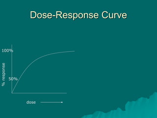
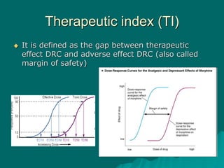
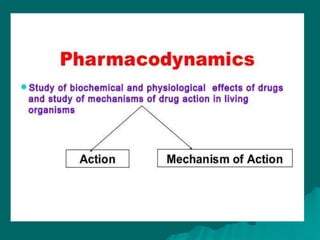
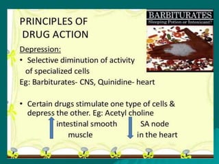
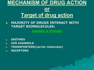
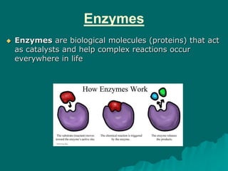
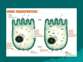
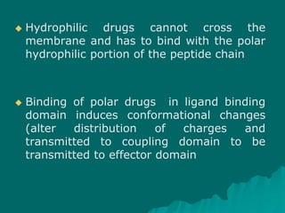
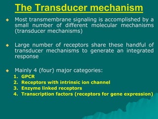
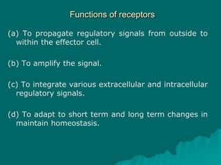
Pharmacodynamics
What is Pharmacodynamics?
What the drug does to the body when it
enters?
Defination.:
It is the study of biochemical and physiological
effects of drug and their mechanism of
action at organ level as well as cellular
level.
MECHANISM
OF DRUG
ACTION
MECHANISM OF DRUG ACTION
or
Target of drug action
 MAJORITY OF DRUGS INTERACT WITH
TARGET BIOMOLECULES:
Usually a Protein
1. ENZYMES
2. ION CHANNELS
3. TRANSPORTERS(carrier molecules)
4. RECEPTORS
1. Enzymes
Enzymes
 Enzymes are biological molecules (proteins) that act
as catalysts and help complex reactions occur
everywhere in life
 Nonspecific inhibition: Denaturation of
proteins e.g. strong acids, heavy metals,
alkalies, alcohol, phenols etc.
 Specific Inhibition:
Competitive Noncompetitive
• equilibrium
• nonequilibrium
Enzyme inhibition – common mode of drug
action
specific enzyme inhibition
 A drug may inhibit a
particular enzyme without
affecting others and
influence that particular
substrate-enzyme reaction
ultimately to influence in
the product formation
Normal
Drug + Enzyme
i) Competitive Inhibition
The drug being structurally similar competes with the
normal substrate for the catalytic binding site of the
enzyme so that the product is not formed or a
nonfunctional product is formed and a new equilibrium
is achieved in the presence of the drug.
(ii) Noncompetitive The inhibitor reacts
with an adjacent site and not with the
catalytic site, but alters the enzyme in such a
way that it loses its catalytic property.
2. Ion Channnel
 Ion channels are pore-forming membrane
proteins whose functions include establishing
a resting membrane potential, shaping action
potentials and other electrical signals by gating the
flow of ions across the cell membrane.
 Ion channels are present in the membranes of all
cells.
 Ion channels are considered to be one of the two
traditional classes of ionophoric proteins, with the
other class known as ion transporters (including
the sodium-potassium pump, sodium-calcium
exchanger, and sodium-glucose transport proteins,
amongst others)
3. Transporters
 This protein also functions as a transporter in the
blood–brain barrier. P-gp transports various
substrates across the cell membrane including.
 Substrates are translocated across membrane by
binding to specific transporters (carriers) – Solute
Carrier Proteins (SLC)
 Pump the metabolites/ions I the direction of
concentration gradient or against it
 Drugs interact with these transport
system
 Examples: Probenecid (penicillin and
uric acid), Furosmide (Na+K+2Cl-
cotransport), Hemicholinium (choline
uptake) and Vesamicol (active
transport of Ach to vesicles)
4. Receptors
 Drugs usually do not bind directly with enzymes,
channels, transporters or structural proteins, but
act through specific macromolecules – RECEPTORS
 Definition: It is defined as a macromolecule or
binding site located on cell surface or inside the
effector cell that serves to recognize the signal
molecule/drug and initiate the response to it, but
itself has no other function, e.g. G-protein coupled
receptor
 Agonist: An agent which activates a receptor to
produce an effect similar to a that of the
physiological signal molecule, e.g. Muscarine and
Nicotine)
 Antagonist: an agent which prevents the action
of an agonist on a receptor or the subsequent
response, but does not have an effect of its own,
e.g. atropine and muscarine
Some Definitions
 Inverse agonist: an agent which activates receptors to
produce an effect in the opposite direction to that of the
agonist,
 Partial agonist: An agent which activates a receptor to
produce submaximal effect but antagonizes the action of a full
agonist, e.g. pentazocine.
 Ligand: any molecule which attaches selectively to particular
receptors or sites (only binding or affinity)
 Affinity: Ability of a substrate to bind with
receptor
 Intrinsic activity (IA): Capacity to induce
functional change in the receptor
D + R DR Complex
Affinity – measure of tendency of a drug to
bind receptor; the attractiveness of drug and
receptor
– Covalent bonds are stable and essentially
irreversible
– Electrostatic bonds may be strong or
weak, but are usually reversible
Drug - Receptor Binding
Affinity
Drug Receptor Interaction
Efficacy (or Intrinsic Activity) – ability
of a bound drug to change the
receptor in a way that produces an
effect; some drugs possess affinity
but NOT efficacy
DR Complex Effect (E)
Receptors – contd.
 Two essential functions:
– Recognition of specific ligand molecule
– Transduction of signal into response
 Two Domains:
– Ligand binding domain
– Effectors Domain – undergoes functional
conformational change
Receptors – contd.
 Cell surface receptors remain floated in cell
membrane lipids
 Functions are determined by the interaction
of lipophillic or hydrophillic domains of the
peptide chain with the drug molecule
 Non-polar hydrophobic portion of the amino
acid remain buried in membrane while polar
hydrophilic remain on cell surface
 Hydrophilic drugs cannot cross the
membrane and has to bind with the polar
hydrophilic portion of the peptide chain
 Binding of polar drugs in ligand binding
domain induces conformational changes
(alter distribution of charges and
transmitted to coupling domain to be
transmitted to effector domain
Receptors – contd.
 Drugs act on Physiological receptors
and mediate responses of
transmitters, hormones, autacoids
and others – cholinergic, adrenergic
or histaminergic etc.
 Drugs may act on true drug receptors
- Benzodiazepine receptors
The Transducer mechanism
 Most transmembrane signaling is accomplished by a
small number of different molecular mechanisms
(transducer mechanisms)
 Large number of receptors share these handful of
transducer mechanisms to generate an integrated
response
 Mainly 4 (four) major categories:
1. GPCR
2. Receptors with intrinsic ion channel
3. Enzyme linked receptors
4. Transcription factors (receptors for gene expression)
A) G-protein Coupled
Receptors
 Large family of cell membrane receptors
linked to the effector
(enzyme/channel/carrier proteins) through
one or more GTP activated proteins (G-
proteins)
 All receptors has common pattern of
structural organization
 The molecule has 7 α-helical membrane
spanning hydrophobic amino acid segments
– 3 extra and 3 intracellular loops
GPCR
G Protein – these are proteins that bind to the
guanine nucleotide (GTP – guanosine
triphosphate, GDP – guanosine diphosphate)
Hydrolysis of GTP releases a phosphate group
which can act on other molecules – transmits
the signal
GTP > GDP + P
G proteins have three subunits – α (alpha),
β (beta), and γ (gamma).
β and γ subunits are tightly bound together.
α binds to GTP
Ligand binding to the transmembrane protein causes a
conformational change and release of the α subunit
The α subunit exchanges GDP > GTP and becomes
active
The α subunit meets a target and phosphorylates it
(adds a phosphate group from GTP converting it to
GDP – this is hydrolysis of GTP)
Hydrolysis = cleavage
Phosphorylation = addition of a phosphate group
Ligands
• Monoamines e.g. dopamine, histamine,
noradrenaline, acetylcholine (muscarinic)
• Nucleotides
• Lipids
• Hormones
• Glutamate
• Ca++
G-protein-coupled receptors (7-TM receptors)
Now the α subunit is bound to GDP, it
becomes inactive again and re-
associates with the transmembrane
protein and the β and γ subunits
Second Messengers
Second messengers – these are molecules
that relay signals from receptors on the cell
surface to target molecules inside the cell
Examples include IP3, Ca2+, cAMP
Allows for amplification of the signal
3.6 Signal transduction pathway
a) Interaction of receptor with Gs-protein
GS-Protein - membrane bound protein of 3 subunits (a, b, g)
- aS subunit has binding site for GDP
-GDP bound non covalently
b g
a
GDP
3. G-protein-coupled receptors (7-TM receptors)
G-proteins and Effectors
 Large number can be distinguished
by their α-subunits
GPCR - 3 Major Pathways
1. Adenylyl cyclase:cAMP pathway
2. Phospholipase C: IP3-DAG
pathway
3. Channel regulation
Adenylyl cyclase:cAMP
pathway
2. Phospholipase C:IP3-DAG pathway
 Physiological function likes,
mediates/modulates contraction,
secretion/transmission
release,eicosenoides synthesis,
neuronal excitability, intracellular
movements, membrane function,
metabolism, cell proliferation etc.
3. Channel regulation
 Activated G-proteins can open or close ion channels – Ca++,
Na+ or K+ etc.
 These effects may be without intervention of any of
above mentioned 2nd messengers – cAMP or
IP3/DAG.
 Bring about depolarization, hyperpolrization or Ca
++ changes etc.
 Gs – Ca++ channels in myocardium and skeletal
muscles
 Go and Gi – open K+ channel in heart and muscle
and close Ca+ in neurons.
 Physiological responses like changes in
inotropy, chronotropy, transmitter
release, neuronal activity and smooth
muscle relaxation follow.
 Receptors found to regulate ionic channels
through G-proteins
B) Intrinsic Ion Channel
Receptors
Intrinsic Ion Channel
Receptors
 Most useful drugs in clinical medicine act by
mimicking or blocking the actions of
endogenous ligands that regulate the flow of
ions through plasma membrane channels
 The natural ligands include acetylcholine,
serotonin, aminobutyric acid (GABA), and the
excitatory amino acids (eg, glycine,
aspartate, and glutamate)
 These cell surface receptors, also called ligand
gated ion channels, enclose ion selective channels
(for Na*, K*, Ca2* or Cl-) within their molecules.
 Agonist binding opens the channel and
causes depolarization/hyperpolarization
/changes in cytosolic ionic composition,
depending on the ion that flows through.
 The nicotinic cholinergic, GABA-A, glycine
(inhibitory), excitatory AA (kainate, NMDA or N-
methylD-aspartate, quisqualate) and 5HT3
receptors fall in this category
3. Enzyme-linked
receptors
 The agonist binding site and the catalytic
site lie respectively on the outer and inner
face of the plasma membrane
 These two domains are interconnected
through a single transmembrane stretch of
peptide chain.
 There are two major subgroups of such
receptors.
Enzyme Linked Receptors
 2 (two) types of receptors:
1. Intrinsic enzyme linked receptors
 Protein kinase or
 guanyl cyclase domain
2. JAK-STAT-kinase binding receptor
a. Intrinsic enzyme receptors
The intracellular
 Domain is either a protein kinase or guanyl
cyclase.
 In most cases the protein kinase specifically
phosphorylates tyrosine residues on
substrate proteins,
 e.g. insulin, epidermal growth factor (EGF),
nerve growth factor (NGF) receptors, but in
few it is a serine or threonine protein kinase.
 In the monomeric state, the kinase
remains inactive.
 Agonist binding induces dimerization of
receptor molecules and activates the
kinase to autophosphorylate tyrosine
residues on each other, increasing
their affinity for binding substrate
proteins and carrying forward the
cascade of tyrosine phosphorylations.
 Activated receptors catalyze phosphorylation of
tyrosine residues on different target signaling
proteins, thereby allowing a single type of activated
receptor to modulate a number of biochemical
processes
 Examples:
– Insulin - uptake of glucose and amino acids and regulate
metabolism of glycogen and triglycerides
– Trastuzumab, antagonist of a such type receptor – used in
breast cancer
JAK-STAT-kinase binding receptor
 Mechanism closely resembles that of receptor
tyrosine kinases
 Only difference - protein tyrosine kinase activity
is not intrinsic to the receptor molecule
 Uses Janus-kinase (JAK) family
 Also uses STAT (signal transducers and activators
of transcription)
 Examples – cytokines, growth hormones,
interferones etc.
JAK-STAT-kinase Receptors
4. Receptors regulating gene expression
(Transcription factors)
 In contrast to the above 3 classes of receptors,
these are intracellular (cytoplasmic or nuclear)
soluble proteins which respond to lipid soluble
chemical messengers that penetrate the cell
 The receptor protein (specific for each hormone/
regulator) is inherently capable of binding to specific
genes/ but is kept inhibited till the hormone binds
near its carboxy terminus and exposes the DNA
binding regulatory segment located in the middle of
the molecule.
 All steroidal hormones (glucocorticoids,
mineralocorticoids, androgens, estrogens,
progesterone), thyroxine, vit D and vit A function
in this manner.
 Different steroidal hormones affect different
target cells and produce different effects because
each one binds to its own receptor and directs a
unique patten of symthesis of specific proteins.
 This transduction mechanism is the slowest
in its time course of action (takes hours).
Functions of receptors
(a) To propagate regulatory signals from outside to
within the effector cell.
(b) To amplify the signal.
(c) To integrate various extracellular and intracellular
regulatory signals.
(d) To adapt to short term and long term changes in
maintain homeostasis.
Summary of Transducers
Dose-Response Relationship
 Dose-plasma concentration
 Plasma concentration (dose)-
response relationship
E =
Emax X [D]
KD + [D]
E is observed effect of drug dose [D], Emax = maximum response,
Kd = dissociation constant of drug receptor complex
When a drug is administered systemically, the
dose-response relationship has two components:
Dose-Response Curve
dose
%response
100%
50%
Dose-Response Curve
 Advantages:
– A wide range of drug doses can easily be
displayed on a graph
– Potency and efficacy can be compared
– Comparison of study of agonists and
antagonists become easier
Potency and efficacy
 Potency: It is the amount of drug required to produce
a certain response
 Efficacy: Maximal response that can be elicited by a
drug
Response
Drug in log conc.
1 2 3 4
Therapeutic index (TI)
 Therapeutic Index =
Median Lethal Dose (LD50)
Median Effective dose (ED50)
Idea of margin of safety Margin of Safety
Therapeutic index (TI)
 It is defined as the gap between therapeutic
effect DRC and adverse effect DRC (also called
margin of safety)
Combined Effects of Drugs
When two or more drugs are given simultaneously or
in quick succession may be either indifferent to each
other or exhibit synergism or antagonism.
The interaction may take place at pharmacokinetic
level or at pharmacodynamic level.
– Additive effect (1 + 1 = 2)
 Aspirin+paracetamol,
 amlodipine+atenolol
– Supraadditive effect (1 + 1 = 4)
 Sulfamethoxazole+trimethoprim,
 levodopa+carbidopa,
 acetylcholine+physostigmine
Drug Synergism (Greek: Syn-together; ergon-work)
when the action of one drug is facilitated or increased by the other,
they are said to be synergistic.
In a synergistic pair, both the drugs can have action in the same
direction or given alone one may be inactive but still enhance the
action of the other when given together.
Synergism can be:
 Drug Antagonism:
1. Physical: Charcoal
2. Chemical: KMNO4, Chelating agents
3. Physiological antagonism: Histamine and
adrenaline in bronchial asthma, Glucagons
and Insulin
4. Receptor antagonism
…. Contd.
 Receptor antagonism:
1. Competitive antagonism (equilibrium)
2. Competitive (non equilibrium)
3. Non-competitive antagonism
Drug antagonism DRC
Drug antagonism DRC – non-
competitive antagonismResponse
Shift to the right
and lowered response
Drug in log conc.
Agonist
Agonist
+ CA (NE)

Pharmacodynamics

  • 2.
  • 3.
    What is Pharmacodynamics? Whatthe drug does to the body when it enters? Defination.: It is the study of biochemical and physiological effects of drug and their mechanism of action at organ level as well as cellular level.
  • 12.
  • 13.
    MECHANISM OF DRUGACTION or Target of drug action  MAJORITY OF DRUGS INTERACT WITH TARGET BIOMOLECULES: Usually a Protein 1. ENZYMES 2. ION CHANNELS 3. TRANSPORTERS(carrier molecules) 4. RECEPTORS
  • 14.
  • 15.
    Enzymes  Enzymes arebiological molecules (proteins) that act as catalysts and help complex reactions occur everywhere in life
  • 18.
     Nonspecific inhibition:Denaturation of proteins e.g. strong acids, heavy metals, alkalies, alcohol, phenols etc.  Specific Inhibition: Competitive Noncompetitive • equilibrium • nonequilibrium Enzyme inhibition – common mode of drug action
  • 19.
    specific enzyme inhibition A drug may inhibit a particular enzyme without affecting others and influence that particular substrate-enzyme reaction ultimately to influence in the product formation Normal Drug + Enzyme
  • 20.
    i) Competitive Inhibition Thedrug being structurally similar competes with the normal substrate for the catalytic binding site of the enzyme so that the product is not formed or a nonfunctional product is formed and a new equilibrium is achieved in the presence of the drug.
  • 21.
    (ii) Noncompetitive Theinhibitor reacts with an adjacent site and not with the catalytic site, but alters the enzyme in such a way that it loses its catalytic property.
  • 23.
  • 24.
     Ion channelsare pore-forming membrane proteins whose functions include establishing a resting membrane potential, shaping action potentials and other electrical signals by gating the flow of ions across the cell membrane.
  • 26.
     Ion channelsare present in the membranes of all cells.  Ion channels are considered to be one of the two traditional classes of ionophoric proteins, with the other class known as ion transporters (including the sodium-potassium pump, sodium-calcium exchanger, and sodium-glucose transport proteins, amongst others)
  • 27.
    3. Transporters  Thisprotein also functions as a transporter in the blood–brain barrier. P-gp transports various substrates across the cell membrane including.  Substrates are translocated across membrane by binding to specific transporters (carriers) – Solute Carrier Proteins (SLC)  Pump the metabolites/ions I the direction of concentration gradient or against it
  • 28.
     Drugs interactwith these transport system  Examples: Probenecid (penicillin and uric acid), Furosmide (Na+K+2Cl- cotransport), Hemicholinium (choline uptake) and Vesamicol (active transport of Ach to vesicles)
  • 30.
    4. Receptors  Drugsusually do not bind directly with enzymes, channels, transporters or structural proteins, but act through specific macromolecules – RECEPTORS  Definition: It is defined as a macromolecule or binding site located on cell surface or inside the effector cell that serves to recognize the signal molecule/drug and initiate the response to it, but itself has no other function, e.g. G-protein coupled receptor
  • 31.
     Agonist: Anagent which activates a receptor to produce an effect similar to a that of the physiological signal molecule, e.g. Muscarine and Nicotine)  Antagonist: an agent which prevents the action of an agonist on a receptor or the subsequent response, but does not have an effect of its own, e.g. atropine and muscarine Some Definitions
  • 32.
     Inverse agonist:an agent which activates receptors to produce an effect in the opposite direction to that of the agonist,  Partial agonist: An agent which activates a receptor to produce submaximal effect but antagonizes the action of a full agonist, e.g. pentazocine.  Ligand: any molecule which attaches selectively to particular receptors or sites (only binding or affinity)
  • 34.
     Affinity: Abilityof a substrate to bind with receptor  Intrinsic activity (IA): Capacity to induce functional change in the receptor
  • 35.
    D + RDR Complex Affinity – measure of tendency of a drug to bind receptor; the attractiveness of drug and receptor – Covalent bonds are stable and essentially irreversible – Electrostatic bonds may be strong or weak, but are usually reversible Drug - Receptor Binding Affinity
  • 36.
    Drug Receptor Interaction Efficacy(or Intrinsic Activity) – ability of a bound drug to change the receptor in a way that produces an effect; some drugs possess affinity but NOT efficacy DR Complex Effect (E)
  • 37.
    Receptors – contd. Two essential functions: – Recognition of specific ligand molecule – Transduction of signal into response  Two Domains: – Ligand binding domain – Effectors Domain – undergoes functional conformational change
  • 38.
    Receptors – contd. Cell surface receptors remain floated in cell membrane lipids  Functions are determined by the interaction of lipophillic or hydrophillic domains of the peptide chain with the drug molecule  Non-polar hydrophobic portion of the amino acid remain buried in membrane while polar hydrophilic remain on cell surface
  • 39.
     Hydrophilic drugscannot cross the membrane and has to bind with the polar hydrophilic portion of the peptide chain  Binding of polar drugs in ligand binding domain induces conformational changes (alter distribution of charges and transmitted to coupling domain to be transmitted to effector domain
  • 40.
    Receptors – contd. Drugs act on Physiological receptors and mediate responses of transmitters, hormones, autacoids and others – cholinergic, adrenergic or histaminergic etc.  Drugs may act on true drug receptors - Benzodiazepine receptors
  • 41.
    The Transducer mechanism Most transmembrane signaling is accomplished by a small number of different molecular mechanisms (transducer mechanisms)  Large number of receptors share these handful of transducer mechanisms to generate an integrated response  Mainly 4 (four) major categories: 1. GPCR 2. Receptors with intrinsic ion channel 3. Enzyme linked receptors 4. Transcription factors (receptors for gene expression)
  • 44.
    A) G-protein Coupled Receptors Large family of cell membrane receptors linked to the effector (enzyme/channel/carrier proteins) through one or more GTP activated proteins (G- proteins)  All receptors has common pattern of structural organization  The molecule has 7 α-helical membrane spanning hydrophobic amino acid segments – 3 extra and 3 intracellular loops
  • 46.
  • 47.
    G Protein –these are proteins that bind to the guanine nucleotide (GTP – guanosine triphosphate, GDP – guanosine diphosphate) Hydrolysis of GTP releases a phosphate group which can act on other molecules – transmits the signal GTP > GDP + P
  • 49.
    G proteins havethree subunits – α (alpha), β (beta), and γ (gamma). β and γ subunits are tightly bound together. α binds to GTP
  • 50.
    Ligand binding tothe transmembrane protein causes a conformational change and release of the α subunit The α subunit exchanges GDP > GTP and becomes active The α subunit meets a target and phosphorylates it (adds a phosphate group from GTP converting it to GDP – this is hydrolysis of GTP) Hydrolysis = cleavage Phosphorylation = addition of a phosphate group
  • 51.
    Ligands • Monoamines e.g.dopamine, histamine, noradrenaline, acetylcholine (muscarinic) • Nucleotides • Lipids • Hormones • Glutamate • Ca++ G-protein-coupled receptors (7-TM receptors)
  • 52.
    Now the αsubunit is bound to GDP, it becomes inactive again and re- associates with the transmembrane protein and the β and γ subunits
  • 54.
    Second Messengers Second messengers– these are molecules that relay signals from receptors on the cell surface to target molecules inside the cell Examples include IP3, Ca2+, cAMP Allows for amplification of the signal
  • 55.
    3.6 Signal transductionpathway a) Interaction of receptor with Gs-protein GS-Protein - membrane bound protein of 3 subunits (a, b, g) - aS subunit has binding site for GDP -GDP bound non covalently b g a GDP 3. G-protein-coupled receptors (7-TM receptors)
  • 56.
    G-proteins and Effectors Large number can be distinguished by their α-subunits
  • 57.
    GPCR - 3Major Pathways 1. Adenylyl cyclase:cAMP pathway 2. Phospholipase C: IP3-DAG pathway 3. Channel regulation
  • 58.
  • 59.
  • 60.
     Physiological functionlikes, mediates/modulates contraction, secretion/transmission release,eicosenoides synthesis, neuronal excitability, intracellular movements, membrane function, metabolism, cell proliferation etc.
  • 61.
    3. Channel regulation Activated G-proteins can open or close ion channels – Ca++, Na+ or K+ etc.
  • 62.
     These effectsmay be without intervention of any of above mentioned 2nd messengers – cAMP or IP3/DAG.  Bring about depolarization, hyperpolrization or Ca ++ changes etc.
  • 63.
     Gs –Ca++ channels in myocardium and skeletal muscles  Go and Gi – open K+ channel in heart and muscle and close Ca+ in neurons.
  • 64.
     Physiological responseslike changes in inotropy, chronotropy, transmitter release, neuronal activity and smooth muscle relaxation follow.
  • 65.
     Receptors foundto regulate ionic channels through G-proteins
  • 67.
    B) Intrinsic IonChannel Receptors
  • 68.
    Intrinsic Ion Channel Receptors Most useful drugs in clinical medicine act by mimicking or blocking the actions of endogenous ligands that regulate the flow of ions through plasma membrane channels  The natural ligands include acetylcholine, serotonin, aminobutyric acid (GABA), and the excitatory amino acids (eg, glycine, aspartate, and glutamate)
  • 69.
     These cellsurface receptors, also called ligand gated ion channels, enclose ion selective channels (for Na*, K*, Ca2* or Cl-) within their molecules.
  • 70.
     Agonist bindingopens the channel and causes depolarization/hyperpolarization /changes in cytosolic ionic composition, depending on the ion that flows through.
  • 71.
     The nicotiniccholinergic, GABA-A, glycine (inhibitory), excitatory AA (kainate, NMDA or N- methylD-aspartate, quisqualate) and 5HT3 receptors fall in this category
  • 72.
  • 73.
     The agonistbinding site and the catalytic site lie respectively on the outer and inner face of the plasma membrane  These two domains are interconnected through a single transmembrane stretch of peptide chain.  There are two major subgroups of such receptors.
  • 74.
    Enzyme Linked Receptors 2 (two) types of receptors: 1. Intrinsic enzyme linked receptors  Protein kinase or  guanyl cyclase domain 2. JAK-STAT-kinase binding receptor
  • 75.
    a. Intrinsic enzymereceptors The intracellular  Domain is either a protein kinase or guanyl cyclase.  In most cases the protein kinase specifically phosphorylates tyrosine residues on substrate proteins,  e.g. insulin, epidermal growth factor (EGF), nerve growth factor (NGF) receptors, but in few it is a serine or threonine protein kinase.
  • 77.
     In themonomeric state, the kinase remains inactive.  Agonist binding induces dimerization of receptor molecules and activates the kinase to autophosphorylate tyrosine residues on each other, increasing their affinity for binding substrate proteins and carrying forward the cascade of tyrosine phosphorylations.
  • 80.
     Activated receptorscatalyze phosphorylation of tyrosine residues on different target signaling proteins, thereby allowing a single type of activated receptor to modulate a number of biochemical processes  Examples: – Insulin - uptake of glucose and amino acids and regulate metabolism of glycogen and triglycerides – Trastuzumab, antagonist of a such type receptor – used in breast cancer
  • 81.
    JAK-STAT-kinase binding receptor Mechanism closely resembles that of receptor tyrosine kinases  Only difference - protein tyrosine kinase activity is not intrinsic to the receptor molecule  Uses Janus-kinase (JAK) family  Also uses STAT (signal transducers and activators of transcription)  Examples – cytokines, growth hormones, interferones etc.
  • 82.
  • 83.
    4. Receptors regulatinggene expression (Transcription factors)  In contrast to the above 3 classes of receptors, these are intracellular (cytoplasmic or nuclear) soluble proteins which respond to lipid soluble chemical messengers that penetrate the cell  The receptor protein (specific for each hormone/ regulator) is inherently capable of binding to specific genes/ but is kept inhibited till the hormone binds near its carboxy terminus and exposes the DNA binding regulatory segment located in the middle of the molecule.
  • 85.
     All steroidalhormones (glucocorticoids, mineralocorticoids, androgens, estrogens, progesterone), thyroxine, vit D and vit A function in this manner.  Different steroidal hormones affect different target cells and produce different effects because each one binds to its own receptor and directs a unique patten of symthesis of specific proteins.
  • 86.
     This transductionmechanism is the slowest in its time course of action (takes hours).
  • 87.
    Functions of receptors (a)To propagate regulatory signals from outside to within the effector cell. (b) To amplify the signal. (c) To integrate various extracellular and intracellular regulatory signals. (d) To adapt to short term and long term changes in maintain homeostasis.
  • 88.
  • 89.
    Dose-Response Relationship  Dose-plasmaconcentration  Plasma concentration (dose)- response relationship E = Emax X [D] KD + [D] E is observed effect of drug dose [D], Emax = maximum response, Kd = dissociation constant of drug receptor complex When a drug is administered systemically, the dose-response relationship has two components:
  • 90.
  • 91.
    Dose-Response Curve  Advantages: –A wide range of drug doses can easily be displayed on a graph – Potency and efficacy can be compared – Comparison of study of agonists and antagonists become easier
  • 92.
    Potency and efficacy Potency: It is the amount of drug required to produce a certain response  Efficacy: Maximal response that can be elicited by a drug Response Drug in log conc. 1 2 3 4
  • 93.
    Therapeutic index (TI) Therapeutic Index = Median Lethal Dose (LD50) Median Effective dose (ED50) Idea of margin of safety Margin of Safety
  • 94.
    Therapeutic index (TI) It is defined as the gap between therapeutic effect DRC and adverse effect DRC (also called margin of safety)
  • 95.
    Combined Effects ofDrugs When two or more drugs are given simultaneously or in quick succession may be either indifferent to each other or exhibit synergism or antagonism. The interaction may take place at pharmacokinetic level or at pharmacodynamic level.
  • 96.
    – Additive effect(1 + 1 = 2)  Aspirin+paracetamol,  amlodipine+atenolol – Supraadditive effect (1 + 1 = 4)  Sulfamethoxazole+trimethoprim,  levodopa+carbidopa,  acetylcholine+physostigmine Drug Synergism (Greek: Syn-together; ergon-work) when the action of one drug is facilitated or increased by the other, they are said to be synergistic. In a synergistic pair, both the drugs can have action in the same direction or given alone one may be inactive but still enhance the action of the other when given together. Synergism can be:
  • 100.
     Drug Antagonism: 1.Physical: Charcoal 2. Chemical: KMNO4, Chelating agents 3. Physiological antagonism: Histamine and adrenaline in bronchial asthma, Glucagons and Insulin 4. Receptor antagonism
  • 101.
    …. Contd.  Receptorantagonism: 1. Competitive antagonism (equilibrium) 2. Competitive (non equilibrium) 3. Non-competitive antagonism
  • 102.
  • 103.
    Drug antagonism DRC– non- competitive antagonismResponse Shift to the right and lowered response Drug in log conc. Agonist Agonist + CA (NE)