
Recommended
More Related Content
What's hot
What's hot (20)
Similar to Atomic absorption spectroscopy (AAS)
Similar to Atomic absorption spectroscopy (AAS) (20)
Recently uploaded
Recently uploaded (20)
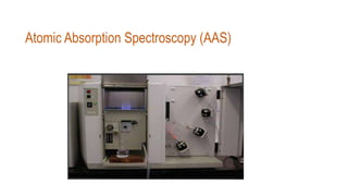
Atomic absorption spectroscopy (AAS)
- 1. Atomic Absorption Spectroscopy (AAS)
- 2. Content: • Introductions • Principal of AAS • Its Instrumentation & • Application
- 3. Introduction • Atomic Absorption Spectroscopy developed by Alan Walsh in the mid 1950s • Various categories of samples can be analysed for their elemental composition. • Concentration up to ppm (parts per million) can be determined.
- 4. • In analytical chemistry, Atomic absorption spectroscopy (AAS) is a technique for determining the concentration of a particular metal element (e.g. Fe, Cu, Al, Pb, Ca, Zn)in a sample ( water, medicine, food) . • Atomic absorption spectroscopy can be used to analyze the concentration of over 62 different metals in a solution.
- 5. Principal • The technique uses basically the principle that free atoms (gas) generated in an atomizer can absorb radiation at specific frequency. • Atomic-absorption spectroscopy quantifies the absorption of ground state atoms in the gaseous state . • The atoms absorb ultraviolet or visible light and make transitions to higher electronic energy levels. The analyte concentration is determined from the amount of absorption.
- 6. • An “absorption spectrum” is the absorption of light as a function of wavelength. • The spectrum of an atom depends on its energy level structure. • Absorption spectra are useful for determination of elements.
- 7. Instrumentation • Hollow Cathode Lamp are the most common radiation source in AAS. • It contains a tungsten anode and a hollow cylindrical cathode made of the element to be determined. • These are sealed in a glass tube filled with an inert gas (neon or argon ) . • Each element has its own unique lamp which must be used for that analysis . Radiation source HCL
- 8. • When a dc voltage of 300–500 V is put across the anode and cathode,the atoms of the inert gas undergo ionisation and rapidly attracted by the cathode. • The fast moving ions strike the surface of the cathode and physically displace the surface metal atoms of the cathode and produce an atomic cloud. This process called sputtering. • A portions of the sputtering metal atoms are in exited states.
- 9. • They emit their characteristics radiation as they return to the ground state. • Eventually,the metal atoms diffuse back to the cathode surface or to the glass walls of tube and are redeposited.
- 10. Nebulizer: It creates a fine spray of the sample in which the sample, fuel & oxidant are mixed thoroughly for the introducing the same into the flame. Automizer Flame automizer : This sample solution is first nebulized. Then,the derosol containing only the droplets of the appropriate size is projected on the flame where the solvent get vapourized and finally dissociated to give free atoms ( Ground state).
- 11. • Intense beam of the analytical light ( radiation) For the element of interest which is passed through the free atoms originating from the automizer. • The wavelength of the light corresponds to the amount of energy required to excite an electron from the ground .
- 12. Monochromator: In monochromator scattered light of undesirable wave lengths are removed and only the photons of analytical light line of desired wave length passing through the flame are isolated. As a result only a narrow spectral line impinges on the detector ( photomultiplier tube ). Detecter— photomultiplier tube (PMT): PMT determine the intensity of Photon of the analytical light line coming out from the monochromator. The detector records that reduction as absorption. That absorption is shown on output device by the data system
- 14. • Estimation of trace elements in biological fluid like blood, urine, etc. • Estimation of trace elements like Copper, Nickle and Zinc in food products. • Estimation of Magnesium, Zinc in blood. • Estimation of Zinc in Zinc insulin solution. • Estimation of Lead in Calcium carbonate and petrol. • Estimation of elements in soil samples, water supply, effluents, ceramics, etc. Application
- 15. Referance: Techniques and methods in biology by K.L. Ghatak Instrumental methods of chemical analysis by Gurdeep R. Chatwal and K. anand https://en.m.wikipedia.org/wiki/Atomic_absorption_spectroscopy