
Recommended
More Related Content
What's hot
What's hot (20)
Similar to Helicobacter pylori
Similar to Helicobacter pylori (20)
Recently uploaded
Recently uploaded (20)
Helicobacter pylori
- 1. Done By B.Saraswathi I M.Sc Applied Microbiology
- 2. Scientific Classification Domain: Bacteria Phylum: Proteobacteria Class: Epsilonproteobacteria Order: Campylobacterales Family: Helicobacteraceae Genus: Helicobacter Species: H. pylori Binomial name: Helicobacter pylori
- 3. Previously called Campylobacter pylori. 1800s-Spirochetes Identified in 1982 by Australian Scientists, Barry Marshal and Robin Warren in Chronic gastritis and gastric ulcers. 2005-Nobel Prize in Medicine for the discovery. About 50% of worlds population have this bacteria in the upper IT.
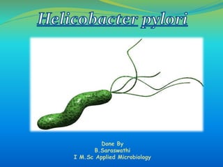
- 4. Morphology Gram Negative Helical or Spiral rod Motile with Unipolar tuft of lophotrichous flagella(4-6) Size - 2-4 µm x 0.5-1.0 µm Smooth cell wall with unusual fatty acids Found in Acidic environments of pH 2 or less.
- 5. Gram Staining-Gram Negative rod
- 6. They are micro-aerophilic, require 5-10% CO2 and high humidity. They are fastidious organism. Blood agar and chocolate agar after incubation for 2-5 days. Colonies are circular, convex and translucent and grow bigger than 2 mm in diameter. On Egg Yolk Emulsion Agar – give large (~ 3mm) and red color colony against yellow background.
- 7. BIOCHEMICALS CATALASE- +VE OXIDASE- +VE UREASE- +VE
- 10. Route Of Entry – Oral Transmission- Not clear Site – Gastric Mucosa(Gastric Antrum) Adhesion - Adhesins bind to membrane associated Carbohydrates and lipids and helps the bacteria to adhere to the epithelial cells. Acidity – Urea is broken down into Ammonia and CO2,so the pH levels increase thereby reducing the acidity for survival of the Bacteria.
- 11. Inflammation- Following attachment to the epithelial cells, the Type IV secretion system expressed by cag injects the inflammation agent- Peptidoglycan from their own cell to the host cells. The PG is recognized by the cytoplasmic pattern recognition receptor(Nod 1) to stimulate cytokines which lead to inflammation. The type IV secretion system also injects cagA into the stomach’s epithelial cells where it disrupts the cellular activities. Other toxic factors- Ammonia produced and other products of H.pylori including proteases,vacA and certain phospholipases damage the cells.
- 13. 80% of individuals affected are asymptomatic. Gastritis - abdominal pain, acid reflux, nausea and vomiting. If untreated for a long time leads to several complications like Gastro esophageal reflux disease(GERD), MALT associated Lymphoma,Peptic ulcers(common) and cancers of Esophagus and Stomach. It can also lead to urticaria sometimes.
- 14. Peptic Ulcer MALT Lymphoma Gastric Adenocarcinoma Mild Gastritis
- 16. Lab diagnosis Samples:Stool,Blood,Biopsy Diagnosis Endoscopic methods Rapid Urease Test Histology Culture PCR Non endoscopic methods Serology Urea breath test Fecal Antigen test
- 17. Rapid Urease Test Rapid diagnostic test Biopsy sample is placed in a medium containing Urea and Phenol red. Urease if produced by the bacteria hydrolyses urea to ammonia which changes the color of the medium from yellow to Red.
- 18. Hematoxylin Eosin Staining Giemsa Stain Genta Stain Immunohistochemical Stain Warthin Starry Silver Stain
- 20. Culture Egg yolk Emulsion Agar (EYE) Skirrows and Dents selective media On Modified Columbia urea agar (MCUA) – give small-middle size rounded and creamy color colony. Change in the color of the slant MCUA tube from orange to pink. On Marshall’s Brain Heart Infusion medium along with Vancomycin, Nalidixic acid and Amphotericin – give discrete dome shaped colonies.
- 21. Columbia Blood Agar Skirrows and Dent Selective Medium
- 22. IgG is detected in the patients serum sample. Sandwich ELISA Method is common.
- 23. UREA Breath Test
- 24. •Stool is collected in tubes with buffer solution to stabilize the antigens •Test cassette contains colloid gold particles conjugated to the antibodies •Below, NC membrane with result and control region •If positive, Pink band is formed.