
Recommended
More Related Content
What's hot
What's hot (20)
Similar to Lower respiratory tract disorders
Similar to Lower respiratory tract disorders (20)
More from SanjaiKokila
More from SanjaiKokila (12)
Recently uploaded
Recently uploaded (20)
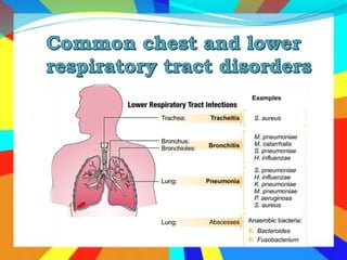
Lower respiratory tract disorders
- 3. Acute inflammation of the tracheobronchial tree Self-limiting Caused by infections or irritants Etiology: Acute infectious bronchitis often part an acute (URTI or with secondary bacterial infection Acute irritative bronchitis - various mineral and vegetable dusts, volatile solvents, tobacco or other smoke.
- 4. Acute Bronchitis cont… Clinical presentation Acute infectious bronchitis is often preceded by symptoms of URTI, coryza, malaise, chilliness, slight fever, back and muscle pain and sore throat. Onset of cough usually signals onset of bronchitis- initially dry but progresses to be productive. Fever to 38.80c may be present up to 3-5 days Persistent fever may suggest complication like pneumonia. Few pulmonary signs: scattered rhonchi and wheezes
- 5. Acute Bronchitis cont… Treatment: General management Often symptomatic. • Rest until fever subsides. • Oral fluids should be taken more:- this facilitates sputum expectoration
- 6. Acute Bronchitis cont… Anti-microbial treatment is indicated: High-grade fever and Purulent sputum For cough: Drug treatment should not be routinely employed Dextromethorphan hydrobromide, 15 – 30 mg p.o. 3 to 4 times a day. For children: 6-12 yrs, 7.5-15 mg; 2-6 yrs, 7.5 mg 3-4 times a day. Codeine phosphate, 10 - 20 mg p.o. 3 – 4 times a day. For children: 0.5 mg/kg p.o. qid.
- 7. Acute Bronchitis cont… For productive cough Guaifenesin, 200- 400 mg P.O. QID; for children: 6- 12 yrs, 100-200 mg; 2 years, 50-100 mg P.O. QID. Antibiotic treatment is indicated when bronchitis is complicated by bacterial infections and choice of antibiotics should be based on gram stain result.
- 8. Acute Bronchitis cont… First line: Amoxicillin, 250- 500 mg P.O. TID, for children: 20 – 40 mg/kg/day P.O. in 3 divided doses or Ampicillin, 500 mg P.O. daily, in 4-divided dose for 5-7 days Alternative: Erythomycin, 250-500 mg qid p.o., in 4 divided doses for 7 days. For children: 30-50 mg/kg/day p.o. in 4 divided doses; 15-20 mg/kg/day i.v over 5 minutes in 3-4 divided doses. or Tetracycline, 250-500 mg qid, for 5-7 days or Sulfamethoxazole + trimethoprim, 800mg/160 mg. p.o. bid for 7 days. For children 6 weeks – 5 months, 100/20 mg; 6 months – 5 yrs, 200/40 mg; 6 – 12 yrs, 400/80 mg bid.
- 9. 9
- 10. An inflammation and infection of lung and lung parenchyma Inflammatory infiltrate in alveoli (consolidation) Common illness which occurs in all age group Leading cause of Morbidity & Mortality in Infants and Older People & people who are chronically & terminal ill. 10
- 11. Pneumonia cont’d… Normally, lungs are well protected For pneumonia to occur, at least one of the following three conditions must occur: Failure or defect in host defenses Exposure to very virulent pathogens Exposure to an overwhelming load of pathogens 11
- 12. Pneumonia cont’d… Host Defenses Natural barriers of the body Antibodies or immunoglobulin production Cellular immunity by phagocytosis 12
- 13. Pneumonia cont’d… Failure of Host Defenses Hair of nares Absence of cough or epiglottic (gag) reflex Dysfunctional muco-ciliary blanket Local production of secretory IGA is reduced Normal flora adhering to mucosal cells of the oro- pharynx 13
- 14. Pneumonia cont’d… Failure of Host Defenses cont’d Immune response stunted: Defective neutrophil function Decreased immunoglobulin production Prior viral infection (common cold) Compromises overall immunity Systemic sepsis weakens immune response 14
- 15. Pneumonia cont’d… Failure of Host Defenses cont’d Immunosuppressive drugs decrease host response Cigarette/second-hand exposure or other toxic fume also weaken lung system Change in mental status (coma, seizure, drug intoxication) 15
- 16. Pneumonia cont’d… Predisposing factors Decrease level of consciousness Tracheal intubation Malnutrition Alcohol, cigarette smoking, Stroke, seizures Advanced age Asthma, cystic fibrosis, anemia etc Increased gram-negative bacilli in oro-pharynx 16
- 17. Pneumonia cont’d… Cause Pneumonia can result from a variety of causes: Bacterial (Gram-positive bacteria, Gram-negative bacteria, "Atypical" bacteria) Virus Fungi Parasites Chemical or physical injury to the lungs 17
- 18. Pneumonia cont’d… Streptococcus pneumoniae Most common cause of uncomplicated in all age groups Atypical Bacteria Mycoplasma Chlamydia Legionella 18
- 19. Pneumonia cont’d… Classification Based on causative agent Bacterial or typical pneumonia, Atypical pneumonia According to the X-ray appearance Lobar pneumonia- homogeneous consolidation of one or more lung lobes Broncho- pneumonia- multiple patchy shadows in a localized or segmental area. 19
- 21. Bronchopneumonia: Lobar pneumonia: • Distributed in a patchy fashion, having originated in one or more localized areas within the bronchi and extending to the adjacent surrounding lung parenchyma • Usually 2ry to other conditions - Viral infections - Obstruction of a bronchus (foreign body or neoplasm) - Inhalation of irritant gases - Major surgery - Chronic debilitating diseases, malnutrition More common than lobar One or more lobes is involved S. Pneumoniae. Previously healthy individuals. Abrupt onset. Unilateral stabbing chest pain on inspiration (due to fibrinous pleurisy).
- 23. Pneumonia cont’d… According to the setting or clinical and environmental Community-acquired pneumonia(CAP) Hospital-acquired pneumonia(HAP) Aspiration pneumonia Pneumonia in immuno-compromised host 23
- 24. Pneumonia cont’d… Community-acquired pneumonia (CAP) Infectious pneumonia in a person who has not recently been hospitalized Common type of pneumonia. Infection usually spread by droplet inhalation. 24
- 25. CAP cont’d… Risk factors include: Cigarette smoking Alcohol intake Pre-existing lung disease Old age, etc. 25
- 26. CAP cont’d… Common causes Streptococcus pneumoniae (the most common cause) Viruses The atypical bacteria Haemophilus influenza 26
- 27. CAP cont’d… Streptococcus pneumoniae ( pneumococcal pneumonia ) commonest cause Mycoplasma pneumoniae Chlamydia pneumoniae Haemophilus influenza Oral anaerobic bacteria Staphylococcus aureus Legionella pneumophila Mycobacterium tuberculosis
- 28. Severe forms of CAP Seriously ill should be treated as inpatient. Criteria for hospitalization of patients with pneumonia are: RR of >28/min , tachycardia >140/min SBP <90mm Hg (hypotension) Hypoxemia (arterial PO2 < 60mm hg) New onset of confusion or ALOC Unstable /significant co-morbidity (e.g. HF , uncontrolled diabetes, chronic renal insufficiency ,alcoholism , immunosuppresion ) Multilobar pneumonia Pleural effusion and complicated result
- 29. Other conditions in which inpatient management may be advisable: • Elderly patient >65 yrs of age • Leukopenia <5000 WBC/ml • Pneumonia caused by st. Aureus or gram negative bacilli • Suppurative complications e.g. Empyema, arthritis, meningitis, endocarditis • Failure of outpatient treatment • Inability to take oral medication or persistent vomiting
- 30. Pneumonia cont’d… Hospital-acquired pneumonia (HAP) Also called nosocomial pneumonia, that acquired during or after hospitalization - at least 48 hours after admission. Up to 5% of patients admitted to a hospital for other causes subsequently develop pneumonia. The presence of a new or progressive infiltrates of chest x-ray , plus at least two of the following • Other findings: dyspnea, hypoxemia and chest pain 30
- 31. Pneumonia cont’d…Nosocomial pneumonia is the 2nd most common hospital-acquired infections. Nosocomial pneumonia is the leading cause of death from hospital-acquired infections. 31
- 32. Pneumonia cont’d… Hospitalized patients may have many risk factors for pneumonia: Mechanical ventilation (VAP) Prolonged malnutrition Underlying heart and lung diseases Decreased amounts of stomach acid Immune disturbances. 32
- 33. Pneumonia cont’d… Causative agent More commonly by G(-) organisms, esp. P. aeruginosa Enterobacteriaceae (klebsiella, Enterobacter, Serratia) or MRSA. E.coli H. influenza 33
- 34. Pneumonia cont’d… Methicillin-resistant Staphylococcus aureus (MRSA) seen more commonly in patients Received corticosteroids Undergone mechanical ventilation >5 days Presented with chronic lung disease Had prior antibiotics therapy 34
- 35. Aspiration pneumonia This occurs when large amount of oropharyngeal or gastric contents are aspirated into the LRT. Aspiration occurs more frequently in patients with: • Decreased level of consciousness (alcoholism, seizure, strokes or general anesthesia) • Neurologic dysfunction of oropharynx and swallowing disorders. • People with periodontal disease are affected more. Common Etiologic agents : It is often polymicrobial • Anerobic organisms in the oral cavity • Enterobateriacae • S. pneumoniae • S.aureus
- 36. Pneumonia in Immunocompromised hosts such as transplant recipients, HIV infected patients, and patients on Chemotherapy etc. are prone to develop pneumonia. etiologic agents are Common bacterial causes of CAP : St. Pnumoniae , H.influenzae, Mycoplasma Gram negative organisms : enterobacteriaceae Funguses such as Pneumocystis carinii ( jerovecii ), C. neoformans , Histoplasmosis , Aspergillus Mycobaterium tuberculosis Viruses : HSV , CMV
- 37. Pathophysiology of Bacterial pneumonia Port of Entry of bacteria to Lungs Inhalation Aspiration -Contiguous extension -Hematogenous
- 38. Pneumonia cont’d… Pathophysiology…… Once inside, bacteria may invade the spaces b/n cells and b/n alveoli through connecting pores. Invasion triggers the immune system to send neutrophils Neutrophils & macrophages engulf and kill the offending organisms The alveolar macrophages also initiate the inflammatory response 38
- 39. Pneumonia cont’d… Pathophysiology…… It releases cytokines, causing a general activation of the immune system. Neutrophils, bacteria and fluid from surrounding blood vessels fill the alveoli Interrupt normal oxygen transportation and venous blood entering the lungs passes through the under ventilated area. 39
- 40. Pneumonia cont’d… Pathophysiology…… Bacteria often travel from an infected lung into the bloodstream, causing serious or even fatal illness such as septic shock Bacteria can also travel to the area between the lungs and the chest wall (the pleural cavity) causing a complication called an empyema. 40
- 41. Pneumonia cont’d… Generally Four stage of pathophysiological change occur due to pneumonia 1. Congestion- occurs during the first 24 hrs Out pouring of fluid from tissue to alveoli- b/se of inflammatory process. Only a few neutrophils are seen at this stage. 41
- 42. Pneumonia cont’d… 2. Red hepatization - Lungs look like the liver There is massive capillary dilation Characterized microscopically by the presence of many RBC, neutrophils, micro-organisms , fibrins in the alveolar spaces 42
- 43. Pneumonia cont’d… 3. Gray hepatization The lung is dry, friable and gray-brown to yellow as a consequence of a persistent fibrinopurulent exudates WBC and fibrin consolidate the alveoli and lung Second and third stages last for 2 to 3 days each 43
- 44. Pneumonia cont’d… 4. Resolution Characterized by enzymatic digestion of the alveolar exudate; Resorption, phagocytosis or coughing up of the residual debris and Restoration of the pulmonary architecture. 44
- 45. Pneumonia cont’d… Clinical manifestations Cough producing greenish or yellow sputum High fever that may be accompanied by shaking chills Shortness of breath Tachy pnea Pleuritic chest pain Headaches 45
- 46. Pneumonia cont’d… Clinical manifestations… Sweaty and clammy (moist) skin, Loss of appetite Fatigue Blueness of the skin Nausea, vomiting Mood swings Joint pains or muscle aches 46
- 47. Pneumonia cont’d… Diagnosis History Physical examination Inspection Increase respiratory rate cyanosis Palpation Increase vibration of the chest when speaking The way of chest expands 47
- 48. Pneumonia cont’d… Percussion Dullness Auscultation A lack normal breath sounds Crackle sounds Increase loudness of whispered speech Ego phony 48
- 49. Pneumonia cont’d… Chest x-ray Chest x-rays can reveal areas of opacity (seen as white) which represent consolidation. Blood tests- a CBC may show a high WBC count. Sputum cultures Chest CT scan or other tests may be needed to distinguish pneumonia from other illness. 49
- 50. Pneumonia cont’d… Medical management Most cases of pneumonia can be treated without hospitalization. Typically, oral antibiotics, rest, fluids and home care are sufficient for complete resolution People with pneumonia who are having trouble breathing, other medical problems & the elderly may need more advanced treatment. 50
- 51. Pneumonia cont’d… Medical management… Initially be treated with a broad-spectrum antibiotic regimen aimed at covering all likely bacterial pathogen This regimen should subsequently be narrowed, according to the result of culture 51
- 52. Pneumonia cont’d… Medical management… For community acquired ambulatory pts (mild pneumonia):- Amoxicillin OR Erythromycin OR Doxycyciline For community acquired hospitalized pts (severe pneumonia):- 52
- 53. Pneumonia cont’d… Non-Drug treatment: Bed rest Frequent monitoring of temperature, blood pressure and pulse rate. Give attention to fluid and nutritional replacements. Administer Oxygen Analgesia for chest pain 53
- 54. Pneumonia cont’d… Drug treatment: Benzyl penicillin PLUS Gentamicin OR Ceftriaxon. Pneumonia due to staphylococcus aureus should be treated as follows: Cloxacillin 1-2 gm, IV or IM QID for 10-14 days. 54
- 55. Pneumonia cont’d… HAP (nosocomial pneumonias) Antimicrobials effective against gram-negative & gram- positive should be given combination. Suitable combination is: Cloxacillin plus Gentamicin OR Ceftriaxon plus Gentamicin Ciprofloxacin Pneumocytis pneumonia responds to Trimethoprin + Sulfamethoxazole 55
- 56. Pneumonia cont’d… Complications Shock and respiratory failure Empyema Pleural effusion Atelectasis Abscess formation Bacteremia 56
- 57. Pneumonia cont’d… Nursing management The nurse should monitor: Changes in temperature and pulse Amount, odor, and color of secretions Frequency and severity of cough Degree of tachypnea or shortness of breath Changes in physical assessment & chest x-ray findings unusual behavior, altered mental status, dehydration, fatigue 57
- 58. Pneumonia cont’d… Nursing diagnoses Ineffective airway clearance related to copious tracheo- bronchial secretions Activity intolerance related to impaired respiratory function Risk for deficient fluid volume related to fever and dyspnea Imbalanced nutrition: less than body requirements 58
- 59. Pneumonia cont’d… Nursing intervention Encourages hydration Lung expansion maneuvers- deep breathing, direct coughing Administers oxygen therapy as prescribed. Encourage increased fluid intake (at least 2 L/day) Limited activity and encourage rest proper diet and hygiene 59
- 61. Global Initiative for Chronic Obstructive Lung Disease (GOLD) has defined chronic obstructive pulmonary disease (COPD) as “a preventable and treatable disease with some significant extrapulmonary effects that may contribute to the severity in individual patients. Pulmonary component is characterized by airflow limitation that is not fully reversible.
- 62. COPD…. COPD may include diseases that cause airflow obstruction • emphysema, • Chronic bronchitis)or • Any combination of these disorders. Other diseases such as : Cystic fibrosis, Bronchiectasis, Asthma COPD can coexist with asthma. Both of these diseases have the same major symptoms; however, symptoms are generally more variable in asthma than in COPD. Previously classified as types of COPD now classified as chronic pulmonary disorders.
- 63. Pathophysiology of COPD Airflow limitation is both progressive and associated with an abnormal inflammatory response of the lungs to noxious particles or gases Chronic inflammation and the body’s attempts to repair it, changes and narrowing occur in the airways. Proximal airways (trachea and bronchi greater than 2 mm in diameter), changes include increased numbers of goblet cells and enlarged submucosal glands, both of which lead to hypersecretion of mucus. In the peripheral airways (bronchioles less than 2 mm diameter), inflammation causes thickening of the airway wall, peribronchial fibrosis, exudate in the airway, and overall airway narrowing (obstructive bronchiolitis).
- 64. Pathophysiology of COPD…. Over time, this ongoing injury-and-repair process causes scar tissue formation and narrowing of the airway lumen Inflammatory and structural changes also occur in the lung parenchyma Alveolar wall destruction leads to loss of alveolar attachments and a decrease in elastic recoil. Finally, the chronic inflammatory process affects the pulmonary vasculature and causes thickening of the lining of the vessel and hypertrophy of smooth muscle, which may lead to pulmonary hypertension
- 67. Chronic Bronchitis A disease of the airways, is defined as the presence of cough and sputum production for at least 3 months in each of 2 consecutive years. Etiology/ risk factors Bronchial irritants (e.g. cigarette smoke, exposure to pollution) Genetic predisposition (alpha-1 antitrypsin deficiency) Secondary bacterial or viral infections
- 68. Chronic Bronchitis: Pathophysiology Chronic inflammation Hypertrophy & hyperplasia of bronchial glands that secrete mucus Increase number of goblet cells Cilia are destroyed
- 69. Chronic Bronchitis: PP….Narrowing of airway airflow resistance work of breathing Hypoventilation & CO2 retention hypoxemia & hypercapnea
- 70. Chronic Bronchitis: PP…. Bronchial walls thickened, bronchial lumen narrowed, and mucus may plug in the airway Alveoli become damaged and fibrosed, Altered function of the alveolar macrophages. The patient becomes more susceptible to respiratory infection.
- 73. Chronic Bronchitis: PP…. Mucus plug Normal lumen
- 74. Chronic Bronchitis: PP…. It is characterized by:- An increase in the size and number of sub mucous glands in the large bronchi An increased number of goblet cell Impaired cilliar function Bronchial mucosa inflammation Bronchial smooth muscle hyper reactivity
- 75. Chronic Bronchitis: cont… Clinical manifestations In early stages Clients may not recognize early symptoms Symptoms progress slowly May not be diagnosed until severe episode with a cold or flu Productive cough (copious) Cyanosis Dyspnea Tachypnea Wheezing
- 76. Chronic Bronchitis cont’d… Diagnoses Hx (e.g., smoking, occupation, environmental exposure) Physical exam Radiology Sputum culture ABG analysis ( PaCo2, decrease PaO2)
- 77. Chronic Bronchitis: Management The treatment is complex and depends on the stage of bronchitis and whether other health problems are present. Lifestyle changes, such as quitting smoking or polluted air, controlled regular exercise. Supplemental oxygen Treat other respiratory infections Nutritional support, Fluid intake ~3 lit/day
- 78. Chronic Bronchitis cont’d… Medical management Medications Inhaled bronchodilators Short acting B2-agonists Long-acting B2-agonists Methylxanthines Anti-inflammatory Corticosteroids
- 79. Chronic Bronchitis cont’d… Expectorants for cough Codeine phosphate Dextromethorphan hydro bromide Antipyretics for fever Mucolytics, e.g. Acetylcysteine
- 80. Chronic Bronchitis cont’d… Nursing management Rest Encourage Increased fluid intake Smoking cessation Maximize self-management and improved coping ability, Adherence to the therapeutic program and home care, Promoting absence of complications.
- 81. Chronic Bronchitis cont’d… Prognosis The progression of chronic bronchitis may be slowed, and an initial improvement in symptoms may be achieved. However, there is no cure for chronic bronchitis, and the disease can often lead to or coexist with emphysema.
- 82. Emphysema An abnormal distention of the air spaces beyond the terminal bronchioles, with destruction of the alveoli result in impaired gas exchange.
- 83. Emphysema cont’d… Types of emphysema Two main types, both may occur in the same patient. 1. Pan lobular- there is destruction of the respiratory bronchioles, alveolar duct and alveoli. 2. Centrilobular- pathologic changes take place mainly in the center of the secondary lobule, preserving the peripheral portions or alveoli unchanged.
- 85. Emphysema cont’d… Etiology /risk factors Actual cause is unknown Tobacco smoking (80%) Other percentages is caused by inhaling too many air pollutants (especially in occupational setting) Underlying respiratory disease Congenital-alpha 1-antitrypsin deficiency
- 86. Emphysema cont’d… Pathophysiology Smoking damages cleansing mechanism of lung Airflow is obstructed and Air becomes trapped behind the obstruction. Affects alveolar membrane Destruction of alveolar wall Loss of elastic recoil Over distended alveoli Smoking also irritates the goblet cells and mucus glands- infection and damage to the lung.
- 87. Emphysema cont’d… Walls of the alveoli are destroyed that causing An increase in dead space & Impaired oxygen diffusion. In later stages of disease, carbon dioxide elimination is impaired Resulting in increase carbon dioxide tension in arterial blood and causing respiratory acidosis.
- 90. Fig. Scanning electron microscopy (SEM) of lung tissue indicating emphysema.
- 91. Emphysema cont’d… Clinical manifestation Early stages Barell chest Central cyanosis Finger clubbing Dyspnea Wheezing Chronic fatigue Difficult in sleeping Hypoxia Polycythemia Cough & sputum production
- 92. Emphysema cont’d… Later stages Hypercapnea Purse-lip breathing Use of accessory muscles to breathe Underweight No appetite & increase breathing workload
- 94. A-P diameter 1 Transverse diameter = 2 A-P diameter 2 Transverse diameter = 1 Normal chest wall Barrel-shaped chest of emphysema
- 95. Emphysema cont’d… Typical posture of a person with COPD—primarily emphysema. The person tends to lean forward and uses the accessory muscles of respiration to breathe, forcing the shoulder girdle upward and causing the supraclavicular fossae to retract on inspiration.
- 96. Emphysema cont’d… Diagnoses Hx (smoking, occupational exposure), Physical exam Chest-X-ray ABG analysis Normal in moderate disease Later: hypercapnia and respiratory acidosis CBC Increase RBC Leukocytes
- 97. Emphysema cont’d… Management Medications Anti-inflammatory- Corticosteroids Bronchodilators Beta-adrenergic agonist Methylxanthines Anticholinergics Mucolytics Expectorants
- 98. Emphysema cont’d… Anti-infective drugs Steroid medications- Prednisolone. Antitrypsin replacement therapy -prolastin Oxygen Surgery : in advanced emphysema. Lung-reduction surgery, Transplantation of either one or both lungs
- 99. Emphysema cont’d… Client teaching Support to stop smoking Conservation of energy Breathing exercises Pursed lip breathing Chest physiotherapy Percussion, vibration Postural drainage
- 100. Emphysema cont’d… Nursing diagnosis Ineffective Air way clearance related to broncho- spasm evidenced by statement of difficulty breathing Imbalanced nutrition, less than body requirements related to dyspnea evidenced by weight loss.
- 101. Emphysema cont’d… Nursing interventions Maintain patient air way Assist patient to assume position of comfort Keep environmental pollution to a minimum Encourage/assist with abdominal or pursed-lip breathing exercises Increase fluid intake to 300 ml/day within cardiac tolerance.
- 102. Emphysema cont’d… Maintain adequate diet intake Assess dietary habits, recent food intake. Give frequent oral care, remove expectorated secretions promptly Encourage a rest period of 1 hr before and after meals. Provide frequently small feedings Avoids gas-producing foods and carbonated beverages, Avoid very hot or very cold foods.
- 103. Emphysema cont’d… Prognosis It is a serious and chronic disease that cannot be reversed. Overall, the prognosis for patients with emphysema is poor If detected early, the effects and progression can be slowed. Complications of emphysema include: higher risks for pneumonia and acute bronchitis. However, individual cases vary and many patients can live much longer with supplemental oxygen and other treatment
- 104. Summary of COPD
- 106. Bronchiectasis It is one of the suppurativa lung diseases A pathologic, chronic, irreversible dilation of the bronchi and bronchioles Dilation of the bronchial walls results airflow obstruction impaired clearance of secretions Causing sputum to pool inside the dilated areas instead of being pushed upward
- 107. Bronchoectasis cont’d…. The pooled sputum provides an environment conducive to the growth of infectious pathogens, and these areas of the lungs are thus very vulnerable to infection. Bronchiectasis is usually localized, affecting a segment or lobe of a lung, most frequently the lower lobes
- 108. Bronchoectasis cont’d…. Causes/risk factors Air way obstruction Diffuse air way injury Pulmonary infection Genetic disorder such as cystic disorder Inhalation of noxious gases Repeated pneumonia
- 109. Bronchoectasis cont’d…. Respiratory irritation- smoking Repeated URTI Underlying disease (e.g. cystic fibrosis, immunodeficiency, TB) Complications of measles & pertussis (whooping cough)
- 110. Bronchoectasis cont’d…. Pathophysiology Inflammations associated with pulmonary infection damages bronchial wall and causing loss of its supportive structure Diminish cilia function Retention of secretion& obstruction Collapse of alveoli distal to obstruction Increase mucus production, reduced elasticity
- 111. Bronchoectasis cont’d…. Pathophysiology… Permanent dilation of anxious areas in the tracheobrnchial tree. Inflammatory scaring or fibrosis replaces functioning lung tissue. Respiratory insufficiency, decrease ventilation, increase the ratio of residual volume Ventilation- perfusion imbalance, hypoxemia
- 112. Bronchoectasis cont’d…. Clinical manifestation Chronic productive cough Copious purulent sputum Foul smelling/ Offensive The sputum typically forms three layers when collected in a glass container: - upper layer is foam (mucus), - middle one is liquid -lower one is sediment.
- 113. Bronchoectasis cont’d…. Clinical manifestation ….. Hemoptysis Clubbing of the fingers Repeated episodes of pulmonary infection Fever, malaise, wheezing Night sweating, weight loss, anorexia
- 114. Bronchoectasis cont’d…. Diagnosis History Physical examination Radiology peribronchial fibrosis with honeycomb appearance, Segmental lung collapse CT scan detects cystic fibrosis and rule out neoclassic obstruction Bronchography -Definitive diagnosis
- 115. Bronchoectasis cont’d…. Medical Management Postural drainage (drainage by gravity ) Mucopurulent sputum must be removed by bronchoscopy. Antibiotic to treat of bacterial Bronchi dilators Vaccination against influenza & pneumococcus pneumonia Surgery (resection or lung transplantation)
- 116. Bronchoectasis cont’d…. Nursing Management Encourage to stop smoking & other factors that increase the production of mucus Teaching the patient and family How perform postural drainage Encourage to increase fluid intake Assess patients’ nutritional status/appetite Teach the patient about early signs of respiratory infection and the progression of the disorder
- 119. A chronic inflammatory disease of the airways that characterized by reversible airflow obstruction which causes: Airway hyper responsiveness Mucosal edema Mucus production Patients with asthma may experience symptom-free periods alternating with acute exacerbations, which last from minutes to hours or days.
- 120. Asthma cont’d… Asthma is characterized as: Reversible inflammation and obstruction Intermittent attacks Sudden onset Varies from person to person Severity varies from shortness of breath to death
- 121. Asthma cont’d… Etiology/ predisposing factors Allergens Genetic predisposition Common allergens Seasonal Perennial Common triggers for asthma symptoms and exacerbations:- Extrinsic agents Intrinsic agents
- 122. Asthma cont’d…
- 124. Asthma cont’d… Clinical classification of severity Severity in patients ≥ 12 years of age Symptom frequency Nighttime symptoms %FEV1 of predicted Interference with normal activity Use of short- acting beta2 agonist for symptom control (not for prevention) Intermittent ≤2 per week ≤2 per month ≥80% None ≤2 days per week Mild persistent >2 per week but not daily 3-4 per month ≥80% Minor limitation >2 days/week but not daily Moderate persistent Daily >1 per week but not nightly 60–80% Some limitation Daily Severe persistent Throughout the day Frequent (often 7x/week) <60% Extremely limited Several times per day
- 125. Asthma cont’d… Clinical manifestation The most common three symptoms are:- Cough Dyspnea (Chest tightness) Wheezing (Prolonged expiratory phase) If exacerbation progresses:- Diaphoresis Tachycardia Central cyanosis (hypoxemia)
- 126. Asthma cont’d… C/M… Hypoxia Confusion Increased heart rate & blood pressure Respiratory rate up to 40/minute & pursed lip breathing Use of accessory muscles Flaring nostrils
- 127. Asthma cont’d… Diagnosis Hx Physical examination Chest X-ray Sputum increase viscosity CBC- eosinophills Lung Function Tests Arterial blood gas analysis and pulse oximetry
- 128. Asthma cont’d… Medical management Non-drug treatment: Prevention of exposure to known allergens and inhaled irritants. Drug treatment Broncho dilators- 2-adrenergic agonists Methylxanthines Corticosteroids Leukotriene modifiers Anticholinergics
- 129. Asthma cont’d… Initial treatment Salbutamol (metered dose inhaler MDI). Alternatives Aminophylline, 5mg/kg by slow I.V. push over 5 minutes. OR Adrenaline, 0.5ml sc.
- 130. Asthma cont’d… Maintenance therapy for chronic asthma in adults: Requires prolonged use of anti-inflammatory drugs mainly in the form of steroid inhalers Intermittent asthma: Salbutamol, inhaler 200 microgram/puff,1-2 puffs to be taken as needed but not more than 3-4 times a day Alternative Ephedrine + Theophylline
- 131. Asthma cont’d… Persistent mild asthma Salbutamol, inhaler, 200 micro gram/puff 1-2 puffs to be taken, as needed but not more than 3-4 times/day PLUS Beclomethasone, oral inhalation 1000mcg QD for two weeks Alternative Ephedrine + Theophylline (11mg + 120mg), P.O. two to three times a day PLUS Beclomethasone oral inhalation 1000mcg QD for two weeks.
- 132. Asthma cont’d… Persistent moderate asthma Salbutamol, inhalation 200microgram/puff 1-2 puffs as needed PRN not more than 3-4 times a day. PLUS Beclomethasone, 2000mcg, oral inhalation QD for two weeks and reduce to 1000 mcg if symptoms improve.
- 133. Asthma cont’d… Severe persistent asthma Salbutamol, inhalation , 200 micro gram/puff 1-2 puffs not more than 3-4 times a day PLUS Beclomethasone, 2000 mcg, oral inhalation daily
- 134. Asthma cont’d…Nursing management Assessing the patients respiratory status Obtaining history of allergic reaction to medication before administering Fluid administration if the patient is dehydrated Antibiotics administration for underlying respiratory infections The purpose and action of each medication Triggers to avoid, and how to do so
- 136. Asthma cont’d… Status asthmatics Severe & persistent asthma that does not respond to conventional therapy can be experienced as a complication. Pts aware of increasing chest tightness, wheezing, and dyspnea that are often not or poorly relieved The attacks can last longer than 24 hours.
- 137. Clinical Manifestations The clinical manifestations are the same as those seen in severe asthma: Labored breathing, Prolonged exhalation, Engorged neck veins, and Wheezing As the obstruction worsens, wheezing may disappear, a sign of impending respiratory failure.
- 138. Asthma cont’d… Mgt of status asthmatics Pts requires supplemental Oxygen administration and IV fluid administration Oxygen therapy is initiated to treat dyspnea, central cyanosis, and hypoxemia. Initially pts treated with high dose of short acting beta- adrenergic agonist & corticosteroids If there is no response to repeated treatments, hospitalization is required.
- 139. Asthma cont’d… Nursing management Constantly monitors the patient for the first 12 to 24 hours. Assesses the patient’s skin turgor to identify signs of dehydration. Fluid intake is essential to combat dehydration, to loosen secretions & to facilitate expectoration. Room should be quiet and free of respiratory irritants
- 141. Lung abscess Necrosis of the pulmonary parenchyma caused by microbial infection Generally caused by aspiration of anaerobic bacteria. Chest x-ray demonstrates a cavity of at least 2 cm.
- 142. Lung abscess… Patients who are at risk for lung abscess include Impaired cough reflexes Central nervous system disorders (eg, seizure, stroke), drug addiction, alcoholism, esophageal disease, or compromised immune function; patients without teeth and those receiving nasogastric tube feedings; and patients with an altered state of consciousness due to anesthesia.
- 143. Lung abscess… Cause : Organisms frequently associated with lung abscesses are S. Aureus, Klebsiella, and Other gram-negative specie
- 144. Lung abscess… Lung abscesses can be as Complication of bacterial pneumonia or Secondary to mechanical or functional obstruction of the bronchi by a tumor, foreign body, or bronchial stenosis, or from necrotizing pneumonias, TB, pulmonary embolism (PE), or chest trauma. Site of the lung abscess is related to gravity and is determined by position. Posterior segment of an upper lobe and the superior segment of the lower lobe are the most common areas.
- 145. Lung abscess… Pathophysiology Initially, the cavity in the lung may or may not extend directly into a bronchus. Eventually, the abscess becomes surrounded, or encapsulated, by a wall of fibrous tissue. Necrotic process may extend until it reaches the lumen of a bronchus or the pleural space and establishes communication with the respiratory tract, the pleural cavity, or both. If the bronchus is involved, the purulent contents are expectorated continuously in the form of sputum. If the pleura is involved, an empyema results. A communication or connection between the bronchus and pleura is known as a bronchopleural fistula.
- 146. Lung abscess… Clinical manifestations May vary from a mild productive cough to acute illness. Insidiously fever and a productive cough with moderate to copious amounts of foul-smelling, sometimes bloody, sputum. Leukocytosis may be present. Pleurisy or dull chest pain, dyspnea, weakness, anorexia, and weight loss are common.
- 147. Lung abscess… Diagnostic findings Physical examination dullness on percussion and decreased or absent breath sounds with an intermittent pleural friction rub (grating or rubbing sound) on auscultation. Crackles may be present. Chest x-ray if confirmatory, Sputum culture, Fiberoptic bronchoscopy.
- 148. Lung abscess… Prevention Reduce the risk of lung abscess: Appropriate antibiotic therapy before any dental procedures Adequate dental and oral hygiene, appropriate antimicrobial therapy for patients with pneumonia
- 149. Lung abscess… Medical Management Adequate drainage of the lung abscess through postural drainage and chest physiotherapy. Some may require insertion of a percutaneous chest catheter for long-term drainage of the abscess A diet high in protein and calories Pharmacologic Therapy IV antimicrobial based on results sputum culture and sensitivity of organism
- 151. Pneumothorax Pneumothorax occurs when the parietal or visceral pleura is breached and the pleural space is exposed to positive atmospheric pressure Normally the pressure in the pleural space is negative or sub atmospheric; this negative pressure is required to maintain lung inflation. When either pleura is breached, air enters the pleural space, and the lung or a portion of it collapses.
- 152. Pneumothorax… Types of Pneumothorax Include : Simple, Traumatic, and Tension pneumothorax.
- 153. Types of Pneumothorax…. Simple Pneumothorax/ spontaneous, Air enters the pleural space through a breach of either the parietal or visceral pleura ( bronchopleural fistula) It may be associated with diffuse interstitial lung disease and severe emphysema.
- 154. Types of Pneumothorax…. Traumatic Pneumothorax Air escapes from a laceration in the lung itself and enters the pleural space or from a wound in the chest wall. Causes: • Blunt trauma (e.g., rib fractures), • Penetrating chest or abdominal trauma • Diaphragmatic tears. • Invasive thoracic procedures (ie, thoracentesis, transbronchial lung biopsy, insertion of a subclavian line Accompanied by hemothorax
- 155. Traumatic Pneumothorax…. Often both blood and air are found in the chest cavity (hemopneumothorax) after major trauma. Open pneumothorax is one form of traumatic pneumothorax (occurs when a wound in the chest wall is large enough to allow air to pass freely in and out of the thoracic cavity with each attempted respiration.)
- 156. Open pneumothorax
- 157. Types of Pneumothorax…. Tension pneumothorax Occurs when air is drawn into the pleural space from a lacerated lung or through a small opening or wound in the chest wall. It may be a complication of other types of pneumothorax.
- 159. Clinical manifestations Signs and symptoms associated with pneumothorax depend on its size and cause. Pain is usually sudden and pleuritic. Only minimal respiratory distress with slight chest discomfort and tachypnea Pneumothorax is large and the lung collapses totally, acute respiratory distress occurs.
- 160. Clinical Manifestations…. In a simple pneumothorax, the trachea is midline, expansion of the chest is decreased, breath sounds may be diminished percussion of the chest may reveal normal sounds or hyperresonance. In a tension pneumothorax, trachea is shifted away from the affected side, chest expansion may be decreased or fixed in a hyperexpansion state, breath sounds are diminished or absent percussion to the affected side is hyperresonant.
- 161. Management Medical management Depends on its cause and severity. Goal of treatment is to evacuate the air or blood from the pleural space Small chest tube (28fr) is inserted near the second intercostal space Hemothorax, a large-diameter chest tube Opened surgically (thoracotomy) if more than 1500 ml of blood is aspirated initially by thoracentesis (or is the initial chest tube output) or if chest tube output continues at greater than 200 ml/h.
- 162. Management… Medical Management….. In an emergency situation, a tension pneumothorax can be decompressed or quickly converted to a simple pneumothorax by inserting a large-bore needle (14- gauge) at the second intercostal space, midclavicular line on the affected side.
- 163. Tuberculosis
- 164. Tuberculosis Tuberculosis (TB) is an infectious disease caused by Mycobacterium tuberculosis Occasionally the disease can also be caused by Mycobacterium bovis and Mycobacterium africanum.
- 165. Tuberculosis ….. Transmission: Inhalation most commonly Droplet nuclei (the dried residua of larger respiratory droplets) while talking, sneezing, spitting or singing Single cough can produce 3,000 droplet nuclei and they can remain suspended in the air for several hours. Consumption of raw milk containing m.Bovis
- 166. Tuberculosis ….. Risk of infection depends on: Extent of exposure to droplet nuclei - determine by concentration of droplet nuclei in contaminated air and length of time spent breathing that air. Susceptibility to infection- determined by the proximity and duration of contact with an infectious source case
- 167. Tuberculosis ….. TB affects individuals of all ages and both sexes. More vulnerable to develop the disease. Poverty, Malnutrition and Over-crowded living conditions HIV infection Age group mainly affected is between 15 and 54 years, and this leads to grave socio-economic consequences in a country with a high prevalence of the disease.
- 169. Pathogenesis: Pulmonary infection occurs when TB bacilli, contained in a small infectious aerosol droplet, reaches a terminal airway A localized granulomatous inflammatory process occurs within the lung and this is called the primary (ghon) focus. From the ghon focus, bacilli drain via lymphatics to the regional lymph nodes. The ghon focus with associated tuberculous lymphangitis and involvement of the regional lymph nodes is called the primary (ghon) complex.
- 170. Pathogenesis: From the regional lymph nodes bacilli enter the systemic circulation directly or via the lymphatic duct. This occult haematogenous spread occurs during the incubation period, before adequate immune responses contain the disease. After dissemination, bacilli may survive in target organs for prolonged periods. The future course of the disease at each of these sites depends on the dynamic balance between host immunity and the pathogen.
- 171. Tuberculosis ….. Natural history: 90-95% of persons infected with M. Tuberculosis, keeps them suppressed (silent focus) causing latent Tuberculosis infection. Only a 5-10% of such infected persons (primary infection) develop active disease. Post primary TB usually affects the lungs (>85%)
- 172. Natural history…. If untreated, TB leads to deaths within 5 years in at least half of the patients. Without treatment, about 20 to 25% would have natural healing and 25 to 30% would remain chronically ill,
- 173. Tuberculosis ….. Classification of TB Cases of TB are also classified according to the: 1. Anatomical site of disease; 2. Bacteriological results (including drug resistance); 3. History of previous treatment; 4. HIV status of the patient.
- 174. Classification of TB ... 1. Anatomical site of TB disease Pulmonary tuberculosis (PTB) -refers to a case of TB involving the lung parenchyma Extrapulmonary tuberculosis (EPTB) - refers to a case of TB involving organs other than the lungs such as pleura and larynx. A patient with both pulmonary and extrapulmonary tb should be classified as a case of pulmonary TB.
- 175. Classification of TB ... Bacteriologic classification - smear status of pulmonary cases and the identification of M. tuberculosis for any cases by culture or newer method Smear-positive pulmonary TB (PTB+) Smear-negative pulmonary TB (PTB-) Extra-pulmonary TB (EPTB)
- 176. Bacteriologic classification of TB… a. Smear-positive pulmonary TB (PTB+) A patient with at least two initial sputum smear examinations positive for AFB by direct microscopy, Or A patient with one initial smear examination positive for AFB by direct microscopy and culture positive, Or A patient with one initial smear examination positive for AFB by direct microscope and radiographic abnormalities consistent with active TB as determined by a clinician.
- 177. Bacteriologic classification of TB… b. Smear-negative pulmonary TB (PTB-) A patient having symptoms suggestive of TB with at least 3 initial smear examinations negative for AFB by direct microscopy, And 1. No response to a course of broad-spectrum antibiotics, And 2. Again three negative smear examinations by direct microscopy, And 3. Radiological abnormalities consistent with pulmonary tuberculosis, And 4. Decision by a clinician to treat with a full course of anti- tuberculosis Or A patient whose diagnosis is based on culture positive for M. tuberculosis but three initial smear examinations negative by direct microscopy
- 178. Bacteriologic classification of TB… c. Extra-pulmonary TB (EPTB) TB in organs other than the lungs, proven by one culture- positive specimen from an extra-pulmonary site or histo-pathological evidence from a biopsy, Or TB based on strong clinical evidence consistent with active EPTB and the decision by a clinician to treat with a full course of anti-TB therapy.
- 179. Classification of TB…. 3. History of previous treatment: patient registration group - At the time of registration, each patient meeting the case definition is also classified according to whether or not he or she has previously received TB treatment and, if so, the outcome (if known). identify previously treated patients because they are at increased risk of drug resistance, including MDR-TB. New patients Previously treated patients
- 180. History of previous treatment… New case (N): A patient who never had treatment for TB, or has been on anti-TB treatment for less than four weeks. Relapse (R): A patient declared cured or treatment completed of any form of TB in the past, but who reports back to the health service and is now found to be AFB smear-positive or culture positive.
- 181. History of previous treatment…. Treatment after Failure (F): A patient who, while on treatment, is smear or culture positive at the end of the fifth month or later, after commencing. Return after default (D): A patient previously recorded as defaulted from treatment and returns to the health facility with smear-positive sputum.
- 182. History of previous treatment… Transfer in (T): A patient who is transferred-in to continue treatment in a given treatment unit after starting treatment in another treatment unit. The receiving treatment unit should register such patients as “transfer in” or “T” in the unit TB registers. Other (O): A patient who does not fit in any of the above mentioned categories, e.g. smear-negative PTB case who returns after default, EPTB case returning after default, previously treated TB patients with an unknown outcome of that previous treatment and who have returned to treatment with smear-negative PTB or bacteriologically negative EPTB.
- 183. Classification of TB…. 4. HIV status- Classifications of TB Cases in HIV positive individuals Smear-positive PTB: • One sputum smear examination positive for Acid-fast bacilli(AFB) and • Laboratory confirmation of HIV infection
- 184. HIV status-… Smear-negative PTB: • At least three sputum specimens negative for AFB, and • Radiologic abnormalities consistent with active tuberculosis, and • Laboratory confirmation of HIV infection, and • Decision by a clinician to treat with full course of Anti-TB chemotherapy, or • A patient with AFB smear-negative sputum which is culture- positive for MTB.
- 185. HIV status-… Extrapulmonary TB: • One specimen from an extrapulmonary site culture or smear Positive for AFB, or • Histological or strong clinical evidence consistent with active extrapulmonary TB. • Laboratory confirmation of HIV infection, and • Decision by a clinician to treat with full course of Anti-TB chemotherapy.
- 186. Clinical presentation of tuberculosis Symptoms of TB are grouped in to general( non- specific)and symptoms associated with organ specific TB. General symptoms of TB(pulmonary or extra- pulmonary) include: Weight loss, Fatigue Malaise Fever Night sweats loss of appetite
- 187. Clinical presentation… Symptoms of pulmonary tuberculosis: Cough with or without sputum production, chest pain, haemoptysis, and breathlessness. Symptoms of extra-pulmonary TB: in addition to the general symptoms of TB, patients with extrapulmonary TB present with features related to the pathology of the affected organ.
- 188. Symptoms of extra-pulmonary TB… Tuberculous lymphadenitis - painless Cervical Lymph node Tuberculous pleurisy: pain while breathing in, dull lower chest pain, intermittent cough, breathlessness on exertion. TB of bones and/or joints: localized pain and/or swelling of insidious onset, discharge of pus, muscle weakness, paralysis, and stiffness of joints. Intestinal TB: loss of appetite and weight, chronic abdominal pain, diarrhoea or constipation, mass in the abdomen, fluid in the abdominal cavity (ascites). Tuberculous meningitis: Headache, fever, vomiting, neck stiffness and mental confusion of insidious onset.
- 189. Diagnostic methods 1. Bacteriological methods A. Direct light smear microscopy/conventional microscopy Mainstay of diagnostic methods for TB in Ethiopia. Most efficient and applicable in peripheral laboratories. Three sputum specimens must be collected and examined in two consecutive days (spot-early morning-spot). Day 1:the first "on-the-spot" sample is collected. Day 2:the early morning sample (sample 2) is submitted, and thenthe second "on- the-spot" sample (sample 3)is collected.
- 190. Diagnostic methods…. B. Light emitting diode (LED) fluorescent microscopy Newly introduced Recommended for centers with high case load as it saves time and improves sensitivity. Requires additional training. C. Culture: to isolate mycobacterium, is a highly sensitive , gold standard, with DST used for the diagnosis and management of drug-resistant TB ,
- 191. Diagnostic methods…. 2. Molecular Tests for TB Diagnosis Line Probe Assay (LPA): GeneXpert MTB/RIF: (Mycobacterium tuberculosis and rifampicin resistance by polymerase chain reaction) new rapid DNA test for TB 3. Histo-Pathological Examination Samples can be collected Fine needle aspiration from accessible mass Aspiration of effusions from serous membranes Tissue biopsy 4. Radiological Examination X-ray
- 192. Tuberculin skin test
- 193. Treatment of TB The aims of TB treatment: • To Cure the TB patient and restore quality of life and productivity • To prevent death from active TB or its late effects • To prevent relapse of TB • To prevent the development and transmission of drug resistance • To decrease TB transmission to others.
- 194. Treatment of TB… To achieve the aims of TB treatment, the patient should receive adequate chemotherapy: • Rapidly and substantially reduces the number of actively multiplying bacteria. • Cures patients. • Prevents relapse of the disease • prevents the development of resistance to the drugs.
- 195. Treatment of TB… The requirements for adequate chemotherapy are therefore: • An appropriate combination of drugs • Prescribed in the correct dosage • Taken regularly by the patient • For a sufficient period of time
- 196. Drugs used for the chemotherapy of TB First line drugs for the treatment of TB in Ethiopia include: • Rifampicin(R) • Ethambutol (E) • Isoniazid (H) • Pyrazinamide (Z) • Streptomycin (S) Second line : Fluoroquinolones: Moxifloxacin (Mfx); Levofloxacin (Lfx) Oral bacteriostatic - Ethionamide (Eto); Cycloserine (Cs); para- aminosalicylic acid (PAS) Injectable Kanamycin(Km); Amikacin(Am); Capreomycin(Cm); S Unclear role in DR-TB: Clofazimine (Cfz); Linezolid (Lzd); Amoxicillin/clavulanate(Amx/Clv); Thioacetazone (Thz); Imipenem/cilastatin (Ipm/Cln); High-dose isoniazid (High-dose H).
- 197. Treatment of TB… Fixed dose combination (FDC): • RHZE 150/75/400/275 mg • RHZ 150/75/400 mg • RH 150/75 mg • EH 400/150mg Loose form: • ethambutol 400 mg • isoniazid 300 mg • streptomycin sulphate vials 1 gm (injectable) Pediatric FDC: • Rifampicin + Isoniazid(RH): 60mg + 30mg tab/dispersible • Rifampicin + Isoniazid(RH): 60mg + 60mg tab/dispersible • Rifampicin + Isoniazid(RH) + Pyrazinamide: 60mg + 30mg + 150mg tab/dispersible loose form: • Ethambutol: 100mg tab
- 198. Phases of Chemotherapy Intensive (initial) phase 8 weeks Five drugs for the first eight weeks followed by four drugs for the next four weeks for re-treatment cases. Make non-infectious within 2-3 weeks except in case of drug resistance. Continuation phase Immediately follows the intensive phase Two drugs, to be taken for 4 months for new cases and treatment with a combination of three drugs for re- treatment cases for 5months. Ensure cure or completion of treatment.
- 199. Treatment of TB… TB patient categories and how to select the correct treatment regimen Before you put patients on anti TB drugs: • Determine the type of TB: PTB+, PTB- and EPTB • Determine previous treatment history: New patient, Previously treated • Select based on the three standard treatment regimens: i. New patient regimen ii. Previously treated patient regimen iii. MDR-TB regimen
- 200. Treatment of TB… New TB Patients will be treated with 2RHZE/ 4RH. Previously treated TB cases will be re-treated with 2S(RHZE) / 1(RHZE) / 5 (RH)E (streptomycin CI for pregnant women and max.Dose 0.75g for age >60 yrs) Others will be treated with 2RHZES/RHZE/5RHE
- 201. 202 S. No TB Patient Type Recommended Regimen Action 1 NEW Rx as new 2 RHZE/4RH Send sputum for culture & DST if contact of known MDR- TB case. End of 2nd, 5th , and 6th month AFB
- 202. 203 S.N o TB Patient Type Recommended Regimen Action 2 PREVIOUSLY TREATED •Rx after failure •Rx after default or •Relapse after one course of RX Rx as retreatment 2RHZES/1RHZE/ 5RHE Send sputum for culture & DST while treating the pt End of 3rd , 5th & 8th months AFB • Others Default pt come with smear –ve PTB,EPTB or previously treated pt come with unknown outcome Rx as retreatment 2RHZES/1RHZE/ 5RHE Send sputum for culture & DST while treating the pt. End of 3rd , 5th & 8th months AFB
- 203. 204 S.N o TB Patient Type Recommended Regimen Action PREVIOUSLY TREATED Relapse after second or subsequent course of RX Wait for DST result Send culture & DST and refer to MDR treatment initiating center
- 204. Anti-TB Drug Dosages Drugs Recommended Dose Dose and range (mg/kg Bwt) Maximum (mg) Isoniazid 5 (4–6) 300 Rifampicin 10 (8–12) 600 Pyrazinamide 25 (20–30) 2,000 Ethambutol 15 (15–20) 1600 Streptomycina 15 (12–18) 1000
- 205. Anti-TB drugs dosage for new TB cases Patient’s weight in kgs Treatment regimen and dose Intensive phase 2RHZE Continuation phase 4RH 20-29* 1 ½ 1 ½ 30-39 2 2 40-54 3 3 ≥55 4 4
- 206. Anti-TB drugs dosage for previously treated cases Patient’s weight in kgs Treatment regimen and dose Intensive Phase 2SRHZE/1RHZE Continuation Phase 5 (RH)E S* RHZE RH E 20-29* ½ (0.5 g) 1 ½ 1 ½ 1 ½ 30-39 ½ (0.5 g) 2 2 1 ½ 40-54 ¾ (0.75g) 3 3 2 ≥55 1g 4 4 3
- 207. Treatment of TB in Special Situations Pregnancy- streptomycin (permanent deafness in the baby ) Pyridoxine supplementation for INH Oral Contraception – interaction with Rifampicin • Take an oral contraceptive pill containing a higher dose of estrogen • Another form of contraception. Breastfeeding-INH preventive for baby then, give BCG if not given, Pyridoxine for all breast feeding on INH Patients with TB & Leprosy -Rifampicin common to both&must be given with the required dose for TB. Once anti-TB course is completed, patients should continue their anti-leprosy treatment.
- 208. Special Situations….. Patient with Renal Failure Consult expert, otherwise avoid Streptomycin & Ethambutol. recommended regimen is therefore 2RHZ/4RH. Patients with Liver Disorder- avoid Pyrazinamide 2SERH/6RH, 9RHE or 2SEH/10EH. HIV Patients on Anti- retroviral drugs temporary worsening of symptoms and signs after starting TB treatment
- 209. Indications for Admission of TB Patients In the majority of cases, admission is not necessary for TB patients. However, indicated when there is: • Severe clinical deterioration • Tuberculosis related complications like massive hemoptysis, pneumothorax, empyema; • Serious side effects • Severe co-morbid conditions
- 210. Outcomes….. Treatment failure: A patient whose sputum smear or culture is positive at 5 months or later during treatment. Or Patients found to harbor a multidrug-resistant (MDR) strain at any point of time during the treatment, whether they are smear-negative or -positive. Died: A patient who dies for any reason during the course of TB treatment.
- 211. Outcomes….. Defaulter: A patient who has been on treatment for at least four weeks and whose treatment was interrupted for eight or more consecutive weeks. Transfer out: A patient who has been transferred to another recording and reporting unit and whose treatment outcome is unknown.
- 212. Drug-Resistant Tuberculosis Man-made problem - consequence of human error Management of drug supply, Patient management, Prescription of chemotherapy, and Patient adherence Poor infection control practice
- 213. MDR-TB….. Terms Mono-resistance: Resistance to only one first line anti-TB drugs. Poly-resistance: Resistance to more than one first line anti-TB drugs, but not to both isoniazid and rifampicin. Multidrug-resistance (MDR): Resistance to at least isoniazid and rifampicin. Extensive drug-resistance (XDR): Resistance to isoniazid and rifampicin (i.e. MDR) as well as any fluoroquinolone, and any of the second line injectable Anti TB drugs (capreomycin, kanamycin, and amikacin).
- 214. MDR-TB….. The diagnosis of DR-TB is made only by laboratories performing DST. Treatment of MDR-TB is more complicated and longer than treatment of drug susceptible TB. It is important to treat MDR-TB patients both to prevent morbidity, mortality and to limit the spread of drug- resistant TB in the community.
- 215. MDR-TB….. Increased risk for drug resistance • previous exposure to anti-tb treatment • Exposure to a known MDR-TB case • History of using poor or unknown quality TB drugs • Treatment in poorly-performing control program • Co-morbid conditions associated with mal-absorption • HIV/AIDS
- 216. MDR-TB Suspects Treatment failure of previously treated cases Symptomatic close contacts of confirmed MDR-TB cases Symptomatic individuals from known high risk groups (ex: healthcare workers ( hcws) Previously treated cases (treatment failure of new cases, return after relapse, return after default)
- 217. MDR-TB Treatment Regimens in Ethiopia The standardized treatment regimen addresses 5 patient categories: 1. Patients with MDR-TB confirmation, but no full DST results available yet: regimen: e-z-km(am)-lfx-eto-cs 2. Ethambutol (E), Pyrazinamide (Z) , Kanamycin(Km);, Amikacin(Am); 3. Levofloxacin (Lfx) , Ethionamide (Eto); Cycloserine (Cs); 2. MDR-TB patients susceptible to both kanamycin and quinolone: Regimen is the same as above.
- 218. MDR-TB Treatment Regimens… 3. MDR-TB patients susceptible to Kanamycin, but resistant to Quinolone: Regimen: E-Z-Km(Am)-Mfx-Eto-Cs-PAS Ethambutol (E), Pyrazinamide (Z) , Kanamycin(Km);, Amikacin(Am); Moxifloxacin (Mfx); , Ethionamide (Eto); Cycloserine (Cs); para-aminosalicylic acid (PAS) 4. MDR-TB patients susceptible to Quinolone, but resistant to Kanamycin: Regimen: E-Z-Cm-Lfx-Eto-Cs Ethambutol (E), Pyrazinamide (Z) Capreomycin(Cm); Levofloxacin (Lfx) Ethionamide (Eto); Cycloserine (Cs);
- 219. 5. XDR-TB Cases (i.e.: MDR-TB and resistance to Quinolone and Kanamycin) Regimen: E-Z-Cm-Mfx-Eto- Cs-PAS Ethambutol (E), Pyrazinamide (Z) Capreomycin(Cm); Moxifloxacin (Mfx); Ethionamide (Eto); Cycloserine (Cs); para-aminosalicylic acid (PAS)
- 221. Atelectasis/lung collapse Closure or collapse of alveoli May be acute or chronic ranging from microatelectasis to macroatelectasis Excess secretions or mucous plugs may also cause obstruction of airflow and result in atelectasis in an area of the lung. Atelectasis also is observed in COPD that impedes or blocks air flow to an area of the lung (eg, obstructive atelectasis)
- 222. Atelectasis…. High risks for atelectasis Postoperative Monotonous, low tidal breathing Supine positioning, splinting of the chest wall because of pain, or abdominal distention Secretion retention, Airway obstruction, and an impaired cough reflex
- 223. Atelectasis…. Types of atelectasis Absorptive/resorptive – when bronchial lumen is obstructed Compressive –excess fluid in plural cavity and pushes the lungs(push mediatnium to contra-lateral lung, tracheal shift = cardiopulmonary embarrassment ) Contraction – when excess fibrosis
- 225. Clinical Manifestations Increasing dyspnea, cough, and sputum production. In large lung tissue involvement (lobar atelectasis), marked respiratory distress. Tachycardia, tachypnea, pleural pain, and central cyanosis Difficulty breathing in the supine position and are anxious. In chronic atelectasis, signs and symptoms are similar to those of acute atelectasis,
- 226. Atelectasis…. Management Goal : to improve ventilation & remove secretions In case of bronchial obstruction from secretions, the secretions must be removed Chest physical therapy (postural drainage) If the cause of atelectasis is compression of lung tissue, the goal is to decrease the compression Thoracentesis
- 227. Atelectasis…. Prevention: first-line measures Frequent turning, Early ambulation, Lung volume expansion maneuvers (eg, deep- breathing exercises, incentive spirometry), and Coughing,
- 228. Cor pulmonale Cor pulmonale is a condition in which the right ventricle of the heart enlarges as a result of diseases that affect the structure or function of the lung Type of pulmonary arterial hypertension due to a known cause Any disease affecting the lungs and accompanied by hypoxemia may result in cor pulmonale. Causes Severe COPD(most) Deformities of the thoracic cage, massive obesity) Conditions reduce the pulmonary vascular bed
- 229. Cor pulmonale…. Pathophysiology Any condition that deprives the lungs of oxygen can cause hypoxemia and hypercapnia, Ventilatory insufficiency. pulmonary arterial vasoconstriction Reduction of the pulmonary vascular bed Increased resistance in the pulmonary circulatory system, Right ventricular hypertrophy
- 230. Cor pulmonale…. Clinical manifestations Symptoms of cor pulmonale are usually related to the underlying lung disease, such as COPD. With right ventricular failure, Edema of the feet and legs, distended neck veins, an enlarged palpable liver, pleural effusion, ascites, and heart murmurs. Headache, confusion, result of increased levels of carbon dioxide (hypercapnia). Increasing shortness of breath, wheezing, cough, and fatigue.
- 231. Management Medical Management Objective of treatment to improve ventilation and to treat both the underlying lung disease and the manifestations of heart disease. Oxygen- 24-hour oxygen in severe hypoxemia Chest physical therapy heart failure mg’t Nursing Management underlying disorder Intubation and mechanical ventilation if required
- 232. Pulmonary embolism Pulmonary embolism (PE) refers to the obstruction of the pulmonary artery or one of its branches by a thrombus (or thrombi) that originates somewhere in the venous system or in the right side of the heart. common disorder and often is associated with trauma, surgery (orthopedic, major abdominal, pelvic, gynecologic), pregnancy, heart failure, age older than 50 years, hypercoagulable states, and prolonged immobility.
- 233. Pulmonary embolism…. Clinical manifestations Depend on the size of the thrombus Dyspnea common Chest pain - sub sternal and may mimic angina pectoris or a myocardial infarction. Anxiety, fever, tachycardia, apprehension, cough, diaphoresis, hemoptysis, and syncope.
- 234. Management Medical management Often a medical emergency Stabilize the cardiopulmonary system Nasal oxygen Intravenous infusion Hemodynamic measurements Digitalis glycosides, IV diuretics, and antiarrhythmic Pharmacologic Therapy Anticoagulation Therapy Thrombolytic Therapy Surgical Management •Embolectomy
- 235. Management…. Nursing Management: • Minimizing the Risk of Pulmonary Embolism • Preventing Thrombus Formation • Assessing Potential for Pulmonary Embolism • Managing Pain • Managing Oxygen Therapy • Relieving Anxiety • Monitoring for Complications
- 236. Pleural conditions Involves : Membranes covering the lungs (visceral pleura) Surface of the chest wall (parietal pleura) Pleural space.
- 237. Pulmonary edema Pulmonary edema is defined as abnormal accumulation of fluid in the lung tissue, the alveolar space, or both. It is a severe, life-threatening condition.
- 238. Pulmonary edema…. Clinical manifestations Increasing respiratory distress, with dyspnea, air hunger, and central cyanosis Usually very anxious and often agitated. Coughs up foamy, frothy, and often blood-tinged secretions. The patient experiences acute respiratory distress and may become confused. crackles in the lung bases (especially in the posterior bases)
- 239. Management Medical Management focuses on correcting the underlying disorder. Cardiac origin Vasodilators, inotropic medications, afterload or preload agents, or contractility medications may be administered. Additional If the problem is fluid overload, diuretics and fluids are restricted Oxygen and pain control Nursing management Administration of oxygen and intubation and mechanical ventilation if respiratory failure occurs. Administers medications
- 240. Pleurisy (pleuritis) Refers to inflammation of both layers of the pleurae (parietal and visceral). Develop in conjunction with pneumonia or URTIS ,TB, or collagen disease; after trauma to the chest, pulmonary infarction; and after thoracotomy. The parietal pleura has nerve endings, and the visceral pleura does not. When the inflamed pleural membranes rub together during respiration (intensified on inspiration), the result is severe, sharp, knifelike pain.
- 241. Pleurisy (pleuritis)… Clinical manifestations Pleuritic pain: Related to respiratory movement. Taking a deep breath, coughing, or sneezing worsens the pain. Minimal or absent when the breath is held. It may be localized( usually occurs only on one side )or radiate to the shoulder or abdomen.
- 242. Pleurisy (pleuritis)… Medical management Treatment of underlying condition Monitor for signs and symptoms of pleural effusion, such as shortness of breath, pain, assumption of a position that decreases pain, and decreased chest wall excursion. Analgesic Indomethacin Nonsteroidal anti-inflammatory agent Nursing management Since pain is on inspiration, enhance comfort, such as turning frequently onto the affected side. Teaches to use the hands or a pillow to splint the rib cage while coughing.
Editor's Notes
- Natural history: 90-95% of persons infected with M. Tuberculosis, the immunological defence either kills the inhaled or ingested bacilli or perhaps more often, keeps them suppressed (silent focus) causing latent Tuberculosis infection. Only about 5-10% of such infected persons (primary infection) develop active disease in their lifetime. Active TB disease arises from progression of the primary lesion as a continuous process within a year or so after infection, or from endogenous reactivation of latent foci, which remained dormant since the initial infection or exogenous re-infection. Post primary TB usually affects the lungs (more than 85%) but can involve any part of the body. The characteristic features of post-primary pulmonary TB are the following: extensive lung destruction with cavitation; positive sputum smear; upper lobe involvement; and usually no intra-thoracic lymphadenopathy.
- Grouping of Anti-tuberculosis Agents Group 1: First-line oral agents: Isoniazid (H);Rifampicin (R);Ethambutol (E); Pyrazinamide (Z); Rifabutin (Rfb) Group 2: Injectable agents: Kanamycin(Km); Amikacin(Am);Capreomycin(Cm); Streptomycin (S) Group 3: Fluoroquinolones: Moxifloxacin (Mfx); Levofloxacin (Lfx) Group 4: Oral bacteriostatic second-line agents: Ethionamide (Eto); Cycloserine (Cs); para-aminosalicylic acid (PAS) Group 5: Agents with unclear role in DR-TB treatment: Clofazimine (Cfz); Linezolid (Lzd); Amoxicillin/clavulanate(Amx/Clv); Thioacetazone (Thz); Imipenem/cilastatin (Ipm/Cln); High-dose isoniazid (High-dose H).
- from microatelectasis (not detectable on chest x-ray) to macroatelectasis with loss of segmental, lobar, or overall lung volume.
- Pleural conditions Pleural effusion, a collection of fluid in the pleural space empyema is an accumulation of thick, purulent fluid within the pleural space Pulmonary edema is defined as abnormal accumulation of fluid in the lung tissue,