SlideShare a Scribd company logo
1 of 68
EXERCISE PHYSIOLOGY - ADVANCED
What is Exercise Physiology:
 The study of how the body adapts physiologically to the acute stress of exercise or the
chronic stress of physical training.
Acute: physiological response to single or short term bout of exercise.
Chronic: Physiological adaptation when body is exposed to repeated bouts of exercise.
Response: sudden / temporary change in function (disappears shortly after exercise).
Adaptation: Persistent change in function or structure following exercise. Allows the
body to respond more efficiently and easily to an exercise stimulus.
Energy and Energy Systems:
Energy: the capacity to do work
 Breaking of chemical bonds  Heat + Free Energy
 Basic Unit of Energy is ATP  Adenosine Triphosphate
 ATP is High Energy Phosphate Bonds
 ATP comes from a number of energy producing systems that are “never shut off”
 3 main fundamental systems: phosphagen, anaerobic, aerobic. (all energy systems
contribute to some degree)
 Relative contribution of factors depends on frequency, intensity, time/type of
activity, status of tissue, age, sex, nutritional status and level of conditioning.
Bioenergetics
 Energy changes seen with biochemical reactions.
 Reactions liberate energy as they move from a higher to a lower energy state.
 In order to make energy you need energy
 Exergonic Reaction: give up energy from high e intermediate.
 Endergonic Reaction: requires energy coupled to exergonic
 Endergonic reactions cannot occur by themselves (driven by exergonic reactions)
 But combined exer / endo reactions must be exergonic (ie) muscle contraction
(1) PHOSPHAGEN ENERGY SYSTEM:
 “cash / energy on hand
 ATP stored in the tissue that can be used immediately (ATP  ADP + Pi)
Creatine Phosphate (CP or phosphocreatine):
 High energy phosphate reserve stored in the tissue and used to immediately
resynthesize ATP when it gets broken down.
ATP  ADP + Pi “CP gets broken down and combines with ADP to
↓ maintain ATP stores”
CP + ADP  ATP + Creatine  must have creatine kinase enzyme.
Creatine Kinase
 Note: (CP) 3-4 times > (ATP) – CP readily available to help regenerate ATP
immediately after being broken down.
 When high rates of ATP breakdown are occurring ADP can accumulate and if it does
not react with CP, accumulated ADP promotes the formation of ATP via myokinase.
ADP + ADP ------ ATP + AMP
Myokinase
 Other high energy phosphates may contribute to ATP generation via substrate level
phosphorolation.
GTP (guanosine Triphosphate) + ADP  ATP +GDP (Kinase rx’n)
 The phosphagen energy system needs anaerobic and aerobic metabolism to
regenerate/ restore normal ATP and CP stores.
(2) ANAEROBIC ENERGY SYSTEM (GLYCOLYSIS)
 ATP production without utilizing oxygen (sarcoplasm)
 Represents the incomplete breakdown of carbohydrates (CHO) to the end product
of anaerobic metabolism lactic acid (LA)
 Not a very efficient process as high energy bonds still remain in its end product
LA.
 Produces limited amounts of ATP b/c end product (LA) inhibits glycolysis.
 LA generated via anaerobic process also termed anaerobic glycolysis (from
glucose) and anaerobic glycogenolysis (from skeletal glycogen).
(3) AEROBIC ENERGY SYSTEM
 ATP production with the use of oxygen in the mitochondria.
 Use of oxygen to produce ATP for all processes.
 Breaks down CHO, fats and proteins to allow the production of ATP
 Oxidative production involves 3 processes: glycolysis, krebs cycle and ETC.
Overview:
Which is better Carbs or Fats?
 Long run  fats is better!
 Workout (weights)  carbs is better!
 therefore, it all depends on the transitions (the rate and state of the tissue at work)
Capacity:
 the amount of ATP independent of time of O2 in greatest capacity during aerobic
 the body has more fat reserves than CHO reserves
 Therefore, CHO is used when oxygen is limited (ie. High intensity weight lifting)
 At the start of exercise as the cardiovascular system catches up to supply more
oxygen the body will utilize CHO rather than fats to produce ATP during aerobic
Power Of Energy Systems:
 Rate of ATP production per unit time.
 When time is a factor and intensity is high, and assume adequate )2 and substrates
 phosphagen > anaerobic > aerobic
 Aerobic system and also the anaerobic metabolism are too slow to meet energy
demands of tissue.
Enzymes:
 Proteins that speed up a reaction to improve biochemical r’xns and energy
 Critical to allow optimal high energy production.
 3 main basic types: Kinases, Dehydrogenases, Phosphatases.
 Amount of enzyme varies in fast (white), fast oxidative glycotic (FOG, mixture)
and slow (red) twitch skeletal muscle.
 Sensitive to temperatures, pH.
Temperature: pH
 Increase in Body Temperature  increase in H+ ion and therefore
with xcise elevates enzyme activity a decrease in pH may increase or
but may also denature enzymes. decrease enzyme activity
 Decrease in temp reduces lactate (lactic acid) dissociates and H+ ions
enzyme activity and too high are released reducing the pH, enzyme
a body temp with destroy enzymes. activity and actin and myosin interaction.
Dehydration also reduces heat loss
and elevates internal temperature
 thus, decreasing efficiency.
Lactate Vs. Lactic Acid:
 Lactic acid: chemical formula C3H6O3
 Anaerobic glycolysis produces LA
 L.A releases H+ remaining compound joins with Na+ or K+ to form salt.
 Lactate: salt of LA.
3 Classifications of Enzymes:
(1)Kinases:
(a) transfers a phosphate from ATP to a substance or
(b) transfers a phosphate off a substance to generate a high energy compound.
 dephosphorylations or phosphorylations
 hexokinase (HK), pyruvate kinase (PK)
(2) Dehydrogenases (DH): transfer of H+ electrons in oxidation and reduction rxns.
Oxidation: removal of H+ electrons from a substance.
 In this rxn a coenzyme called nicotinamide adenine dinucleotide (NAD+) is
reduced meaning it gains H+ and electrons “LEO”
 NAD+  NADH + H+
 Glyceraldehydes 3 phosphate dehydrogenase (G3PDH)
Therefore coenzyme is being reduced but you are gaining H+ electrons
Reduction: addition of H+ electrons to a substance
 In this rxn the coenzyme nicotinamide adenine dinucleotide (reduced form,
NADH + H+) loses + electrons  NAD+
 lactate dehydrogenase.
Therefore, coenzyme is being oxized but you are losing H+ electrons.
(3) Phosphatase: removes a phosphate from a substance – dephosphorylation.
ANAEROBIC GLYCOLYSIS:
 skeletal muscle breakdown/utilization of blood glucose (BG) and/or glucose 6
phosphate (G6P) from glycogenolysis (glycogen breakdown) to generate adenosine
triphosphate (ATP) without the use of oxygen.
 Incomplete breakdown of CHO to lactate (lactic acid) in the sarcoplasm
(cytoplasm) of tissue.
 Not very efficient as energy producing bonds are still found in lactate.
 Critical for high intensity or power events with shorter performance times.
 Insulin facilitates glucose entry but BG can be transported w/o insulin.
Step By Step Breakdown of Enzymes (Glycolysis)!
Hexokinase:
 aids in transporting blood glucose from the liver or diet to skeletal muscles.
 Uses ATP to Phosphorylate glucose at #6 carbon to form GSP.
 Activated (+) by low intracellular glucose 6 Phosphate levels.
 Inhibited (-) by high intracellular glucose 6 phosphate levels.
Phopshohexose isomerase –isomerase:
 Changes G6P to F6P – to more useful shapes (doesn’t change molecular formula)
Phosphofructokinase:
 Rate limiting enzyme of glycolysis: irreversible reaction
 Activated by high levels of AMP, ADP, Pi – by-products of ATP breakdown
 Inhibited by high levels of ATP, CP, citrate (from aerobic metabolism in mitro)
Aldolase:
 Splits the 6C Fructose 1,6 diphosphate into 2-3 C units – DHAP and G3P
Isomerase:
 Converts DHAP into G3P – forms 2nd molecule of G3P
 Changes the shape to a more useful configuration, not the molecular formula.
Glyceraldehyde 3 Phosphate Dehydrogenase:
 Oxidation of G3P (removal of H+ electrons) to form 1,3 diphosphorglycerate
 Coenzyme is reduced – NAD gains H+ and electrons
 NAD+  NADH + H+
 Also attaches a free inorganic phosphate (Pi) which is found free in the
cytoplasm. “Involved in the process of Cycling.”
3 phosphoglycerate Kinase:
 Transfers phosphate from 1,3 DPG to ATP to form 1st anaerobically produced
ATP.
Phosphoglyceromutase – mutase
 Moves phosphate to a new position.
Enolase
 Configuration change – inhibited by fluoride.
Pyruvate Kinase:
 2nd anaerobically produced ATP as phosphoenolpyruvate changed into pyruvate.
 Irreversible reaction
 Inhibited by ATP, CP
 Activated by high levels of PEP
Lactate Dehydrogenase: (lactate DH)
 Reduction of Pyruvate (addition of H+ electrons) to form lactate (lactic acid)
 Uses NADH + H+ losses H+ electrons and becomes NAD+
 Coenzyme is oxidized NADH + H+ looses H+ and becomes NAD+.
 NAD+ is formed and is used back at the G3PDH rxn – called cycling.
 Allows glycolysis to continue with the consequence being LA accumulation
(inhabitation)
 LA can diffuse out of the muscle and can be taken up by the liver and other tissue.
 LA can reduce intracellular pH (increase free H+ which have dissociated from
LA) which can alter enzyme rxns reducing ATP production and actin/myosin
interaction.
ATP Production From Anaerobic Use of Glucose:
Glycolysis: Uses Produces
Hexokinase - 1 ATP
Phosphofructokinase - 1 ATP
Glyceraldehydes 3 phosphate DH +2 NADH + H+
Phosphoglycerate kinase +2 ATP
Pyruvate kinase +2 ATP
Total ATP Made ANAEROBICALLY from Glucose/ glucose 6 phosphate:
= (-2 ATP) + (4 ATP) = 2ATP …….NET GAIN
 Note: (G3PDH) produces +2 NADH + H
EXERCIZE PHYSIOLOGY #2
“Cycling” term used to describe the importance/dependancy of an oxidation and
reduction reaction and the enzyme termed dehydrogenase (DH) in anaerobic Glycolysis.
- oxidation – removal of H+ electrons from a substance
Glyveraldehyde 3 phosphate dehydrogenase (G3PDH)
 Note: Pi is a free inorganic phosphate!
- reduction – addition of H+ electrons to a substance.
Lactate Dehydrogenase (LDH)
● Combination of oxidation & reduction reaction allow anaerobic glycolysis to continue
unimpeded.
 slowing of one of these reactions during anaerobic glycolysis slows anaerobic ATP
generation.
 both reactions depend on production of “by-product” from the other rxn to operate
normally – G3PDH needs NAD+ from the LDH while LDH requires NADH + H+ from
the G3PDH rxn..
Factors that regulate “cycling”
 When ATP demand is greater than aerobic/oxygen delivery system can provide the
anaerobic energy system is highly activated.
- Therefore we see an increase in pyruvate to lactate conversion in an attempt to
generate more ATP at a faster rate.
- This leads to increase of LA within the sarcoplasm which by simply end product
inhibition will reduce the activity of LDH as the (LA) increases in sarcoplasm
thereby reducing ATP production anaerobically.
- Increase in LA will also increase rate of H+ ion dissociation from LA and
subsequently lead to a rise in fee (H+) ions which reduces pH (more acidic) and
reduces anaerobic enzyme efficiency as well as reducing actin, myosin
interaction.
 LA will also increase in sarcoplasm as there may be an increase in (blood LA) as well
as a reduced removal of LA by the liver – liver converts LA back to glucose. (maybe a
result of decrease in LA use by the liver during exercize.
- LA will rise due to decrease in LA use by other tissue – (concentration gradient).
- LA increase also represents an increased use of fast twitch muscle fibers.
LA Accumulation:
 At rest (LA) is 10X greater concentration than pyruvate
LA accumulation can reduce pH (increase the (free H+) that can dissociate from LA.
Note: high accumulation of LA usually means there is also an accumulation of pyruvate
(pyruvic acid) which can also increase the free H+.
 LA diffuses out of the muscle fiber due to the concentration gradient and can:
(1) Enter the blood stream and go to the liver for gluconeogenesis.
 Formation of glucose from CHO and non-CHO sources in the liver during rest under
the stress of starvation and / or during prolonged exhaustive exercise to help maintain
blood glucose levels and to help spare/reduce dependency on normal liver glycogen
stores to maintain glucose levels.
(2) Enter the blood stream and be taken up by the heart muscle for oxidation.
 LA converted back to pyruvate via LDH and converted to acetyl COA by PDH for an
aerobic fuel.
(3) Transported into the skeletal fibers mitrocondria by an intracellular lactate shuttle
called a monocarboxylate transporter.
(4) LA can be shuttled/ diffuse to adjacent skeletal muscle fibers with a lower (LA)
 ie. Hi LA in FT fiber can diffuse to an adjacent ST fiber with lower (LA) for aerobic
metabolism any removal of LA slows the change in pH due to LA accumulation.
Aerobic Metabolism of CHO:
 How CHO are used aerobically to help generate ATP.
 When there is limited amounts of oxygen or a high demand for ATP pyruvate is
converted dominately to lactate.
 When there is a lot of oxygen available pyruvate is converted to acetyl CoA
which enters in the TCA cycle.
Aerobic metabolism also Krebs Cycle or TCA (tricarboxylic acid) cycle
SUBSTRATE ENZYME/ R’XN PRODUCT
Pyruvate Pyruvate dehydrogenase (PDH)
Oxidation rxn - NAD  NADH + H+
Decarboxylase– C removal from (3C)
pyruvate as CO2 and helps to form (2C)
molecule acetyl COA (AcCOA).
Transferase: transfers on a COA group to
form acetyl CoA.
Acetyl CoA
Acetyl CoA
oxalacetate
Citrate Synthase
-Committed step to aerobic metabolism
-Acetyl COA (2C) + OAA (4C)  (6C)
Citrate
isocitrate Isocitrate DH
Oxidation rxn – NAD+  NADH + H+
Decraboxylation: CO2 removal to (5C)
ketogluterate
Ketoglutarate
Ketoglutarate Ketoglutarate DH
Oxidation rxn – NAD+  NADH + H+
 Addition of CoA from CoA – SH
Decraboxylation: removal of Co2 to form
(4C) succinyl CoA
Succinyl COA
Succinyl CoA Succinyl CoA Synthetase
-substrate level phosphorylation
-only enough enrgy to convert GDP into GTP
-GTP + ADP yields an ATP
- Addition of free Pi and removal of CoA
group
Succinate
Succinate Succinate DH
Oxidation rx’n: FAD+  FADH2
Fumerate
Fumerate Fumerase:
 Adds an H20 molecule
Malate
Malate Malate DH
Oxidation rxn – NAD+  NADH + H+
Oxaloacetate
 The reducing equivalents NADH + H+ and FADH2 from oxidation rx’n enter
intot he mitrocondrial electron transport system to generate ATP.
ATP Production From Aerobic Metabolism of Glucose:
Glycolysis: Uses: Produces:
Hexokinase -1 ATP
Phosphofructokinase -1 ATP
Glyceraldehyde 3 Phosphate DH +2 NADH + H+
Phosphoglycerate Kinase +2 ATP
Pyruvate Kinase +2 ATP
Pyruvate DH +2 NADH + H+
Krebs Cycle:
Isocitrate DH +2 NADH + H+
Alpha Ketogluterate DH +2 NADH + H+
Synthetase (2 GTP) +2 ATP
Succinate DH +2 FADH2
Malate DH +2 NADH + H+
= (6ATP – 2 ATP) + (10 NADH + H+ x 3 ATP) + (2 FADH2 x 2 ATP)
= 4 ATP + 30 ATP + 4 ATP
Total ATP Generated = 38 ATP
Note: each NADH + H+ yield 3 ATP and each FADH2 yield only 2 ATP.
 NADH + H+ produced in glycolysis at the glyceraldehydes 3 phosphate
dehydrogenase step can be transported/shuttled into the mitochondria for electron
Trasport system and produce ATP aeroblically or be use at the LDH reaction in
glycolysis to produce lactate. “Cycling Process”
Malate-Aspartate Shuttle (HEART)
 Shuttles cytoplasmic NADH + H+ from the cytosol into the mitochondria.
 Cyto Malate DH (MDH) catalyzes cyto oxaloacetate
(OAA) + NAD+  Malate + NADH + H+ and can be used in G3PDH rxn. (cytoplasm)
 Malate readily enerts cytoplasm via transporter.
 Mito MDH catalyzes oxidation rx’n:
Malate + NAD+  OAA + NADH + H+
 NADH + H+ enters the ETS to produce 3 ATP.
 Mito OAA via a transamination rxn converted to apartate which leaves the mito.
 Once outside the Mito a cytoplasmic aspirate transaminated back to cytoplasmic
OAA.
Glycerol Phosphate Shuttle (Muscle)
 Shuttles Cytoplasmic NADH + H+ from cytosol into the mitochondria.
 Cytoplasmic Glyceral 3 Phosphate DH coverts:
DHAP + (NADH + H+)  Glycerol-3-Phosphate.
 Glycerol-3-Phosphate diffuses into the mitochondria.
 Mitochondrial glycerol 3 Phosphate DH allows:
Glycerol-3-Phosphate + FAD+  FADH2 + DHAP
Note: Does not use NAD+ onside the mito, but instead uses FAD+ to form the
reducing equivalent FADH2 which is free to enter the ETS – yield one less ATP
molecule.
 Mitol DHAP readily diffuses out of the mitochondria to react with another NADH
+ H+.
Electron Transport system: (Oxidative Phosphorylation) or Respiratory chain
 NADH + H+ and FADH2 provide H+ and electrons for concentration gradient
and thus provide energy for the phosphorylation of ADP to form ATP.
 3 critical enzymes in ETS that expel H+ ions to create:
(1) electrical chemical gradient (-) charge inside
(2) pH gradient less H+ ions inside provides energy for phosphorylation (pH inside)
3 enzymes:
NADH DH
Cytochrome b-c1 complex  all involved in proton (H+) translocation
Cytochrome oxidase
Note: Cytochrome oxidase – where ½ )2 from the blood accepts 2 electrons and
combines with 2 H+ ions to form H20.
 These enzymes transfer the protons (H+ ions) from the reducing equivalents
across the inner mitochondrial membrane into the intermembrane space.
 More H+ in the intra mitomembrane space (or less (+) charges or more (-)
charges) in the inside of mito.
 This transfer of H+ protons creates the electrochemical gradient which provides
the energy for the synthesis of ATP from mito ADP and Pi.
Note: NADH + H+ enters the ETS at a level higher then FADH2 does and thus is able to
generate 3 ATP versus only 2 for an FADH2.
Final Steps of ETC:
 ATP is brought into the intra mito membrane space via the adenine nucleotide
translocase and it is converted to ADP via the creatine phosphate kinase (CPK).
 Creatine gets a free Pi from the breakdown of ATP in the previous step which
creates Cratine Phosphate in the sarcoplasm.
 Creatine phosphate is then broken down with the help of CPK and ADP becomes
ATP in the sarcoplasm.
Overview:
 Standard Calculation of 1 molecule of Glucose! How Many ATP Produced?
(4 Cycles need to be reviewed)
(1) Anaerobic Glycolysis (stop at pyruvate)
 1 NADH + H+ (G3PDH rxn)
(2) Krebs Cycle:
 (pyruvate goes through TCA)
(3) Are The Shuttles Used? YES
 NADH=H+ from anaerobic glycolysis (G3PDH) is shuttled to the mitochondria.
(4) ETC – NADH + H+ is now in mitochondria it will undergo oxidative
phosphorylation to produce ATP.
HOMEWORK:
(1) 3 Molecules of 2 PG: How many ATP Produced?
(2) From 1 molecule of Lactate: How many ATP produced?
LECTURE 1: STUDY NOTES
Physiology:
Acute:
 A response, sudden and temporary.
Chronic
 An adaptation and persistent change.
3 Types of Muscles:
Smooth:
 Involuntary, unconsciously controlled, found in blood vessels/ internal organs.
Cardiac:
 Controls itself with assistance of nervous and endocrine systems, found in heart.
Skeletal:
 Voluntary, consciously controlled, over 600 in body.
Muscular Control:
Muscle Fiber: made of myofibrils
 Bundles of fibers are called fasciculli.
 1000s per muscle (bicep 200, 000); 20-100um thick – largest same as human hair
 Polygonal shape allow greater packing density
Myofibrils: contain actin and myosin filaments which
 Makes up 85% of fiber;1-2um in diameter
 Run whole length of muscle fiber.
Thin Filament: (actin)
 Troponin: a protein inhibits myosin ATPase activity until it is inactivated by
calcium ion release from the SR.
 Tropomyosin: protein which blocks attachment sites on actin.
Thick Filament:
 Two protein strands twisted together
 1 end globular head
 binding site for actin
enzymatic site
The Sacromere:
 Contains 2 sets of thin filaments – 1 at each end.
 1 end of each thin filament is anchored to the Z-line the other end overlaps a
portion of the thick filaments.
 Two successive Z-lines define the limits of the sacromere.
Terms:
Transverse Tubules + Sacroplasmic reticulum cross sect to form a terminal cisternae
 Sarcoplasmic Reticulum: stores calcium
I Band:
 only have actin filaments (b/w ends of A band)
 contains portion of thin that doesn’t overlap thick
 Bisected by Z-line.
A Band: only contains thick filaments
H Zone:
 Only myosin filaments; Appears when sacromere is resting
 M-line narrow dark band
 Titan: anchors thick filaments to z-line.
Nebulan: anchors actin to z-disks.
Crossbridges: projections that b/w Thick& thin filaments
Exercise Physiology Lecture #2:
Motor Neurons:
 Innervate skeletal muscle
 Cell bodies are located in the brainstem and spinal cord.
 Axons are myelienated (large diameter)
 Axon of MN divides into many branches forming a single junction with fiber.
 Thus a single MN innervates many muscle fibers even though one muscle fiber
can only be attached to one motor neuron.
Motor Unit:
 A single alpha motor neuron plus the muscle fiber that it innervates.
 Either all fast or all slow twitch fibers within the motor unit (never mixed).
Motor Neuron Pool
 The group of motor neurons innervating a muscle.
Neuromuscular Joint (NMJ)
 The junction of an axon terminal with a motor end plate.
 Sarcolemma region under terminal portion of axon = motor end plate.
 Axon terminal has a membrane bound vesicle that contains Ach
 Action potentional is transferred from the alpha motor neuron to the muscle.
Muscle Contraction:
Excitation-Contraction Coupling:
(1) A motor neuron which signals from the brain or spinal cord, releases
neurotransmitter Ach.
(2) Ach crosses the junction and binds to receptors on the sarcolemma.
(3) This depolarizes the cell and initiates an action potential.
(4) The action potential travels along the sarcolemma and through the T-tubules to
the SR releasing Ca2+.
(5) The Ca2+ binds to troponin on the actin filament and the troponin pulls the
tropomyosin off the actin active sites, allowing myosin heads to attach to the actin
filament.
(6) Once a strong binding site is established with actin, the myosin head tilts, pulling
the actin filament (power stroke)
(7) The myosin head binds with ATP and ATPase found on the head splits ATP into
ADP and Pi, releasing energy.
(8) Muscle action ends when calcium is actively pumped out of the sarcoplasm back
into the SR for storage.
(9) Energy for muscle action is provided when the myosin head binds to ATP.
ATPase on the myosin head splits the ATP into a useable energy source.
Relaxed--------------Contracted----------Fully
See picture below:
The Role of Ca++
 Inhabitation of actin due to troponin and tropomyosin.
 Troponin: holds tropomyosin in position.
 Tropomyosin blocks the ATP binding site.
 Therefore for all XB interactions to occur the binding site on actin must be
opened with Ca++ (pulls tropomyosin of troponin)
 Troponin binds up to 4 Ca++
 Ca acts as a switch for A-M binding
Regulation of Ca++
 Ca++ assoc. with contraction is located in SR.
 Transport pump in SR have a higher affinity for Ca++ than does troponin.
 Network of T-tubules allow spread of the action potential into the interior.
 Increased sarcoplasmic Ca++ also activates SR pumps that rapidly restore the
relaxed state unless another AP is elicited.
 Approx. 90% of the SR is covered in these SR Ca+ pumps.
 Ca++/ATPase provides energy to transport Ca++ back into the SR enabling
relaxation.
 When muscles are relaxed, the [Ca++]is much higher (several hundred times) in
the SR than in the sarcoplasm. This is because of the action of the Ca++ pump
which couples the use of energy in ATP with the movement of Ca++ from the
sarcoplasm to the SR.
 A single AP releases sufficient Ca2+ to fully activate the contractile apparatus,
but the Ca2+ is pumped rapidly back into the SR before muscle has time to
develop tension.
 This results in a twitch.
 Repetitive AP can cause a summation of twitches producing a partial or complete
Tetanus.
The Sliding Filament Theory:
Role of ATP in Cross Bridge Cycle
 Energy released from hydrolysis provides energy for x-bridge movement.
 Binding of ATP to Myosin brinks the link between A-M, allowing the cycle to
repeat.
Cross Bridge power Stroke:
 Always want to result in sacromere shortening.
 Success of this shortening depends on load imposed on muscle.
Cross Bridge Desire vs. Outcome
 CB force > load = concentric (shortening)
 CB force = load = isometric (static)
 CB force < load = eccentric (lengthening)
Power Stroke Statistics:
Force
 Force of power stroke: 3-4 pN
 1 pN= 10-12N; 1kg =10N; 10N=10 trillion pN
 If allow5pN per XB power stroke.
 2 trillion XBs needed for 10N(1kg)
Length:
 Length of power stroke: 10nM
 1 million nm= 1mm; 1000nm = 1um
 2um= 2000nm
 1 power stroke=10nm
 2000/10=200 power strokes needed to shorten sacromere from max. to min. length
Sacromere in Series:
 30um/2um=15sacromeres
 10,000um (=10mm,1cm)
 10,000um/2um=5000sacromeres to produce working range
Working Range of Sacromere= 3.5-1.5=2um
Working Range of Myofibril/Muscle Fiber
 Optimal length 2um x 5000=10mm
 Assume 5,000 sacromeres/myofibril
 2um x 5000 = 10,000 um (10mm)
 Fiber Length Range: 7.5-17.5mm
Muscle fiber Types:
Slow Twitch FOG (FTA) Fast Twitch(FTB)
High Aerobic
(oxidative)
Moderate Aerobic
(oxidative)
Low Aerobic
(oxidative)
High Fatigue
Resistance
Moderate fatigue
Resistance
Low Fatigue
Resistance
Low anaerobic
(glycolytic)
Moderate anaerobic
(glycolytic)
High anaerobic
(glycolytic)
Low contractile
speed 110ms to
Fast Contractile
Speed 50ms to reach
Fast Contractile
Speed 50ms to reach
reach peak tension. peak tension peak tension
10-180 fibers per
motor neuron
300-800 fibers per
motor neuron
300-800 fibers per
motor neuron
Low SR
development
High SR
development
High SR
development (more
Ca2+)
Red Type I aerobic
Ex. distance runner
Pink-Mix Type IIA
Ex. middle distance
runner 400m
White Type IIB
Ex. Sprinter,
explosive activity
Muscle Biopsy:
 Hollow needle is inserted into muscle to take a sample.
 Sample is mounted, frozen, thinly sliced and examined under a microscope.
 Allows study of muscle fibers and the effects of acute exercise and exercise
training on fiber composition.
What determines Fiber Type?
 Genetics determines which type of motor neurons innervates our individual
muscle fibers.
 Muscle fibers become specialized according to the type of neuron that stimulates
them.
 Endurance training, strength training and muscular inactivity may result in small
changes (less than 10% of FT or ST)
 Endurance training has been shown to reduce the percentage of FTb fibers, while
increasing the fraction of FTa fibers.
 Aging may result in changes in the percentage of FT and ST fibers  alpha motor
neurons die off and with no intervention orphan fibers are created, a MN in the
general area will undergo collateral branching with the orphan fibers which
increases the terminal end bulbs and the orphan takes over the new characteristics
of the MN. Therefore if the motor neuron contained slow twitch fibers the new
orphan will also contain ST fibers. (this is the usual case)
 Fast Twitch IIB has more myosin ATPase activity which results in more
glycolyitc potential.
 The Twitch response of a fast twitch is much greater than that of a slow twitch
fiber. However this results in quicker fatigue.
All or None Response:
 For a motor unit to be recruited into activity the motor nerve impulse must meet
or exceed the threshold.
 When this occurs, all muscle fibers in the motor unit act maximally.
 If the threshold is not met no fibers in that unit act.
 More force is produced by activating more motor units.
Orderly Recruitment of Muscle Fibers:
 Principle of orderly recruitment states that Motor units are activated in a fixed
order, based on their ranking in the muscle.
 Henneman’s Size principle states that the order of recruitment is directly related
to their motor neuron size.
 Slow twitch fibers which have smaller motor neurons are recruited before fast-
twitch fibers.
Ramplike Recruitment of Fibers:
Peak Power Generated By Fibers:
 The difference in force development between FT and ST motor units is due to the
number of muscle fibers per motor unit and the large diameter of the FT fibers.
Recruitment Order:
 Henneman’s Size Principle
 Smallest to largest, size of cell body, size of the axon, not size of the muscle unit.
 Thus, slow to fast.
 Achieved by increase in excitatory drive.
 Most gross movements done through recruitment
Discharge Rate:
 2nd means to increase force (behind rec order)
 recruitment order is still followed
 increase in discharge rate
 Muscle can be recruitment or discharge rate controlled
 Generally fine motor movements done through discharge.
Functional Classification of Muscles:
 Agonists: prime movers; responsible for movement. (biceps)
Antagonists: oppose the agonists to prevent overstretching of them. (triceps)
Synergists: assist the agonists and sometimes fine-tune the direction of movement.
(brachioradialis)
Mechanics of Single Fiber Contraction:
 Tension: force exerted on an object by contracting muscle.
 Load: force exerted on a muscle by an object
 In order for shortening and contracting of the whole muscle to occur:
tension>load.
Types of Muscle Action Step 4:
(1) Concentric: X-bridge bound to actin moves to angled position which shortens the
cross bridge.
(2) Eccentric: X-bridge pulled toward z-line by load while still bound to the actin.
(3) Isometric: Bound x-bridge unable to move Actin b/c of load on muscle.
 Steps 1,2,3 are same in all 3 types of contractions.
 Chemical changes are the same for all 3 contractions.
Factors influencing force generation:
(1) number of motor units activated
(2) type of motor units activated (FT or ST)
(3) muscle size
(4) initial muscle length
(5) joint angle
(6) speed of muscle action (shortening or lengthening)
Muscle Length vs, Force Production:
Physiology Lecture #3: Strength, Power and Speed
Strength:
 Peak force or torque of a MVC (maximum volume contraction) against a given
load resistance
 Contraction Type
Velocity
Position on Strength Curve
Speed:
 Velocity of shortening of a MVC against a given load (resistance)
 Vmax= maximum shortening velocity of an unloaded maximal concentric
contraction.
Power:
 Force (strength) x Velocity (speed)
The Bottom Line: XB Mechanism:
 Strength  The number of active crossbridges in the cross sectional area x
force/XB
 Speed XB cycle rate/power stroke speed (Vmax)
Rate of Force Development:
 Applies to all contraction types (E,C,I)
 Most often associated with isometric contraction.
 Rate of activation of XB’s + XB cycle rate/power stroke speed (Vmax)
 Activation + Speed
 Activation of XB’s occurs at the point of stimulation of actin and myosin (ec
coupling) and is dependant on how fast the action potential is traveling
Max Force vs. Max Velocity:
 Max Force: dependant on the # of active XB’s in the CSA
 Max. Velocity: independent of # of active XB’s in the CSA; dependant on the
XB cycle rate/power stroke speed.
Relationship between force, length and velocity:
Fiber CSA vs. Length:
 Peak Force: determined by # of sacromeres (XB’s) in parallel. (CSA)
 Vmax: determined by # of sacromeres (XB’s) in series. (fiber length)
Don’t Confuse Sacromere and Muscle Fiber:
Factors Affecting Concentric Force Decreasing as Velocity Increases:
(1) Slackening of SERIES ELASTIC COMPONENT
 Velocity Increases  Slackening of SEC Increases  Increased Difficulty
in taking up SEC  Force Decreases
The elastic compound stores potential energy
(2) Sliding Filament “Effect”
 Increase Velocity of Sliding  Increase difficulty of XB pulling on actin
Increase difficulty of XB binding on the actin (myosin heads slide my actin
binding site)  Decrease Force
(3) Negative XB Braking Effect:
 Some XBs remain bound to the actin filament too long XB’s then become
stretchedthis braking action opposes shortening  decreases force
Factors Affecting Eccentric Force Increasing as Velocity Increases:
(1) Positive Braking Effect
 Increase velocity of lengthening  Increases stretch of XB while bound to
actin Increase passive resistance to stretch  Increases braking effect
Increase ECC force.
(2) XB’s suspended in bound state, do not use ATP, which reduces energy cost of
contraction.
ECC attachedforcibly detachedreattaches
More Attached XB’s in Eccentric?
 In maximal isometric contraction, 50% of XB’s are attached at any time.
 In maximal ECC > # of XB’s are attached at any time 75%
 More attached XB’s = > force
Factors affecting the amount of SCC potentiation:
(1)Force/Intensity of ECC Contraction
 Increasing force of ECC will lead to great potential energy for CON contraction.
(2) Effect of Velocity on SSC Potentiation
 Increasing velocity during ECC with lead to greater relative (%) potentiation
which will increase CON contraction.
In both cases the ECC has more to give and CON has more to gain. (See diagram)
These are both positive effects on CON contraction.
(3)During of the ISO phase: (coupling time)
 Increased coupling time ill lead to a decreased CON time.
 Any potential energy stored during ECC becomes lost by coupling time
Disadvantages:
 High ECC force decays to lower ISO force.
 XB’s stretched during ECC detach during ISO, positive breaking force lost.
Gender Differences in SCC Potentiation:
(1) Passive Muscle Properties:
 Cytoskeleton:
 Connective tissue
 Females have more; therefore more energy can be stored by the SEC.
(2) Cross-Bridge Attachment Time:
 More ST fibers in females lead to a longer XB cycle.
 This leads to a greater stretch during binding time.
 This results in greater storage of elastic energy.
(3) ECC/CON ratio:
 Females have increased ECC/CON ratio
 Males have greater concentric force but females have a larger ECC
capacity.
(4) Reflex Potentiation:
 Females have greater reflex potentiation
 Enables them to better sense changes in muscle length = a quicker
monosynaptic reflex.
(5) Reflex Inhibition:
 Females have less reflex inhibition.
 Males extra muscle mass limits the amount of stretching that occurs
during the ECC contraction
***Revisit aging and compare to females in correspondence to the SSC****
Muscle Architecture: Roles:
Parallel: Pennate:
Absolute vs. Specific Force:
 Specific Force: force per unit CSA
 Absolute Force: specific force x CSA (isometric measurement)
Factors Affecting Specific Force: (assumes Po at Lo)
 Intrinsic force/XB
 Temperature
 Fatigue
 Fiber Type
Myofilament Density: Unlikely to be altered at Lo (peak isometric force)
Myofibril density (note: myofibril = 85% of fiber contents)
 Glycogen
 Fat Droplets
 Mitochondria
Muscle Architecture
 Affects force desired: (in series or parallel)
 Muscle type (pinnate, parallel)
Force (tension) Velocity Relationship:
 Max shortening velocity is achieved at a zero load.
 Max CON force is equal to max ISO tension (Po)
Horizontal vs. Vertical Axes:
Measurement of Force Velocity:
 (1) Set velocity; measure force
 Isokinetic dynamometer
 Usually concentric
 Velocity limit
 Single vs. multi joint
Curve linear relationship
 At max force = lowest velocity
 At max velocity = lowest force
(2) Set Load (force), measure velocity
 Load-velocity device
 Single vs. multi joint
 Unilateral vs. bilateral
 More relevant to performance
 Single Max lift just before ISO max; then load is too heavy to move
 VMAX would just be the barbell, very light = increased velocity
Twitch Contractile Properties:
 Latent Period: when ca2+ is traveling from the SR to troponin (traveling time)
 Twitch: response to an action potential
 TPT: Time to peak tension; time it takes for the rising phase of the twitch to reach
its’ peak.
 HRT: half relaxation time: half of the time taken for the twitch to fall from its
peak value.
 CD: Contraction Duration = TPT + HRT
 PT: Peak tension: force or height of twitch
 Tetanus the maximum amount of tension that can be generated.
Single Twitch:
 Single AP = single twitch
 Force can be changed by increasing the frequency of stimulation
Summation of Twitches:
 Many AP’s = summation of twitches
 The muscle doesn’t relax completely before the 2nd stimulus occurs; resulting
in a more powerful contraction.
Summation Leading To Unfused Tetanus:
 Many AP’s close together = unfused tetanus
 Will not stay at max tension
Summation Leading to Complete Tetanus:
 Many AP’s close together = fusion of twitches.
 Will stay at max tension
Post activation Potential: (PAP)
Muscle contractile performance is affected by preceding activity
 Activation history may be chronic (training)
 Activation history may become acute
Increase twitch and low frequency force due to prior activation
 MVCPAP
Evoked high frequency tetanusPTP (post tetanic potentiation)
Series of Twitches Staircase
PAP will only have an effect at low frequencies; once tetanus is reached it is useless.
Mechanism of PAP:
(a)Ca2+ release from SR
(b)Ca2+ binds to calmodulin
(c)Calmoldulin activates protein kinase
(d)Kinase binds to phosphate (from ATP splitting to myosin light chains phosphorylation
(e)Myosin more sensitive to Ca2+
(f)Increases Potentiation
Neurological Control of Movement:
STRUCTURE OF A NEURON
 Axon Hillock: adds up the graded potentials, if threshold value is reached the
axon hillock will allow the AP to travel down the axon.
 Nodes of Ranvier: Part of axon not covered by the myelin sheath; the AP jumps
along these nodes.
 Nerve Impulse: an electrical charge that passes from 1 neuron to the next and
finally to an end organ such as a group of muscle
fibers
Resting Membrane Potential:
 Difference between the electrical charges I?S and O/S a cell, caused by separation
of charge across a membrane (I/S is –vely charged compared to O/S)
 Thus, diff. in ion [ ] and difference in permeability determine RMP.
 High [ ] of K+ inside the neuron and high [ ] of Na+ O/S the neuron
 K+ ions can move freely, even O/S the cell to help maintain imbalance. (passive)
 Sodium Potassium pump actively transport K+ and Na+ ions to maintain
imbalance by the ATPase pump (electrogenic active).
Changes in Membrane Potential:
 Depolarization: inside of cell becomes less negative relative to outside
(>-70)
 Repolarization: inside of the cell becomes more negative relative to the outside
(<-70)
 Graded Potentials: localized changes in membrane potential
(Either depolarization or repolarization)
 Action Potential: rapid, substantial depolarization of the membrane
(-70 to +30 to -70)
What is a graded potential?
 Confined to a small region
 Produced =change in environment acting on small region of membrane.
 Graded b/c magnitude of change is variable, magnitude of stimulation.
Temporal Summation:
 Input arrives at same cell but different times
 Potentiation summates b/c greater # of open channels
Spatial Summation:
 2 inputs on the same cell but different locations
What is an Action Potential?
 RMP of -70mv is maintained by Na-K pump
 Changes in RMP occur when ion gates open, allows ions to move from one side
to another
 Starts as a graded potential
 Requires depolarization greater than the threshold value Ex. (-50mv)
 Once threshold is met of exceeded the all or none principle applies.
(1)Resting State (-70)
(2)Depolarization (-50)
(3) Propagation of AP (along axon)
(4)Repolarization (-90mv)
(5)Return to resting state (-70mv) –Na/K facilitates this
Velocity Of Action Potential:
 Myleinated Fibers: allow Saltatory Conduction whereby an AP travels quickly
from one break in myelin (Node of Ranvier) to the next.
 5-150 times faster in myleinated fibers as opposed to non-myleinated.
Diameter of Neuron:
 Large diameter neurons conduct nerve impulses faster because there is less
resistance.
 Therefore a FT fiber creates less resistance and quicker impulses than a ST.
The Synapse:
Site of impulse transmission
(1) The impulse travels to a presynaptic axon terminal where it causes
(2) Synaptic Vesicles on the terminal to release chemicals (neurotransmitters) into the
synaptic cleft.
(3) The neurotransmitters then bind to the postsynaptic receptors.
 Note: Neural impulses can only be transmitted from the dendrite or cell body through
the axon to the adjacent neuron since the neurotransmitter are released only from the
terminal end of the axon.
The Neuromuscular Junction:
 Site where MN communicates with muscle fiber
 Motor axon terminal releases NT’s (acetylcholine) which cross synaptic cleft and
bind to receptors on fiber.
 The binding causes depolarization, thus possibly causing an AP.
 The AP spreads across the sarcolemma causing the muscle fiber to contract.
Note: Unidirectional movement down axon vs. Bidirectional movement in muscle fiber
Refractory Period:
 Period of repolarization
 Muscle fiber is unable to respond to any further stimulation.
 The RP limits a motor units firing frequency.
Organization of Nervous System:
PNS: (peripheral) contains cranial and spinal nerves:
(1)Sensory: carries sensory information from body via afferent fibers to the CNS.
(2)Motor: transmits information from CNS via efferent fibers to target organs.
Our Focus is the Sensory Nervous System:
Types of Sensory Receptors:
 Mechanoreceptors: respond to mechanical forces such as pressure, touch,
vibration, stretch
 Thermoreceptors: respond to changes in temperature.
 Nociceptors: respond to changes in temperature
 Photoreceptors: respond to light to allow vision.
 Chemoreceptors: respond to chemical stimuli from foods, odors, and change in
blood conc.
Muscle and Joint Nerve Ending:
 Joint Kinesthetic receptors in joint capsules sense the position and movement.
Muscle Spindles: sense how much a muscle has stretched.
 Middle of fiber (intrafusal) are activated by gamma motor neurons
 Intrafusal fibers sense the change in length but they cannot contract as it contains
little or no actin/myosin.
 When extrafusal fibers attached to the spindle are stretched, sensory neurons on
the spindle transmit info to the CNS about muscle length.
 Reflexive muscle contractions are then triggered though the alpha motor neuron
to resist further stretching.
Golgi Tendon Organs: detect tension (or strength of muscle contraction)
 Encapsulated sensory organs through which muscle tendon fibers pass.
 Located close to the tendon’s attachment to the muscle.
 Inhibit contraction (agonist) muscles by exciting the antagonist muscles to prevent
injury.
Neuromuscular Adaptations to Resistance Training:
Strength, Power and Speed: Part 2
Strength: the maximal force a muscle of muscle group can generate.
-Evaluating strength:
 1 rep max: max amount of load that you can lift only once (bench press with
trained spotters)
 Measure the maximal force that can be exerted using specialized equipment
 MVC: maximum volumetric force produced from isometric contraction. (lab
setting)
Power: the product of strength + speed of movement (strength x speed)
Evaluating Power:
 The functional application of speed and strength
 The key component of many athletic performances
 Power = force * distance/time or P=FV (velocity = distance/time)
 Ex A: lift 150 kg, 1m, takes 0.5s P=300 watts
 Ex. B lift 200kg , 1m, takes 1s P=200 watts
 Therefore, A has more power.
Muscular Endurance: the capacity to sustain repeated muscle actions over a given
time.
-Evaluating Endurance:
 Can be evaluated by noting the number of repetitions you can perform at a given
percentage of your 1-RM. (ex. # of times you can lift 50kg)
 Is increased through gains in muscular strength.
 Is increased through changes in local metabolic and circulatory function.
 Increased mitochondria, oxidative capacity, capillarization, metabolic rate of
removal of byproducts.
Designing Resistance Training Programs:
1. Consider different dynamic training programs.
 Polymeric (SSC), free weights, circuits etc.
(a) Static (isometric) actions
(b) Dynamic actions: free weights, eccentric training, variable resistance,
isokinetic actions, speed constant (Bicep curl: 100 = 100%; 120 = 98%;
60=67%)
(c) Electrical Stimulation Training: rehab, immobilization, stimulate a nerve to
induce a response
2. Perform a training needs analysis.
 Goals, set intensity, reps (increase intensity and decrease reps)
 What muscles need to be trained
 What method of training should be used?
 What energy system should be stressed
 What are the primary sites of concern for injury prevention?
3. Select appropriate resistance levels/ 4 Decide on single sets vs. multiple sets.
 Strength: few sets and high resistance (6RM)
 Muscular Endurance: many reps and low resistance(20RM)
 Power: several sets of reps and moderate resistance; emphasize speed of
movement.
 Muscle Size: move than 3 sets of 6RM to 12RM; short rest periods.
5. Designa training program using periodization
 Relives boredom; every 6 weeks vary your program
 Change in exercise stimulus over a specific period to keep an
individual from overtraining.
 Cycle of five phases: four active phases followed by one active
recovery phase.
 Each phase gradually decreases volume and gradually increases
intensity.
Variable Phase 1
Hypertrophy
Phase 2
Strength
Phase 3
Power
Phase 4
Peaking
Phase 5 Active
Recovery
Sets 3-5 3-5 3-5 1-3 General activity
or light resistance
training
Reps 8-20 2-6 2-3 1-3
Intensity Low Hi Hi Very Hi
Duration 6 6 6 6 2
6. Assign specific forms of resistance training depending on the sport or desired
results.
 Specifity: a ballerina wont work on a rowing machine!
Forms of Resistance Training:
 Isometric/static resistance training
 Plyometric/jumping training
 Eccentric Training: Increase strength by 30% relative to concentric
 Free weights: (recruits MN in agonists & synergists stabilizers)
 Electrical Stimulation.
Basic Resistance Factors:
 The muscles or muscle groups you want to condition
 Increase intensity of training (amount of weight lifted with each repetition)
 Number of Repitions per set.
 Number of sets per workout.
Mechanisms in gains in Muscle Strength:
Neural Adaptations:
(1) Synchronization and recruitment of additional motor units
(2) Autogenic inhibition
(3) Co-activation of agonist and antagonist muscles
(4) Rate coding: the firing frequency of motor units.
Muscle Size:
 Muscle Hypertrophy: refers to an increase in muscle size.
 Atrophy: refers to a decrease in muscle size
Muscle strength is not dependant on just size.
Resistance training
 Increases muscle size (hypertrophy)
 Alterations of Neural Control of trained muscles
Studies show strength gains can be achieved without changes in muscle size, but not
without neural adaptations
Neural Factors of Strength Gains:
(a) Recruitment of additional motor units for greater force production
(b) Counteraction of autogenic inhibition allowing greater force production
(c) Reduction of Co-activation of agonist and antagonist muscles.
(d) Changes in discharge rates of motor units (firing frequency)
(e) Changes in the NMJ increase or decrease activity.
(1)Recruitment of Additional MU’s
 MU’s usually recruited asynchronously. Controlled by many excitatory
and inhibitory impulses
 Summation of impulses determines whether fibers contract or stay relaxed.
 Contract when excitatory>inhibitory (reach threshold)
Strength may increase if MU act synchronomously (force Increases
 This is due to strength training because there is more connections between
neurons (motor) within the spinal cord (will improve neuromuscular
adaptation)
 More synapses = more communication = synchronized = Increase Force
(2) Autogenic Inhibition:
GTO may prevent too forceful of contraction
(a) increase concentric force (Increase load…biceps)
(b) GTO sense change in tension signal to spinal cord (sensory/afferent)
(c) Interneurons (disynaptic synapse) in SC (inhibitory)
(d) Signals agonists to decrease (efferent/motor)
(e) Signals to antagonist to shorten (excitatory) & signal Agonist (inhibitatory to
lengthen)
GTO sense threshold being exceeded, MN’s to muscle inhibited (autogenic inhibition),
also reticular formation in BS & cerebral cortex initiate inhibitory pulses.
 Training reduces inhibitory pulses, muscles reach greater strength
 Training increases strength by reducing inhibition
 Theory explains super human strength and increase strength w/o hypertrophy
Note: autogenic inhibition can be shut off sue to increases in adrenaline.
(3) Other neural Factors:
 Co-activation of agonist & antagonist muscles
 Decreased Co-activation of antagonists will increase force of agonists (static
situation: antagonists and agonists are equal)
 Rate Coding: (discharge/firing rate) of MU increases force production (seems to
be more likely in older adults, muscle dependant)
 NMJ changes many different components, most likely morphology.
Model of Neural and Hypertrophic Factors:
 8 weeks: no physical increases in size but strength increase due to neural
adaptations.
 >8 weeks: hypertrophy visible and contributes to overall strength gains
(progressive overload; Increase load)
Muscle Hypertrophy:
 Transient: (acute) pumping up of muscle during a single xcise due to fluid
accumulation from the blood plasma into the interstitial space of the muscle
(Edema)
 Chronic: (long term) increase of muscle size after long term resistance training
due to changes in muscle fiber # (fiber hyperplasia) or muscle fiber size (fiber
hypertrophy)
Fiber Hypertrophy-Increase Fiber Size:
 The # of myofibrils and actin & myosin filaments increase, resulting in more
crossbridges; sarcoplasm and connective tissue increase.
 Muscle protein synthesis increases during the post exercise period.
 Muscle protein synthesis increases during the post-exercise period.
 Testosterone plays a role in promoting muscle growth.
 Training at higher intensities appears to cause greater fiber hypertrophy than
training at lower intensities
 Eccentric training is more likely to cause hypertrophy.
Fiber Hyperplasia: Increase in Number
 It has been proven that muscle fibers can split in half with intense weight training
(cats)
 Parent fibers split to daughters, each half then increases in size of the parent fiber.
 Satellite cells (myogenic stem cells) may also be involved in skeletal muscle fiber
generation.
 It has been clearly shown to occur in animal models; only a few studies show this
occurs in humans too (body builders)
Satellite Cells Respond to Injury:
 Injury (ex. due to ECC contraction)
 Satellite cells activation & proliferation
 Cells migrate to the injury.
 Diffuse with original muscle fiber will cause hypertrophy and diffuse with each
other will cause hyperplasia.
Neural Activation and Fiber Hypertrophy
 Early gains in strength seem to be more influenced by neural factors.
 Long Term strength increases are largely the result of muscle fiber hypertrophy.
Effects of Muscular Inactivity:
 Muscular atrophy (decrease in muscle size)
 Cause by lack of muscle use, subsequent loss of protein (heavy resistance trainer
suddenly stops.
 Decrease in muscle protein synthesis (first 6 weeks)
 Strength loss most dramatic in 1st week
 Primarily ST fibers affected 1st (discontinuity of Z-disks, fusion of myofibrils,
mitochondrial damage)
 Recovery when activity resumed.
Strength Changes in Women:
Are Muscle Fiber Type Alterations Possible?
 Early studies no change in fiber type but changes in characteristics of muscle
fibers.
 Cross innervations studies (changing neuron) and chronic stimulation studies
(changing frequencies) demonstrate changes
 Recent Research Performed on animal subjects.
 Possible Change from FTb to FTa and from FTa to ST with endurance training
 Possible change from FTb to FTa with resistance training or FTa-FTb (highly
explosive resistance training.
Changes in Muscle Fiber Type:
Acute Muscle Soreness:
 Results from accumulation of end products of exercise in the muscles.
 Usually disappear within minutes or hours after exercise.
DelayedMuscle Soreness: (DOMS)
 Results primarily from eccentric action.
 Is associated with damage or injury within the muscle
 Disruption of the sarcolemma, contractile filaments (A&M) – pulling apart of Z-
disks.
 May be caused by inflammatory reaction inside damaged muscles, white BC
response
 May be due to an edema (accumulation of fluid) inside muscle compartment.
 Is felt 12-48 hrs after a strenuous bout of exercise.
Armstrong’s Sequence of Events in DOMS
1. structural damage b/c of high contractile tension
2. impaired calcium homeostasis b/c cell membrane damage (necrosis)-peak
48hrs
3. Accumulation of irritants (histamine, kinins, K+) o/s the cell
4. Increased macrophage activity b/c of by-products.
5. Increased inter/intracellular fluid increases tissue pressure which in turn
activates pain receptors in muscle.
DOMS and Performance:
 DOMS causes a reduction in force generation of muscles
 Maximal force generation returns after days or weeks.
 Muscle glycogen synthesis (glycogenesis) is impaired with DOMS. Normal for
first 6-12 hr after exercise but then stops completely as muscle undergoes repair,
thus limiting fuel-storage capacity of injured muscle.
Reducing Muscle Soreness:
 Reduce eccentric component of muscle action during early training
 Start training at a low intensity, increasing gradually
 Or begin with a high intensity, exhaustive bout of eccentric-action exercise to
cause much soreness initially, but decrease future pain.
Exercise Associated Muscle Cramps:
 Due to fluid or electrolyte imbalances and sustained alpha MN activity with
increased muscle spindle activity and decreased GTO activity.
 Rest, passive stretching and holding the muscle in the stretched position can be
effective treatments.
 Proper conditioning, stretching and nutrition are prevention strategies.
Cholesterol Biosynthesis:
 Occurs primarily in the liver
 Dependant upon the availability of acetyl CoA similar to ketone and fatty acid
biosynthesis
 Key regulatory enzyme is 3HMG CoA reductase which forms the 6C unit
called mevalonate normally inhibited by free cholesterol- end product inhibition.
 Cholesterol is a product of animal metabolism – meats, egg yolks, liver, brain etc.
 Cholesterol is found in normal diet and the liver receives cholesterol from
chylomicrons and excretes it into the bile and into plasma transported as VLDL.
 The majority of all cholesterol is in the form of cholesterol esters – cholesterol
bound to fatty acids.
 Adipose tissue removes the TG leaving esterified cholesterol with the VLDL
which is converted to LDL
 Cells can take up cholesterol by making receptors for LDL
 LDL is internalized, degraded by lysosomal enzymes resulting in free cholesterol.
 High levels of free cholesterol also inhibit the synthesis of LDL receptors and the
de novo synthesis of cholesterol
 Excess free cholesterol leaves the cells bound to HDL and is returned to the liver
and excreted in the feces as bile salts and neutral steroids.
 Familial Hypercholesterolemia: defective LDL receptor so there is no
internalization therefore cholesterol biosynthesis is not turned off resulting in
hypercholesterolemia which leads to atherosclerosis and early death.
Cholesterol Synthesized in liver:
1. Converted to bile acids for digestive process
2. Used to synthesize steroid hormones
3. Used to build all membranes
4. Esterified to a cholesterol ester by Acyl CoA Cholesterol Acyl
Transferase (ACAT- within liver while LCAT is on the way to the liver)
and secreted into blood as part of lipoprotein VLDL.
Exercise Effects on Cholesterol metabolism:
 Decrease cholesterol by decreasing the amount of body fat
 Increase the activity of LCAT and LPL but decrease the activity of hepatic LPL
resulting in an increase in circulation time for HDL
 Increase HDL may inhibit the LDL binding to smooth muscle cells and
endothelial cells.
Cholesterol Biosynthesis:
Acid Base Balance:
Why Breath:
1. The exchange of O2 and CO2 at rest and during exercise:
 Max xcise PAO2 (1oommHG) is at or very near normal while PACO2 may
actually decrease
 PAO2+ partial pressure of pulmonary arterial oxygen
 Overall, what this tells us is that no matter how hard we run, PAO2 is at a peak
value (always during rest and xcise)
 So if the PCO2 in the system increases, the response is to breath more, and the
result is a lower CO2 during xcise b/c it is breathed off more at rest.
 During Xcise, Ve matches or exceeds required O2/CO2 exchange
 Gases move from high pressure  low pressure
 CO2 diffuses faster than O2
2. To help Maintain the Acid Base Balance (pH)
 pH helps regulate proper enzyme function and efficient contraction:
CO2 + H2O H2CO3  H+ + HCO3- (Carbonic Acid Equation)
 Under normal circumstances this equation remains in equilibrium
 Xcise increases the CO2/H+ release in FT/ST (always aerobic and anaerobic
metabolism, b/c always forming LA (rest) and always ventilating (O2 needs)
 Ve+ buffering and kidney function to maintain pH or respond to different
situations (ie. Xcise, altitude, vomiting and returning to sea level)
 Ex. Vomiting: lack of breathing from ridding stomach of acid  response is to
slow breathing (to counteract acid losses by keeping CO2 in blood drive equation
to the left, to increase H+ = decrease pH and increase acidity)
What regulates Ve at Rest/Xcise?
 Ve is regulated by CRC (Cardiorespiratory Center in Medulla Oblongata) and is
adjusted so that PAO2 and PACO2 are at acceptable levels and Ve cost is lowest
at rest/xcise (maintenance to function normally)
 Largest variations are seen during altitude changes
 Carbonic acid equation is mostly driven by CO2 changes (best ion to follow b/c it
can pass through the BBB)
 Ve control is the same in the UT vs. TR.
What Mechanisms Change Ve at rest/Xcise
 Peripheral Chemoreceptors
 Carotid (most important, responds better, key is near brain) and Aortic PCR’s
 Stimulated by a decrease in PAO2 (hypoxia)
 Response to O2 potentiated by increase in PACO2 and an increase in arteiol H+
to decrease pH (more acidic)
 With Xcise PAO2, doesn’t decrease unless at altitude but some changes in PAO2
and pH in PCR may alter Ve.
Enzymes:
 Protein catalysts that speed up the rate of reaction
 Most reactions will slow without enzymes
Non Catalytic Proteins:
no catalytic activity
bulk of protein: hemoglobin (not destroyed in process)
 Depends on a combo of 20AA (3D structure)
 Amount of enzyme varies in fast (white), fast oxidative glycolytic (FOG) and
slow (red) twitch
 Active site: binding site and catalytic domain
 Increase reaction rate by lowering activation energy
Most enzymes require additional chemical components for activation:
 Cofactors: inorganic (Mg or Fe)
 Coenzymes: complex, organic molecules (NAD, FAD, vitamins)
Sensitive To:
(1) Temperature:
 Optimal body temperature is the goal
 Exercise may increase the activity of enzymes
 Excessively high temperatures will denature enzymes (lost function)
 Decreases in temperature reduce enzyme activity
Warm up muscles to reach optimal enzymatic activity prior to a competition
 Dehydration reduces heat loss and elevates internal body temperature
(2) pH
 pH alters the rate of an enzyme reaction
 H+ dissociation decreases pH (LA or PY)
 Lactate, Co2 and HCO3 all alter pH
 pH also alters free energy released
 Decrease in pH may reduce enzyme reactions (alter SE binding)
(3) Quantity of Enzyme:
 Characteristics of enzyme are unaltered
 Increased # and size of mitochondria
 Increased protein synthesis (glyconeogenesis)
Acid Base Balance:
 Acid: release or donate H+
 Base: (A) can combine with or accept H+
pH:
 Is a measure of [H+]
 pH=-log [H+]
 pH of 7= [H+] of 10-7 M or o.ooo1mM
 acidy depends on free [H+] ions (protons)
Changes in pH:
 During intense or prolonged exercise there is an increase in protons [H+] that
exceed the buffering capacity, causing acidosis.
 Protons from LA, PA, oxidation and decarboxylation
Buffer Capacity:
Maintaining pH Importance:
 Changes in pH reduce CA++ bind to troponin, altering A-M interaction.
 May alter RMP or AP which alters muscle stimulation or recruitment patterns.
 Changes in RMP/AP will also alter CA++ release from SR.
 Therefore, since pH alters enzyme activity: maintain pH will optimize aerobic and
anaerobic energy during exercise.
Characteristics of Enzyme Reactions:
I:
 Low [S] – linear increase Vi
 Vi dependant on [S]
 Rate of product formation is proportional to [S]
II
 Increase [S] leads to a curvilinear increase in Vi
 First order reaction
III:
 Increase [S] causes no change in the reaction rate
 Vmax is reached; enzyme is saturated (3X dependant on enzyme)
 Zero order kinetics (product formation is independent of [S]
Km: (1X dependant on substrate)
 [S] that gives ½ Vmax
 measure of affinity
 Independent of [E]
 Increase Km value = low affinity = weak binding = max. sensitivity change in [S].
 Decrease Km value = high affinity = strong binding = easily saturated = change in
[S} has little effect.
Enzyme Rate depends on kinetic Properties:
 Glucokinase and Hexokinase both have the same catalytic function.
 BG G6P (ATP ADP + Pi)
 But they have different catalytic activates that can be measured by kinetic
properties.
 In order to Increase high Km you need a lot more substrate to Increase reaction rate.
Ex. Glucokinase (Blood  Liver)
 Much easier to increase low Km with substrate
Ex. Hexokinase (Liver  Blood)
Characteristics of an Enzyme:
(1) Some enzymes are very specific to a particular substrate: carbohydrates, fats,
amino acids.
(2) Broad Specificity to a common structural feature:
(a) Lock & Key Model:
 Substrate and enzyme lock at the active (catalytic) site; it has broad specifity and
a common structural feature allowing binding and metabolism.
(b) Induced Fit:
 Binding of the S-E causes the catalytic site to become positioned correctly
 Enzymes can have a # of independent binding sites with no cooperative
interaction between the actual sites.
(3) Cooperative vs. Non Cooperative binding:
 Binding increases as other substances may have bound onto the enzyme.
 The initial binding may be slow
 When plateau in reaction is reached; there are no more binding sites (saturation)
 An enzyme can have a number of independent active binding sites with
cooperative interaction between the binding sites
 The binding sites are for the same substrate.
Allosteric Enzymes:
 Binding of an enzyme at one site affects binding of the enzyme at another site.
 Often committed step enzymes (PFK)
 Often inhibited by end products of the reaction mechanism which unlike the
initial substance can regulate its own synthesis.
 Characteristically has a sigmoidal plot
 Influences the Km ((-) shift to the left; (+) shift to the right)
Allosteric Regulation of Binding Negative Feedback:
 Reversible binding of the end product of a biochemical pathway that inhibits one
of the key enzymes of the pathway.
 End product binds to an allosteric site remote to the catalytic site of the key or
irreversible enzyme reducing the reaction rate.
 Increase in products allow binding of allosteric enzymes thereby reducing the
reaction rate
 May be termed non competitive inhibition as the end product does not compete
for the substrate binding site
Covalent Modification:
 Reversible modification of Catalytic site by Covalent attachment
 Interconvertible Enzymes
 Phosphate or Dephosphatase can activate or deactivate an enzyme reaction.
 Similar to Feedback:
 Provides short & long term regulation of metabolic flow in response to specific
signals
 Acts on early enzymes without altering gene expression
Ex.
Allosteric Vs. Covalent:
Interaction other than the active site Covalent Modification of amino acid
residues in the enzyme Protein.
Fine Tuning type of enzyme activity
modulation
Rapidly turned on/off attachment of
phosphate group or release of a phosphate
group.
Competitive Inhibitors:
 Resembles substrate and binds to active site
 Substrate is prevented from binding
 Inhibitor is unreactive
Non Competitive Inhibitors:
 Don’t resemble an enzyme substrate (don’t bind to active site)
 Bind to enzyme at another site other than active site (allosteric)
 Causes a change in enzyme structure and impairs ability to catalyze the reaction.
Fatty Acid Biosynthesis:
 Also known as esterfication or denovo lipogenesis
 Occurs mainly in the liver but also the adipose tissue.
 Synthesis takes place in the cytosol and degradation takes place in the
mitochondria
 Acetyl CoA is the primary substrate and comes from CHO or AA
 First step in FA biosynthesis is considered the committed step and also the rate
limiting control enzyme of FA biosynthesis and is called Acetyl Carboxylase
 (+) allosteric = citrate/isocitrate and insulin (b/c at rest not exercising) + inhibits
TGL (reversal type mechanism)
 (-) long chain fatty acyl CoA- end product inhibition.
 Malonyl CoA must be present to permit FA biosynthesis
 Fatty acid biosynthesis allows the sequential addition of 2C units derived from
malonyl CoA (3C)
 End product is always palmitate (C-16); elongases will add carbon groups or
desaturases will insert double bonds into the structure.
 NADPH is the electron donor for reductive biosynthesis (NOT ATP production)
Acetyl CoA for FA biosynthesis comes from:
 Note: NADPH and NADPH+H+ are primarily for biosynthetic processes and come
from reductive synthesis outside the mitochondria- primarily from the:
(1) Hexose monophosphate: (pentose phosphate, HMP)
 some tissues have very high HMP activity and therefore high in
lipogenesis
 liver, adipose tissue, mammary glands.
(2) Isocitrate DH extramitichondrial
(3) Malic (malate) DH enzyme extramitochondrial – pyruvate malate shuttle.
Glucose Alanine Cycle:
 In this process amino acids that are degraded from the skeletal muscle protein
react with pyruvate to form the amino acid alanine via the transamination
reaction.
 Alanine is released into the BS and picked up by the liver to enter the process of
gluconeogenesis to help maintain BG.
Proteins:
 Made of 20 amino acids
 10 amino acids are considered essential b/c they cannot be synthesized by the
body these need to come from our food intake.
 The other 10 non-essential amino acids are synthesized by the body.
ESSENTIAL NON-ESSENTIAL
valine glycine
isoleucine alanine
leucine serine
threonine tyrosine
phenylalanine proline
tryptophan hydroxyproline
histidine Aspartic acid
methionine Glutamic acid
lysine
 While all proteins are made up of amino acids NOT all AA are fuels for
exercising muscle
 Only 6 amino acids are oxidized by the muscle: alanine, aspirate, glutamate.
Leucine, isoleucine and valine (all BCAA)
 The increase in circulating alanine is originally derived from pyruvate which can
come from glycolysis but with prolonged xcise the quantity of glucose and
glycogen would become inadequate to supply pyruvate.
 Therefore, the source of pyruvate or more correctly the carbon backbone of
pyruvate comes from other carbon sources namely proteins which are made of
amino acids.
 Before the AA can be used as a fuel the nitrogen containing amine group must be
removed via the transamination reaction.
Branch Chain Amino Acid Metabolism:
Overview:
Glucose Alanine Cycle:
 Alanine reaching the liver is transaminated back to pyruvate to enter
gluconeogenesis.
 Also produces glutamate which carries the amine group to the urea cycle.
 Elevations in blood alanine are NOT representative of breakdown of alanine rich
proteins
 Alanine serves as a common intermediate of BCAA breakdown
Oxidative Deamination:
 Occurs in the liver to remove nitrogen component of AA now in glutamate form
(NH3)
 Uses Glutamate DH which forms alpha ketogluterate thereby fueling the system.
Urea Cycle:
1. Not very efficient
2. NH4 comes from oxidative deamination
Elevations in urea excretion represent a higher than normal protein breakdown.
 Increased Gluconeogenesis = Increased Urea Cycle
 Within 4+ hours of xcise the output of glucose from alanine can account for 45% of
total glucose
 As much as 15% of energy can come from glucose alanine cycle.
Ketones:
 Alternative use of fats
 Seen only during CHO starvation, fasting, diabetes and prolonged exhaustive
exercise in which there is a decrease in blood glucose and a decrease in insulin
levels.(decrease insulin = activate TGL)
 Fatty acids come from adipose tissue transport across BS via albumin and are
taken up by the liver to produce ketones. (oxidation=fuel)
 Elevated FA = increased beta oxidation and formation of acetyl CoA in liver
 Acetyl CoA is the precursor of ketone bodies.
 Increased H+ dissociation during xcise, leads to a decreased pH and increased
ketoacidosis.
 Ketones elevated under resting conditions only in diabetes patients or under
starvation = acetone breath
 Ketone levels do not increase with exercise in trained athletes due to an increase
in the uptake of ketone bodies as fuel by the exercising tissue.
Overview:
Ketone Body formation:
Ways to help maintain Blood Glucose:
1. Liver Glycogenolysis
2. CHO supercomposition
3. Pre-event meal
4. high FFA utilization/mobilization
5. fluid/glucose intake
6. Amino acid, lactate, pyruvate  gluconeogenesis
7. Ketone body formation
Purine Nucleotide Cycle:
 Uses ADP to combine and go through aerobic metabolism
 Occurs during a heavy bout of exercise in the middle of performance (ex. sprint
during a marathon)
 Goal is to maintain high ADP/ATP ratio
 Occurs in muscle sarcoplasm
Differences b/w beta Oxidation & Fatty Acid Biosynthesis:
Cellular location:
BO- Mitochondria
FAB-cytoplasm
Acyl Group Carrier
BO-CoA
FAB- ACP
Electron Acceptor/Donor
BO- NADH
FAB-NADPH
2 Carbon units Produced/Donated
BO-acetyl CoA
FAB- Malonyl CoA
Triaglycerol (TG) Biosynthesis:
 Occurs in liver
 Formation of TG molecules
 Dependant partially on the availability of glycerol and or DHAP to form glycerol
3 phosphate and fatty acids for acylation.
 Fatty acids incorporated into TG are NOT randomly selected
Regulators:
LiverInsulin increase or decrease in glucagons
Adipose insulin increase or decrease in catecholamine
Muscle Tissue FFA availability, increase in insulin and decrease in catecholamine
Where TG goes:
1. liver synthesizes fatty acids and TG & releases them as VLDL
2. From diet via chylomicron transport.
3. CHO and protein from diet.
 Excess glucose is stored as fat during FAB & TGB when glycogen reservoirs are full
Exercise physiology (complete)

More Related Content

What's hot

Post exercise oxygen consumption
Post exercise oxygen consumptionPost exercise oxygen consumption
Post exercise oxygen consumptionDr Usha (Physio)
 
Chronic Adaptations To Training
Chronic Adaptations To TrainingChronic Adaptations To Training
Chronic Adaptations To TrainingMarian College
 
Effect of exercise on CardioVascular System
Effect of exercise on CardioVascular SystemEffect of exercise on CardioVascular System
Effect of exercise on CardioVascular SystemDr. Prashant Kaushik
 
Energy Expenditure.ppt
Energy Expenditure.pptEnergy Expenditure.ppt
Energy Expenditure.pptdevanshi92
 
EXERCISE PRINCIPLES
EXERCISE PRINCIPLESEXERCISE PRINCIPLES
EXERCISE PRINCIPLESaktaorg
 
Training adaptations
Training adaptationsTraining adaptations
Training adaptationsmdowney69
 
Transfer of energy in human body
Transfer of energy in human bodyTransfer of energy in human body
Transfer of energy in human bodyankitashinde25
 
Neuromuscular adaptations to training
Neuromuscular adaptations to trainingNeuromuscular adaptations to training
Neuromuscular adaptations to trainingdanesmith_1
 
Effects of exercise on endocrine system
Effects of exercise on endocrine systemEffects of exercise on endocrine system
Effects of exercise on endocrine systemPravinRaj54
 
K28. systemic responses to exercise
K28. systemic responses to exerciseK28. systemic responses to exercise
K28. systemic responses to exerciseDaniel Rajkumar
 
Exercise physiology classification of work by energy expenditure
Exercise physiology  classification of work by energy expenditureExercise physiology  classification of work by energy expenditure
Exercise physiology classification of work by energy expenditureDr Usha (Physio)
 
K32. age and sex considerations in exercise
K32. age and sex considerations in exerciseK32. age and sex considerations in exercise
K32. age and sex considerations in exerciseDaniel Rajkumar
 
Influence of exercise on respiratory system
Influence of exercise on respiratory systemInfluence of exercise on respiratory system
Influence of exercise on respiratory systemSoundar Rajan
 
Chronic adaptations
Chronic adaptationsChronic adaptations
Chronic adaptationscjryan
 

What's hot (20)

Human Energy Transfer During Exercise
Human Energy Transfer During ExerciseHuman Energy Transfer During Exercise
Human Energy Transfer During Exercise
 
Cardio respiratory adaptations to exercise training
Cardio respiratory adaptations to exercise training Cardio respiratory adaptations to exercise training
Cardio respiratory adaptations to exercise training
 
Post exercise oxygen consumption
Post exercise oxygen consumptionPost exercise oxygen consumption
Post exercise oxygen consumption
 
Chronic Adaptations To Training
Chronic Adaptations To TrainingChronic Adaptations To Training
Chronic Adaptations To Training
 
Effect of exercise on CardioVascular System
Effect of exercise on CardioVascular SystemEffect of exercise on CardioVascular System
Effect of exercise on CardioVascular System
 
Energy Expenditure.ppt
Energy Expenditure.pptEnergy Expenditure.ppt
Energy Expenditure.ppt
 
EXERCISE PRINCIPLES
EXERCISE PRINCIPLESEXERCISE PRINCIPLES
EXERCISE PRINCIPLES
 
1. Effect of exercise on Body systems
1. Effect of exercise on Body systems1. Effect of exercise on Body systems
1. Effect of exercise on Body systems
 
Training adaptations
Training adaptationsTraining adaptations
Training adaptations
 
Transfer of energy in human body
Transfer of energy in human bodyTransfer of energy in human body
Transfer of energy in human body
 
Neuromuscular adaptations to training
Neuromuscular adaptations to trainingNeuromuscular adaptations to training
Neuromuscular adaptations to training
 
Effects of exercise on endocrine system
Effects of exercise on endocrine systemEffects of exercise on endocrine system
Effects of exercise on endocrine system
 
Recovery and fatigue
Recovery and fatigueRecovery and fatigue
Recovery and fatigue
 
K28. systemic responses to exercise
K28. systemic responses to exerciseK28. systemic responses to exercise
K28. systemic responses to exercise
 
Exercise physiology classification of work by energy expenditure
Exercise physiology  classification of work by energy expenditureExercise physiology  classification of work by energy expenditure
Exercise physiology classification of work by energy expenditure
 
Physiology of movement
Physiology of movementPhysiology of movement
Physiology of movement
 
K32. age and sex considerations in exercise
K32. age and sex considerations in exerciseK32. age and sex considerations in exercise
K32. age and sex considerations in exercise
 
Influence of exercise on respiratory system
Influence of exercise on respiratory systemInfluence of exercise on respiratory system
Influence of exercise on respiratory system
 
Doms
DomsDoms
Doms
 
Chronic adaptations
Chronic adaptationsChronic adaptations
Chronic adaptations
 

Similar to Exercise physiology (complete)

L10 Energy Systems
L10 Energy SystemsL10 Energy Systems
L10 Energy Systemsguest873b3f
 
L10 Energy Systems
L10 Energy SystemsL10 Energy Systems
L10 Energy Systemsguest873b3f
 
Energy system .ppt,(EXERCISE PHYSIOLOGY)
Energy system .ppt,(EXERCISE PHYSIOLOGY)Energy system .ppt,(EXERCISE PHYSIOLOGY)
Energy system .ppt,(EXERCISE PHYSIOLOGY)jeminiparmar2912
 
exercise physiology class 3 by Tesfa.pdf
exercise physiology class 3 by Tesfa.pdfexercise physiology class 3 by Tesfa.pdf
exercise physiology class 3 by Tesfa.pdfeyobkaseye
 
Ex Phys Crash Course 2009 Powerpoint Show
Ex Phys Crash Course 2009 Powerpoint ShowEx Phys Crash Course 2009 Powerpoint Show
Ex Phys Crash Course 2009 Powerpoint ShowMichelle Rockwell
 
High energy compounds
High energy compoundsHigh energy compounds
High energy compoundssushma93
 
Austin baker assignment 1 energy systems
Austin baker assignment 1   energy systemsAustin baker assignment 1   energy systems
Austin baker assignment 1 energy systemsSteve Saffhill
 
Bioenergetics of Exercise
Bioenergetics of ExerciseBioenergetics of Exercise
Bioenergetics of ExerciseMatt Sanders
 
Metabolic functions
Metabolic functionsMetabolic functions
Metabolic functionsfrwalsh33
 
Metabolic functions
Metabolic functionsMetabolic functions
Metabolic functionsfrwalsh33
 
3metabolismofcarbohydrates-130630024656-phpapp01.pdf
3metabolismofcarbohydrates-130630024656-phpapp01.pdf3metabolismofcarbohydrates-130630024656-phpapp01.pdf
3metabolismofcarbohydrates-130630024656-phpapp01.pdfElhamAlwagaa
 
Metabolism of Carbohydrates
Metabolism of CarbohydratesMetabolism of Carbohydrates
Metabolism of CarbohydratesCecille Larong
 
NDD30503: NUTRITION FOR SPORTS AND EXERCISE
NDD30503: NUTRITION FOR SPORTS AND EXERCISENDD30503: NUTRITION FOR SPORTS AND EXERCISE
NDD30503: NUTRITION FOR SPORTS AND EXERCISEwajihahwafa
 
Glycolysis
GlycolysisGlycolysis
GlycolysisAbid Ali
 
Lesson 11[1 St Dec 2008]
Lesson 11[1 St Dec 2008]Lesson 11[1 St Dec 2008]
Lesson 11[1 St Dec 2008]guest30140e
 

Similar to Exercise physiology (complete) (20)

L10 Energy Systems
L10 Energy SystemsL10 Energy Systems
L10 Energy Systems
 
L10 Energy Systems
L10 Energy SystemsL10 Energy Systems
L10 Energy Systems
 
Energy system .ppt,(EXERCISE PHYSIOLOGY)
Energy system .ppt,(EXERCISE PHYSIOLOGY)Energy system .ppt,(EXERCISE PHYSIOLOGY)
Energy system .ppt,(EXERCISE PHYSIOLOGY)
 
exercise physiology class 3 by Tesfa.pdf
exercise physiology class 3 by Tesfa.pdfexercise physiology class 3 by Tesfa.pdf
exercise physiology class 3 by Tesfa.pdf
 
Ex Phys Crash Course 2009 Powerpoint Show
Ex Phys Crash Course 2009 Powerpoint ShowEx Phys Crash Course 2009 Powerpoint Show
Ex Phys Crash Course 2009 Powerpoint Show
 
Fuel for exercise
Fuel for exerciseFuel for exercise
Fuel for exercise
 
High energy compounds
High energy compoundsHigh energy compounds
High energy compounds
 
Austin baker assignment 1 energy systems
Austin baker assignment 1   energy systemsAustin baker assignment 1   energy systems
Austin baker assignment 1 energy systems
 
Sources of energy dr gad
Sources of energy  dr gadSources of energy  dr gad
Sources of energy dr gad
 
Bioenergetics
BioenergeticsBioenergetics
Bioenergetics
 
Bioenergetics of Exercise
Bioenergetics of ExerciseBioenergetics of Exercise
Bioenergetics of Exercise
 
Biological oxidation
Biological oxidationBiological oxidation
Biological oxidation
 
Metabolic functions
Metabolic functionsMetabolic functions
Metabolic functions
 
Metabolic functions
Metabolic functionsMetabolic functions
Metabolic functions
 
3metabolismofcarbohydrates-130630024656-phpapp01.pdf
3metabolismofcarbohydrates-130630024656-phpapp01.pdf3metabolismofcarbohydrates-130630024656-phpapp01.pdf
3metabolismofcarbohydrates-130630024656-phpapp01.pdf
 
Metabolism of Carbohydrates
Metabolism of CarbohydratesMetabolism of Carbohydrates
Metabolism of Carbohydrates
 
NDD30503: NUTRITION FOR SPORTS AND EXERCISE
NDD30503: NUTRITION FOR SPORTS AND EXERCISENDD30503: NUTRITION FOR SPORTS AND EXERCISE
NDD30503: NUTRITION FOR SPORTS AND EXERCISE
 
Ch5 (74 99)
Ch5 (74 99)Ch5 (74 99)
Ch5 (74 99)
 
Glycolysis
GlycolysisGlycolysis
Glycolysis
 
Lesson 11[1 St Dec 2008]
Lesson 11[1 St Dec 2008]Lesson 11[1 St Dec 2008]
Lesson 11[1 St Dec 2008]
 

More from Rohit Paswan

Facial nerve palsy.pptx
Facial nerve palsy.pptxFacial nerve palsy.pptx
Facial nerve palsy.pptxRohit Paswan
 
Skeletal muscle contraction
Skeletal muscle contraction Skeletal muscle contraction
Skeletal muscle contraction Rohit Paswan
 
Cell and organelles
Cell and organellesCell and organelles
Cell and organellesRohit Paswan
 
Neurosensory system
Neurosensory systemNeurosensory system
Neurosensory systemRohit Paswan
 
Functional and anatomy of skeletal muscle
Functional and anatomy of skeletal muscleFunctional and anatomy of skeletal muscle
Functional and anatomy of skeletal muscleRohit Paswan
 
Nervous system (neuron & ion channels)
Nervous system (neuron & ion channels)Nervous system (neuron & ion channels)
Nervous system (neuron & ion channels)Rohit Paswan
 
Pediatric anatomy, physiology & pharmacology
Pediatric anatomy, physiology & pharmacologyPediatric anatomy, physiology & pharmacology
Pediatric anatomy, physiology & pharmacologyRohit Paswan
 
Sensory physiology
Sensory physiologySensory physiology
Sensory physiologyRohit Paswan
 
Respiratory physiology
Respiratory physiologyRespiratory physiology
Respiratory physiologyRohit Paswan
 
Physiology definition
Physiology definitionPhysiology definition
Physiology definitionRohit Paswan
 

More from Rohit Paswan (20)

Facial nerve palsy.pptx
Facial nerve palsy.pptxFacial nerve palsy.pptx
Facial nerve palsy.pptx
 
Blood
BloodBlood
Blood
 
Adrenal Gland
Adrenal GlandAdrenal Gland
Adrenal Gland
 
Blood (2)
Blood (2)Blood (2)
Blood (2)
 
Blood (3)
Blood (3)Blood (3)
Blood (3)
 
Skeletal muscle contraction
Skeletal muscle contraction Skeletal muscle contraction
Skeletal muscle contraction
 
Calcium
CalciumCalcium
Calcium
 
Cell and organelles
Cell and organellesCell and organelles
Cell and organelles
 
Neurosensory system
Neurosensory systemNeurosensory system
Neurosensory system
 
Homeostasis (2)
Homeostasis (2)Homeostasis (2)
Homeostasis (2)
 
Functional and anatomy of skeletal muscle
Functional and anatomy of skeletal muscleFunctional and anatomy of skeletal muscle
Functional and anatomy of skeletal muscle
 
Nervous system (neuron & ion channels)
Nervous system (neuron & ion channels)Nervous system (neuron & ion channels)
Nervous system (neuron & ion channels)
 
Haemoglobin
HaemoglobinHaemoglobin
Haemoglobin
 
Micturition (2)
Micturition (2)Micturition (2)
Micturition (2)
 
Nervous pathway
Nervous pathwayNervous pathway
Nervous pathway
 
Pediatric anatomy, physiology & pharmacology
Pediatric anatomy, physiology & pharmacologyPediatric anatomy, physiology & pharmacology
Pediatric anatomy, physiology & pharmacology
 
Nervous system
Nervous systemNervous system
Nervous system
 
Sensory physiology
Sensory physiologySensory physiology
Sensory physiology
 
Respiratory physiology
Respiratory physiologyRespiratory physiology
Respiratory physiology
 
Physiology definition
Physiology definitionPhysiology definition
Physiology definition
 

Recently uploaded

VIP Call Girls Indore Kirti 💚😋 9256729539 🚀 Indore Escorts
VIP Call Girls Indore Kirti 💚😋  9256729539 🚀 Indore EscortsVIP Call Girls Indore Kirti 💚😋  9256729539 🚀 Indore Escorts
VIP Call Girls Indore Kirti 💚😋 9256729539 🚀 Indore Escortsaditipandeya
 
Vip Call Girls Anna Salai Chennai 👉 8250192130 ❣️💯 Top Class Girls Available
Vip Call Girls Anna Salai Chennai 👉 8250192130 ❣️💯 Top Class Girls AvailableVip Call Girls Anna Salai Chennai 👉 8250192130 ❣️💯 Top Class Girls Available
Vip Call Girls Anna Salai Chennai 👉 8250192130 ❣️💯 Top Class Girls AvailableNehru place Escorts
 
Low Rate Call Girls Ambattur Anika 8250192130 Independent Escort Service Amba...
Low Rate Call Girls Ambattur Anika 8250192130 Independent Escort Service Amba...Low Rate Call Girls Ambattur Anika 8250192130 Independent Escort Service Amba...
Low Rate Call Girls Ambattur Anika 8250192130 Independent Escort Service Amba...narwatsonia7
 
Bangalore Call Girls Majestic 📞 9907093804 High Profile Service 100% Safe
Bangalore Call Girls Majestic 📞 9907093804 High Profile Service 100% SafeBangalore Call Girls Majestic 📞 9907093804 High Profile Service 100% Safe
Bangalore Call Girls Majestic 📞 9907093804 High Profile Service 100% Safenarwatsonia7
 
Call Girl Chennai Indira 9907093804 Independent Call Girls Service Chennai
Call Girl Chennai Indira 9907093804 Independent Call Girls Service ChennaiCall Girl Chennai Indira 9907093804 Independent Call Girls Service Chennai
Call Girl Chennai Indira 9907093804 Independent Call Girls Service ChennaiNehru place Escorts
 
High Profile Call Girls Jaipur Vani 8445551418 Independent Escort Service Jaipur
High Profile Call Girls Jaipur Vani 8445551418 Independent Escort Service JaipurHigh Profile Call Girls Jaipur Vani 8445551418 Independent Escort Service Jaipur
High Profile Call Girls Jaipur Vani 8445551418 Independent Escort Service Jaipurparulsinha
 
Call Girls Doddaballapur Road Just Call 7001305949 Top Class Call Girl Servic...
Call Girls Doddaballapur Road Just Call 7001305949 Top Class Call Girl Servic...Call Girls Doddaballapur Road Just Call 7001305949 Top Class Call Girl Servic...
Call Girls Doddaballapur Road Just Call 7001305949 Top Class Call Girl Servic...narwatsonia7
 
Russian Call Girls in Chennai Pallavi 9907093804 Independent Call Girls Servi...
Russian Call Girls in Chennai Pallavi 9907093804 Independent Call Girls Servi...Russian Call Girls in Chennai Pallavi 9907093804 Independent Call Girls Servi...
Russian Call Girls in Chennai Pallavi 9907093804 Independent Call Girls Servi...Nehru place Escorts
 
Hi,Fi Call Girl In Mysore Road - 7001305949 | 24x7 Service Available Near Me
Hi,Fi Call Girl In Mysore Road - 7001305949 | 24x7 Service Available Near MeHi,Fi Call Girl In Mysore Road - 7001305949 | 24x7 Service Available Near Me
Hi,Fi Call Girl In Mysore Road - 7001305949 | 24x7 Service Available Near Menarwatsonia7
 
Russian Call Girls Chennai Madhuri 9907093804 Independent Call Girls Service ...
Russian Call Girls Chennai Madhuri 9907093804 Independent Call Girls Service ...Russian Call Girls Chennai Madhuri 9907093804 Independent Call Girls Service ...
Russian Call Girls Chennai Madhuri 9907093804 Independent Call Girls Service ...Nehru place Escorts
 
Call Girls Chennai Megha 9907093804 Independent Call Girls Service Chennai
Call Girls Chennai Megha 9907093804 Independent Call Girls Service ChennaiCall Girls Chennai Megha 9907093804 Independent Call Girls Service Chennai
Call Girls Chennai Megha 9907093804 Independent Call Girls Service ChennaiNehru place Escorts
 
Call Girls In Andheri East Call 9920874524 Book Hot And Sexy Girls
Call Girls In Andheri East Call 9920874524 Book Hot And Sexy GirlsCall Girls In Andheri East Call 9920874524 Book Hot And Sexy Girls
Call Girls In Andheri East Call 9920874524 Book Hot And Sexy Girlsnehamumbai
 
Housewife Call Girls Hoskote | 7001305949 At Low Cost Cash Payment Booking
Housewife Call Girls Hoskote | 7001305949 At Low Cost Cash Payment BookingHousewife Call Girls Hoskote | 7001305949 At Low Cost Cash Payment Booking
Housewife Call Girls Hoskote | 7001305949 At Low Cost Cash Payment Bookingnarwatsonia7
 
CALL ON ➥9907093804 🔝 Call Girls Baramati ( Pune) Girls Service
CALL ON ➥9907093804 🔝 Call Girls Baramati ( Pune)  Girls ServiceCALL ON ➥9907093804 🔝 Call Girls Baramati ( Pune)  Girls Service
CALL ON ➥9907093804 🔝 Call Girls Baramati ( Pune) Girls ServiceMiss joya
 
Russian Call Girls in Bangalore Manisha 7001305949 Independent Escort Service...
Russian Call Girls in Bangalore Manisha 7001305949 Independent Escort Service...Russian Call Girls in Bangalore Manisha 7001305949 Independent Escort Service...
Russian Call Girls in Bangalore Manisha 7001305949 Independent Escort Service...narwatsonia7
 
Ahmedabad Call Girls CG Road 🔝9907093804 Short 1500 💋 Night 6000
Ahmedabad Call Girls CG Road 🔝9907093804  Short 1500  💋 Night 6000Ahmedabad Call Girls CG Road 🔝9907093804  Short 1500  💋 Night 6000
Ahmedabad Call Girls CG Road 🔝9907093804 Short 1500 💋 Night 6000aliya bhat
 
Russian Call Girls in Pune Tanvi 9907093804 Short 1500 Night 6000 Best call g...
Russian Call Girls in Pune Tanvi 9907093804 Short 1500 Night 6000 Best call g...Russian Call Girls in Pune Tanvi 9907093804 Short 1500 Night 6000 Best call g...
Russian Call Girls in Pune Tanvi 9907093804 Short 1500 Night 6000 Best call g...Miss joya
 
Sonagachi Call Girls Services 9907093804 @24x7 High Class Babes Here Call Now
Sonagachi Call Girls Services 9907093804 @24x7 High Class Babes Here Call NowSonagachi Call Girls Services 9907093804 @24x7 High Class Babes Here Call Now
Sonagachi Call Girls Services 9907093804 @24x7 High Class Babes Here Call NowRiya Pathan
 

Recently uploaded (20)

Escort Service Call Girls In Sarita Vihar,, 99530°56974 Delhi NCR
Escort Service Call Girls In Sarita Vihar,, 99530°56974 Delhi NCREscort Service Call Girls In Sarita Vihar,, 99530°56974 Delhi NCR
Escort Service Call Girls In Sarita Vihar,, 99530°56974 Delhi NCR
 
VIP Call Girls Indore Kirti 💚😋 9256729539 🚀 Indore Escorts
VIP Call Girls Indore Kirti 💚😋  9256729539 🚀 Indore EscortsVIP Call Girls Indore Kirti 💚😋  9256729539 🚀 Indore Escorts
VIP Call Girls Indore Kirti 💚😋 9256729539 🚀 Indore Escorts
 
Vip Call Girls Anna Salai Chennai 👉 8250192130 ❣️💯 Top Class Girls Available
Vip Call Girls Anna Salai Chennai 👉 8250192130 ❣️💯 Top Class Girls AvailableVip Call Girls Anna Salai Chennai 👉 8250192130 ❣️💯 Top Class Girls Available
Vip Call Girls Anna Salai Chennai 👉 8250192130 ❣️💯 Top Class Girls Available
 
Low Rate Call Girls Ambattur Anika 8250192130 Independent Escort Service Amba...
Low Rate Call Girls Ambattur Anika 8250192130 Independent Escort Service Amba...Low Rate Call Girls Ambattur Anika 8250192130 Independent Escort Service Amba...
Low Rate Call Girls Ambattur Anika 8250192130 Independent Escort Service Amba...
 
Bangalore Call Girls Majestic 📞 9907093804 High Profile Service 100% Safe
Bangalore Call Girls Majestic 📞 9907093804 High Profile Service 100% SafeBangalore Call Girls Majestic 📞 9907093804 High Profile Service 100% Safe
Bangalore Call Girls Majestic 📞 9907093804 High Profile Service 100% Safe
 
Call Girl Chennai Indira 9907093804 Independent Call Girls Service Chennai
Call Girl Chennai Indira 9907093804 Independent Call Girls Service ChennaiCall Girl Chennai Indira 9907093804 Independent Call Girls Service Chennai
Call Girl Chennai Indira 9907093804 Independent Call Girls Service Chennai
 
High Profile Call Girls Jaipur Vani 8445551418 Independent Escort Service Jaipur
High Profile Call Girls Jaipur Vani 8445551418 Independent Escort Service JaipurHigh Profile Call Girls Jaipur Vani 8445551418 Independent Escort Service Jaipur
High Profile Call Girls Jaipur Vani 8445551418 Independent Escort Service Jaipur
 
Call Girls Doddaballapur Road Just Call 7001305949 Top Class Call Girl Servic...
Call Girls Doddaballapur Road Just Call 7001305949 Top Class Call Girl Servic...Call Girls Doddaballapur Road Just Call 7001305949 Top Class Call Girl Servic...
Call Girls Doddaballapur Road Just Call 7001305949 Top Class Call Girl Servic...
 
Russian Call Girls in Chennai Pallavi 9907093804 Independent Call Girls Servi...
Russian Call Girls in Chennai Pallavi 9907093804 Independent Call Girls Servi...Russian Call Girls in Chennai Pallavi 9907093804 Independent Call Girls Servi...
Russian Call Girls in Chennai Pallavi 9907093804 Independent Call Girls Servi...
 
Hi,Fi Call Girl In Mysore Road - 7001305949 | 24x7 Service Available Near Me
Hi,Fi Call Girl In Mysore Road - 7001305949 | 24x7 Service Available Near MeHi,Fi Call Girl In Mysore Road - 7001305949 | 24x7 Service Available Near Me
Hi,Fi Call Girl In Mysore Road - 7001305949 | 24x7 Service Available Near Me
 
Russian Call Girls Chennai Madhuri 9907093804 Independent Call Girls Service ...
Russian Call Girls Chennai Madhuri 9907093804 Independent Call Girls Service ...Russian Call Girls Chennai Madhuri 9907093804 Independent Call Girls Service ...
Russian Call Girls Chennai Madhuri 9907093804 Independent Call Girls Service ...
 
Call Girls Chennai Megha 9907093804 Independent Call Girls Service Chennai
Call Girls Chennai Megha 9907093804 Independent Call Girls Service ChennaiCall Girls Chennai Megha 9907093804 Independent Call Girls Service Chennai
Call Girls Chennai Megha 9907093804 Independent Call Girls Service Chennai
 
Call Girls In Andheri East Call 9920874524 Book Hot And Sexy Girls
Call Girls In Andheri East Call 9920874524 Book Hot And Sexy GirlsCall Girls In Andheri East Call 9920874524 Book Hot And Sexy Girls
Call Girls In Andheri East Call 9920874524 Book Hot And Sexy Girls
 
Housewife Call Girls Hoskote | 7001305949 At Low Cost Cash Payment Booking
Housewife Call Girls Hoskote | 7001305949 At Low Cost Cash Payment BookingHousewife Call Girls Hoskote | 7001305949 At Low Cost Cash Payment Booking
Housewife Call Girls Hoskote | 7001305949 At Low Cost Cash Payment Booking
 
CALL ON ➥9907093804 🔝 Call Girls Baramati ( Pune) Girls Service
CALL ON ➥9907093804 🔝 Call Girls Baramati ( Pune)  Girls ServiceCALL ON ➥9907093804 🔝 Call Girls Baramati ( Pune)  Girls Service
CALL ON ➥9907093804 🔝 Call Girls Baramati ( Pune) Girls Service
 
Russian Call Girls in Bangalore Manisha 7001305949 Independent Escort Service...
Russian Call Girls in Bangalore Manisha 7001305949 Independent Escort Service...Russian Call Girls in Bangalore Manisha 7001305949 Independent Escort Service...
Russian Call Girls in Bangalore Manisha 7001305949 Independent Escort Service...
 
Ahmedabad Call Girls CG Road 🔝9907093804 Short 1500 💋 Night 6000
Ahmedabad Call Girls CG Road 🔝9907093804  Short 1500  💋 Night 6000Ahmedabad Call Girls CG Road 🔝9907093804  Short 1500  💋 Night 6000
Ahmedabad Call Girls CG Road 🔝9907093804 Short 1500 💋 Night 6000
 
Russian Call Girls in Delhi Tanvi ➡️ 9711199012 💋📞 Independent Escort Service...
Russian Call Girls in Delhi Tanvi ➡️ 9711199012 💋📞 Independent Escort Service...Russian Call Girls in Delhi Tanvi ➡️ 9711199012 💋📞 Independent Escort Service...
Russian Call Girls in Delhi Tanvi ➡️ 9711199012 💋📞 Independent Escort Service...
 
Russian Call Girls in Pune Tanvi 9907093804 Short 1500 Night 6000 Best call g...
Russian Call Girls in Pune Tanvi 9907093804 Short 1500 Night 6000 Best call g...Russian Call Girls in Pune Tanvi 9907093804 Short 1500 Night 6000 Best call g...
Russian Call Girls in Pune Tanvi 9907093804 Short 1500 Night 6000 Best call g...
 
Sonagachi Call Girls Services 9907093804 @24x7 High Class Babes Here Call Now
Sonagachi Call Girls Services 9907093804 @24x7 High Class Babes Here Call NowSonagachi Call Girls Services 9907093804 @24x7 High Class Babes Here Call Now
Sonagachi Call Girls Services 9907093804 @24x7 High Class Babes Here Call Now
 

Exercise physiology (complete)

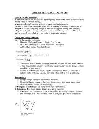
  • 1. EXERCISE PHYSIOLOGY - ADVANCED What is Exercise Physiology:  The study of how the body adapts physiologically to the acute stress of exercise or the chronic stress of physical training. Acute: physiological response to single or short term bout of exercise. Chronic: Physiological adaptation when body is exposed to repeated bouts of exercise. Response: sudden / temporary change in function (disappears shortly after exercise). Adaptation: Persistent change in function or structure following exercise. Allows the body to respond more efficiently and easily to an exercise stimulus. Energy and Energy Systems: Energy: the capacity to do work  Breaking of chemical bonds  Heat + Free Energy  Basic Unit of Energy is ATP  Adenosine Triphosphate  ATP is High Energy Phosphate Bonds  ATP comes from a number of energy producing systems that are “never shut off”  3 main fundamental systems: phosphagen, anaerobic, aerobic. (all energy systems contribute to some degree)  Relative contribution of factors depends on frequency, intensity, time/type of activity, status of tissue, age, sex, nutritional status and level of conditioning. Bioenergetics  Energy changes seen with biochemical reactions.  Reactions liberate energy as they move from a higher to a lower energy state.  In order to make energy you need energy  Exergonic Reaction: give up energy from high e intermediate.  Endergonic Reaction: requires energy coupled to exergonic  Endergonic reactions cannot occur by themselves (driven by exergonic reactions)  But combined exer / endo reactions must be exergonic (ie) muscle contraction
  • 2. (1) PHOSPHAGEN ENERGY SYSTEM:  “cash / energy on hand  ATP stored in the tissue that can be used immediately (ATP  ADP + Pi) Creatine Phosphate (CP or phosphocreatine):  High energy phosphate reserve stored in the tissue and used to immediately resynthesize ATP when it gets broken down. ATP  ADP + Pi “CP gets broken down and combines with ADP to ↓ maintain ATP stores” CP + ADP  ATP + Creatine  must have creatine kinase enzyme. Creatine Kinase  Note: (CP) 3-4 times > (ATP) – CP readily available to help regenerate ATP immediately after being broken down.  When high rates of ATP breakdown are occurring ADP can accumulate and if it does not react with CP, accumulated ADP promotes the formation of ATP via myokinase. ADP + ADP ------ ATP + AMP Myokinase  Other high energy phosphates may contribute to ATP generation via substrate level phosphorolation. GTP (guanosine Triphosphate) + ADP  ATP +GDP (Kinase rx’n)  The phosphagen energy system needs anaerobic and aerobic metabolism to regenerate/ restore normal ATP and CP stores. (2) ANAEROBIC ENERGY SYSTEM (GLYCOLYSIS)  ATP production without utilizing oxygen (sarcoplasm)  Represents the incomplete breakdown of carbohydrates (CHO) to the end product of anaerobic metabolism lactic acid (LA)  Not a very efficient process as high energy bonds still remain in its end product LA.  Produces limited amounts of ATP b/c end product (LA) inhibits glycolysis.  LA generated via anaerobic process also termed anaerobic glycolysis (from glucose) and anaerobic glycogenolysis (from skeletal glycogen).
  • 3. (3) AEROBIC ENERGY SYSTEM  ATP production with the use of oxygen in the mitochondria.  Use of oxygen to produce ATP for all processes.  Breaks down CHO, fats and proteins to allow the production of ATP  Oxidative production involves 3 processes: glycolysis, krebs cycle and ETC. Overview: Which is better Carbs or Fats?  Long run  fats is better!  Workout (weights)  carbs is better!  therefore, it all depends on the transitions (the rate and state of the tissue at work) Capacity:  the amount of ATP independent of time of O2 in greatest capacity during aerobic  the body has more fat reserves than CHO reserves  Therefore, CHO is used when oxygen is limited (ie. High intensity weight lifting)  At the start of exercise as the cardiovascular system catches up to supply more oxygen the body will utilize CHO rather than fats to produce ATP during aerobic
  • 4. Power Of Energy Systems:  Rate of ATP production per unit time.  When time is a factor and intensity is high, and assume adequate )2 and substrates  phosphagen > anaerobic > aerobic  Aerobic system and also the anaerobic metabolism are too slow to meet energy demands of tissue. Enzymes:  Proteins that speed up a reaction to improve biochemical r’xns and energy  Critical to allow optimal high energy production.  3 main basic types: Kinases, Dehydrogenases, Phosphatases.  Amount of enzyme varies in fast (white), fast oxidative glycotic (FOG, mixture) and slow (red) twitch skeletal muscle.  Sensitive to temperatures, pH. Temperature: pH  Increase in Body Temperature  increase in H+ ion and therefore with xcise elevates enzyme activity a decrease in pH may increase or but may also denature enzymes. decrease enzyme activity  Decrease in temp reduces lactate (lactic acid) dissociates and H+ ions enzyme activity and too high are released reducing the pH, enzyme a body temp with destroy enzymes. activity and actin and myosin interaction. Dehydration also reduces heat loss and elevates internal temperature  thus, decreasing efficiency. Lactate Vs. Lactic Acid:  Lactic acid: chemical formula C3H6O3  Anaerobic glycolysis produces LA  L.A releases H+ remaining compound joins with Na+ or K+ to form salt.  Lactate: salt of LA. 3 Classifications of Enzymes: (1)Kinases: (a) transfers a phosphate from ATP to a substance or (b) transfers a phosphate off a substance to generate a high energy compound.
  • 5.  dephosphorylations or phosphorylations  hexokinase (HK), pyruvate kinase (PK) (2) Dehydrogenases (DH): transfer of H+ electrons in oxidation and reduction rxns. Oxidation: removal of H+ electrons from a substance.  In this rxn a coenzyme called nicotinamide adenine dinucleotide (NAD+) is reduced meaning it gains H+ and electrons “LEO”  NAD+  NADH + H+  Glyceraldehydes 3 phosphate dehydrogenase (G3PDH) Therefore coenzyme is being reduced but you are gaining H+ electrons Reduction: addition of H+ electrons to a substance  In this rxn the coenzyme nicotinamide adenine dinucleotide (reduced form, NADH + H+) loses + electrons  NAD+  lactate dehydrogenase. Therefore, coenzyme is being oxized but you are losing H+ electrons. (3) Phosphatase: removes a phosphate from a substance – dephosphorylation. ANAEROBIC GLYCOLYSIS:  skeletal muscle breakdown/utilization of blood glucose (BG) and/or glucose 6 phosphate (G6P) from glycogenolysis (glycogen breakdown) to generate adenosine triphosphate (ATP) without the use of oxygen.  Incomplete breakdown of CHO to lactate (lactic acid) in the sarcoplasm (cytoplasm) of tissue.  Not very efficient as energy producing bonds are still found in lactate.  Critical for high intensity or power events with shorter performance times.  Insulin facilitates glucose entry but BG can be transported w/o insulin.
  • 6. Step By Step Breakdown of Enzymes (Glycolysis)! Hexokinase:  aids in transporting blood glucose from the liver or diet to skeletal muscles.  Uses ATP to Phosphorylate glucose at #6 carbon to form GSP.  Activated (+) by low intracellular glucose 6 Phosphate levels.  Inhibited (-) by high intracellular glucose 6 phosphate levels. Phopshohexose isomerase –isomerase:  Changes G6P to F6P – to more useful shapes (doesn’t change molecular formula) Phosphofructokinase:  Rate limiting enzyme of glycolysis: irreversible reaction  Activated by high levels of AMP, ADP, Pi – by-products of ATP breakdown  Inhibited by high levels of ATP, CP, citrate (from aerobic metabolism in mitro) Aldolase:  Splits the 6C Fructose 1,6 diphosphate into 2-3 C units – DHAP and G3P Isomerase:  Converts DHAP into G3P – forms 2nd molecule of G3P  Changes the shape to a more useful configuration, not the molecular formula. Glyceraldehyde 3 Phosphate Dehydrogenase:  Oxidation of G3P (removal of H+ electrons) to form 1,3 diphosphorglycerate  Coenzyme is reduced – NAD gains H+ and electrons  NAD+  NADH + H+  Also attaches a free inorganic phosphate (Pi) which is found free in the cytoplasm. “Involved in the process of Cycling.” 3 phosphoglycerate Kinase:  Transfers phosphate from 1,3 DPG to ATP to form 1st anaerobically produced ATP. Phosphoglyceromutase – mutase  Moves phosphate to a new position. Enolase  Configuration change – inhibited by fluoride. Pyruvate Kinase:  2nd anaerobically produced ATP as phosphoenolpyruvate changed into pyruvate.  Irreversible reaction  Inhibited by ATP, CP  Activated by high levels of PEP Lactate Dehydrogenase: (lactate DH)  Reduction of Pyruvate (addition of H+ electrons) to form lactate (lactic acid)  Uses NADH + H+ losses H+ electrons and becomes NAD+  Coenzyme is oxidized NADH + H+ looses H+ and becomes NAD+.  NAD+ is formed and is used back at the G3PDH rxn – called cycling.  Allows glycolysis to continue with the consequence being LA accumulation (inhabitation)  LA can diffuse out of the muscle and can be taken up by the liver and other tissue.
  • 7.  LA can reduce intracellular pH (increase free H+ which have dissociated from LA) which can alter enzyme rxns reducing ATP production and actin/myosin interaction. ATP Production From Anaerobic Use of Glucose: Glycolysis: Uses Produces Hexokinase - 1 ATP Phosphofructokinase - 1 ATP Glyceraldehydes 3 phosphate DH +2 NADH + H+ Phosphoglycerate kinase +2 ATP Pyruvate kinase +2 ATP Total ATP Made ANAEROBICALLY from Glucose/ glucose 6 phosphate: = (-2 ATP) + (4 ATP) = 2ATP …….NET GAIN  Note: (G3PDH) produces +2 NADH + H EXERCIZE PHYSIOLOGY #2 “Cycling” term used to describe the importance/dependancy of an oxidation and reduction reaction and the enzyme termed dehydrogenase (DH) in anaerobic Glycolysis. - oxidation – removal of H+ electrons from a substance Glyveraldehyde 3 phosphate dehydrogenase (G3PDH)  Note: Pi is a free inorganic phosphate! - reduction – addition of H+ electrons to a substance. Lactate Dehydrogenase (LDH) ● Combination of oxidation & reduction reaction allow anaerobic glycolysis to continue unimpeded.
  • 8.  slowing of one of these reactions during anaerobic glycolysis slows anaerobic ATP generation.  both reactions depend on production of “by-product” from the other rxn to operate normally – G3PDH needs NAD+ from the LDH while LDH requires NADH + H+ from the G3PDH rxn.. Factors that regulate “cycling”  When ATP demand is greater than aerobic/oxygen delivery system can provide the anaerobic energy system is highly activated. - Therefore we see an increase in pyruvate to lactate conversion in an attempt to generate more ATP at a faster rate. - This leads to increase of LA within the sarcoplasm which by simply end product inhibition will reduce the activity of LDH as the (LA) increases in sarcoplasm thereby reducing ATP production anaerobically. - Increase in LA will also increase rate of H+ ion dissociation from LA and subsequently lead to a rise in fee (H+) ions which reduces pH (more acidic) and reduces anaerobic enzyme efficiency as well as reducing actin, myosin interaction.  LA will also increase in sarcoplasm as there may be an increase in (blood LA) as well as a reduced removal of LA by the liver – liver converts LA back to glucose. (maybe a result of decrease in LA use by the liver during exercize. - LA will rise due to decrease in LA use by other tissue – (concentration gradient). - LA increase also represents an increased use of fast twitch muscle fibers. LA Accumulation:  At rest (LA) is 10X greater concentration than pyruvate LA accumulation can reduce pH (increase the (free H+) that can dissociate from LA. Note: high accumulation of LA usually means there is also an accumulation of pyruvate (pyruvic acid) which can also increase the free H+.  LA diffuses out of the muscle fiber due to the concentration gradient and can: (1) Enter the blood stream and go to the liver for gluconeogenesis.  Formation of glucose from CHO and non-CHO sources in the liver during rest under the stress of starvation and / or during prolonged exhaustive exercise to help maintain blood glucose levels and to help spare/reduce dependency on normal liver glycogen stores to maintain glucose levels. (2) Enter the blood stream and be taken up by the heart muscle for oxidation.  LA converted back to pyruvate via LDH and converted to acetyl COA by PDH for an aerobic fuel. (3) Transported into the skeletal fibers mitrocondria by an intracellular lactate shuttle called a monocarboxylate transporter.
  • 9. (4) LA can be shuttled/ diffuse to adjacent skeletal muscle fibers with a lower (LA)  ie. Hi LA in FT fiber can diffuse to an adjacent ST fiber with lower (LA) for aerobic metabolism any removal of LA slows the change in pH due to LA accumulation. Aerobic Metabolism of CHO:  How CHO are used aerobically to help generate ATP.  When there is limited amounts of oxygen or a high demand for ATP pyruvate is converted dominately to lactate.  When there is a lot of oxygen available pyruvate is converted to acetyl CoA which enters in the TCA cycle.
  • 10. Aerobic metabolism also Krebs Cycle or TCA (tricarboxylic acid) cycle SUBSTRATE ENZYME/ R’XN PRODUCT Pyruvate Pyruvate dehydrogenase (PDH) Oxidation rxn - NAD  NADH + H+ Decarboxylase– C removal from (3C) pyruvate as CO2 and helps to form (2C) molecule acetyl COA (AcCOA). Transferase: transfers on a COA group to form acetyl CoA. Acetyl CoA Acetyl CoA oxalacetate Citrate Synthase -Committed step to aerobic metabolism -Acetyl COA (2C) + OAA (4C)  (6C) Citrate isocitrate Isocitrate DH Oxidation rxn – NAD+  NADH + H+ Decraboxylation: CO2 removal to (5C) ketogluterate Ketoglutarate Ketoglutarate Ketoglutarate DH Oxidation rxn – NAD+  NADH + H+  Addition of CoA from CoA – SH Decraboxylation: removal of Co2 to form (4C) succinyl CoA Succinyl COA Succinyl CoA Succinyl CoA Synthetase -substrate level phosphorylation -only enough enrgy to convert GDP into GTP -GTP + ADP yields an ATP - Addition of free Pi and removal of CoA group Succinate Succinate Succinate DH Oxidation rx’n: FAD+  FADH2 Fumerate Fumerate Fumerase:  Adds an H20 molecule Malate
  • 11. Malate Malate DH Oxidation rxn – NAD+  NADH + H+ Oxaloacetate  The reducing equivalents NADH + H+ and FADH2 from oxidation rx’n enter intot he mitrocondrial electron transport system to generate ATP. ATP Production From Aerobic Metabolism of Glucose: Glycolysis: Uses: Produces: Hexokinase -1 ATP Phosphofructokinase -1 ATP Glyceraldehyde 3 Phosphate DH +2 NADH + H+ Phosphoglycerate Kinase +2 ATP Pyruvate Kinase +2 ATP Pyruvate DH +2 NADH + H+ Krebs Cycle: Isocitrate DH +2 NADH + H+ Alpha Ketogluterate DH +2 NADH + H+ Synthetase (2 GTP) +2 ATP Succinate DH +2 FADH2 Malate DH +2 NADH + H+ = (6ATP – 2 ATP) + (10 NADH + H+ x 3 ATP) + (2 FADH2 x 2 ATP) = 4 ATP + 30 ATP + 4 ATP Total ATP Generated = 38 ATP Note: each NADH + H+ yield 3 ATP and each FADH2 yield only 2 ATP.  NADH + H+ produced in glycolysis at the glyceraldehydes 3 phosphate dehydrogenase step can be transported/shuttled into the mitochondria for electron Trasport system and produce ATP aeroblically or be use at the LDH reaction in glycolysis to produce lactate. “Cycling Process” Malate-Aspartate Shuttle (HEART)  Shuttles cytoplasmic NADH + H+ from the cytosol into the mitochondria.  Cyto Malate DH (MDH) catalyzes cyto oxaloacetate (OAA) + NAD+  Malate + NADH + H+ and can be used in G3PDH rxn. (cytoplasm)  Malate readily enerts cytoplasm via transporter.  Mito MDH catalyzes oxidation rx’n: Malate + NAD+  OAA + NADH + H+  NADH + H+ enters the ETS to produce 3 ATP.  Mito OAA via a transamination rxn converted to apartate which leaves the mito.  Once outside the Mito a cytoplasmic aspirate transaminated back to cytoplasmic OAA.
  • 12. Glycerol Phosphate Shuttle (Muscle)  Shuttles Cytoplasmic NADH + H+ from cytosol into the mitochondria.  Cytoplasmic Glyceral 3 Phosphate DH coverts: DHAP + (NADH + H+)  Glycerol-3-Phosphate.  Glycerol-3-Phosphate diffuses into the mitochondria.  Mitochondrial glycerol 3 Phosphate DH allows: Glycerol-3-Phosphate + FAD+  FADH2 + DHAP Note: Does not use NAD+ onside the mito, but instead uses FAD+ to form the reducing equivalent FADH2 which is free to enter the ETS – yield one less ATP molecule.  Mitol DHAP readily diffuses out of the mitochondria to react with another NADH + H+. Electron Transport system: (Oxidative Phosphorylation) or Respiratory chain
  • 13.  NADH + H+ and FADH2 provide H+ and electrons for concentration gradient and thus provide energy for the phosphorylation of ADP to form ATP.  3 critical enzymes in ETS that expel H+ ions to create: (1) electrical chemical gradient (-) charge inside (2) pH gradient less H+ ions inside provides energy for phosphorylation (pH inside) 3 enzymes: NADH DH Cytochrome b-c1 complex  all involved in proton (H+) translocation Cytochrome oxidase Note: Cytochrome oxidase – where ½ )2 from the blood accepts 2 electrons and combines with 2 H+ ions to form H20.  These enzymes transfer the protons (H+ ions) from the reducing equivalents across the inner mitochondrial membrane into the intermembrane space.  More H+ in the intra mitomembrane space (or less (+) charges or more (-) charges) in the inside of mito.  This transfer of H+ protons creates the electrochemical gradient which provides the energy for the synthesis of ATP from mito ADP and Pi. Note: NADH + H+ enters the ETS at a level higher then FADH2 does and thus is able to generate 3 ATP versus only 2 for an FADH2. Final Steps of ETC:  ATP is brought into the intra mito membrane space via the adenine nucleotide translocase and it is converted to ADP via the creatine phosphate kinase (CPK).  Creatine gets a free Pi from the breakdown of ATP in the previous step which creates Cratine Phosphate in the sarcoplasm.  Creatine phosphate is then broken down with the help of CPK and ADP becomes ATP in the sarcoplasm. Overview:  Standard Calculation of 1 molecule of Glucose! How Many ATP Produced? (4 Cycles need to be reviewed) (1) Anaerobic Glycolysis (stop at pyruvate)  1 NADH + H+ (G3PDH rxn) (2) Krebs Cycle:  (pyruvate goes through TCA) (3) Are The Shuttles Used? YES  NADH=H+ from anaerobic glycolysis (G3PDH) is shuttled to the mitochondria. (4) ETC – NADH + H+ is now in mitochondria it will undergo oxidative phosphorylation to produce ATP. HOMEWORK: (1) 3 Molecules of 2 PG: How many ATP Produced? (2) From 1 molecule of Lactate: How many ATP produced? LECTURE 1: STUDY NOTES Physiology: Acute:  A response, sudden and temporary.
  • 14. Chronic  An adaptation and persistent change. 3 Types of Muscles: Smooth:  Involuntary, unconsciously controlled, found in blood vessels/ internal organs. Cardiac:  Controls itself with assistance of nervous and endocrine systems, found in heart. Skeletal:  Voluntary, consciously controlled, over 600 in body. Muscular Control: Muscle Fiber: made of myofibrils  Bundles of fibers are called fasciculli.  1000s per muscle (bicep 200, 000); 20-100um thick – largest same as human hair  Polygonal shape allow greater packing density Myofibrils: contain actin and myosin filaments which  Makes up 85% of fiber;1-2um in diameter  Run whole length of muscle fiber. Thin Filament: (actin)  Troponin: a protein inhibits myosin ATPase activity until it is inactivated by calcium ion release from the SR.  Tropomyosin: protein which blocks attachment sites on actin. Thick Filament:  Two protein strands twisted together  1 end globular head  binding site for actin enzymatic site The Sacromere:  Contains 2 sets of thin filaments – 1 at each end.  1 end of each thin filament is anchored to the Z-line the other end overlaps a portion of the thick filaments.  Two successive Z-lines define the limits of the sacromere. Terms: Transverse Tubules + Sacroplasmic reticulum cross sect to form a terminal cisternae  Sarcoplasmic Reticulum: stores calcium I Band:  only have actin filaments (b/w ends of A band)  contains portion of thin that doesn’t overlap thick  Bisected by Z-line. A Band: only contains thick filaments H Zone:  Only myosin filaments; Appears when sacromere is resting  M-line narrow dark band  Titan: anchors thick filaments to z-line.
  • 15. Nebulan: anchors actin to z-disks. Crossbridges: projections that b/w Thick& thin filaments Exercise Physiology Lecture #2: Motor Neurons:  Innervate skeletal muscle  Cell bodies are located in the brainstem and spinal cord.  Axons are myelienated (large diameter)  Axon of MN divides into many branches forming a single junction with fiber.  Thus a single MN innervates many muscle fibers even though one muscle fiber can only be attached to one motor neuron. Motor Unit:  A single alpha motor neuron plus the muscle fiber that it innervates.  Either all fast or all slow twitch fibers within the motor unit (never mixed). Motor Neuron Pool  The group of motor neurons innervating a muscle. Neuromuscular Joint (NMJ)  The junction of an axon terminal with a motor end plate.  Sarcolemma region under terminal portion of axon = motor end plate.  Axon terminal has a membrane bound vesicle that contains Ach  Action potentional is transferred from the alpha motor neuron to the muscle. Muscle Contraction: Excitation-Contraction Coupling: (1) A motor neuron which signals from the brain or spinal cord, releases neurotransmitter Ach. (2) Ach crosses the junction and binds to receptors on the sarcolemma. (3) This depolarizes the cell and initiates an action potential. (4) The action potential travels along the sarcolemma and through the T-tubules to the SR releasing Ca2+.
  • 16. (5) The Ca2+ binds to troponin on the actin filament and the troponin pulls the tropomyosin off the actin active sites, allowing myosin heads to attach to the actin filament. (6) Once a strong binding site is established with actin, the myosin head tilts, pulling the actin filament (power stroke) (7) The myosin head binds with ATP and ATPase found on the head splits ATP into ADP and Pi, releasing energy. (8) Muscle action ends when calcium is actively pumped out of the sarcoplasm back into the SR for storage. (9) Energy for muscle action is provided when the myosin head binds to ATP. ATPase on the myosin head splits the ATP into a useable energy source. Relaxed--------------Contracted----------Fully See picture below: The Role of Ca++  Inhabitation of actin due to troponin and tropomyosin.  Troponin: holds tropomyosin in position.  Tropomyosin blocks the ATP binding site.  Therefore for all XB interactions to occur the binding site on actin must be opened with Ca++ (pulls tropomyosin of troponin)  Troponin binds up to 4 Ca++  Ca acts as a switch for A-M binding Regulation of Ca++  Ca++ assoc. with contraction is located in SR.  Transport pump in SR have a higher affinity for Ca++ than does troponin.  Network of T-tubules allow spread of the action potential into the interior.  Increased sarcoplasmic Ca++ also activates SR pumps that rapidly restore the relaxed state unless another AP is elicited.  Approx. 90% of the SR is covered in these SR Ca+ pumps.
  • 17.  Ca++/ATPase provides energy to transport Ca++ back into the SR enabling relaxation.  When muscles are relaxed, the [Ca++]is much higher (several hundred times) in the SR than in the sarcoplasm. This is because of the action of the Ca++ pump which couples the use of energy in ATP with the movement of Ca++ from the sarcoplasm to the SR.  A single AP releases sufficient Ca2+ to fully activate the contractile apparatus, but the Ca2+ is pumped rapidly back into the SR before muscle has time to develop tension.  This results in a twitch.  Repetitive AP can cause a summation of twitches producing a partial or complete Tetanus. The Sliding Filament Theory:
  • 18. Role of ATP in Cross Bridge Cycle  Energy released from hydrolysis provides energy for x-bridge movement.  Binding of ATP to Myosin brinks the link between A-M, allowing the cycle to repeat. Cross Bridge power Stroke:  Always want to result in sacromere shortening.  Success of this shortening depends on load imposed on muscle. Cross Bridge Desire vs. Outcome  CB force > load = concentric (shortening)  CB force = load = isometric (static)  CB force < load = eccentric (lengthening) Power Stroke Statistics: Force  Force of power stroke: 3-4 pN  1 pN= 10-12N; 1kg =10N; 10N=10 trillion pN  If allow5pN per XB power stroke.
  • 19.  2 trillion XBs needed for 10N(1kg) Length:  Length of power stroke: 10nM  1 million nm= 1mm; 1000nm = 1um  2um= 2000nm  1 power stroke=10nm  2000/10=200 power strokes needed to shorten sacromere from max. to min. length Sacromere in Series:  30um/2um=15sacromeres  10,000um (=10mm,1cm)  10,000um/2um=5000sacromeres to produce working range Working Range of Sacromere= 3.5-1.5=2um Working Range of Myofibril/Muscle Fiber  Optimal length 2um x 5000=10mm  Assume 5,000 sacromeres/myofibril  2um x 5000 = 10,000 um (10mm)  Fiber Length Range: 7.5-17.5mm Muscle fiber Types: Slow Twitch FOG (FTA) Fast Twitch(FTB) High Aerobic (oxidative) Moderate Aerobic (oxidative) Low Aerobic (oxidative) High Fatigue Resistance Moderate fatigue Resistance Low Fatigue Resistance Low anaerobic (glycolytic) Moderate anaerobic (glycolytic) High anaerobic (glycolytic) Low contractile speed 110ms to Fast Contractile Speed 50ms to reach Fast Contractile Speed 50ms to reach
  • 20. reach peak tension. peak tension peak tension 10-180 fibers per motor neuron 300-800 fibers per motor neuron 300-800 fibers per motor neuron Low SR development High SR development High SR development (more Ca2+) Red Type I aerobic Ex. distance runner Pink-Mix Type IIA Ex. middle distance runner 400m White Type IIB Ex. Sprinter, explosive activity Muscle Biopsy:  Hollow needle is inserted into muscle to take a sample.  Sample is mounted, frozen, thinly sliced and examined under a microscope.  Allows study of muscle fibers and the effects of acute exercise and exercise training on fiber composition. What determines Fiber Type?  Genetics determines which type of motor neurons innervates our individual muscle fibers.  Muscle fibers become specialized according to the type of neuron that stimulates them.  Endurance training, strength training and muscular inactivity may result in small changes (less than 10% of FT or ST)  Endurance training has been shown to reduce the percentage of FTb fibers, while increasing the fraction of FTa fibers.  Aging may result in changes in the percentage of FT and ST fibers  alpha motor neurons die off and with no intervention orphan fibers are created, a MN in the general area will undergo collateral branching with the orphan fibers which increases the terminal end bulbs and the orphan takes over the new characteristics of the MN. Therefore if the motor neuron contained slow twitch fibers the new orphan will also contain ST fibers. (this is the usual case)  Fast Twitch IIB has more myosin ATPase activity which results in more glycolyitc potential.  The Twitch response of a fast twitch is much greater than that of a slow twitch fiber. However this results in quicker fatigue. All or None Response:  For a motor unit to be recruited into activity the motor nerve impulse must meet or exceed the threshold.  When this occurs, all muscle fibers in the motor unit act maximally.  If the threshold is not met no fibers in that unit act.  More force is produced by activating more motor units. Orderly Recruitment of Muscle Fibers:
  • 21.  Principle of orderly recruitment states that Motor units are activated in a fixed order, based on their ranking in the muscle.  Henneman’s Size principle states that the order of recruitment is directly related to their motor neuron size.  Slow twitch fibers which have smaller motor neurons are recruited before fast- twitch fibers. Ramplike Recruitment of Fibers: Peak Power Generated By Fibers:  The difference in force development between FT and ST motor units is due to the number of muscle fibers per motor unit and the large diameter of the FT fibers. Recruitment Order:  Henneman’s Size Principle  Smallest to largest, size of cell body, size of the axon, not size of the muscle unit.  Thus, slow to fast.  Achieved by increase in excitatory drive.  Most gross movements done through recruitment Discharge Rate:  2nd means to increase force (behind rec order)
  • 22.  recruitment order is still followed  increase in discharge rate  Muscle can be recruitment or discharge rate controlled  Generally fine motor movements done through discharge. Functional Classification of Muscles:  Agonists: prime movers; responsible for movement. (biceps) Antagonists: oppose the agonists to prevent overstretching of them. (triceps) Synergists: assist the agonists and sometimes fine-tune the direction of movement. (brachioradialis) Mechanics of Single Fiber Contraction:  Tension: force exerted on an object by contracting muscle.  Load: force exerted on a muscle by an object  In order for shortening and contracting of the whole muscle to occur: tension>load. Types of Muscle Action Step 4: (1) Concentric: X-bridge bound to actin moves to angled position which shortens the cross bridge. (2) Eccentric: X-bridge pulled toward z-line by load while still bound to the actin. (3) Isometric: Bound x-bridge unable to move Actin b/c of load on muscle.  Steps 1,2,3 are same in all 3 types of contractions.  Chemical changes are the same for all 3 contractions. Factors influencing force generation: (1) number of motor units activated (2) type of motor units activated (FT or ST) (3) muscle size (4) initial muscle length (5) joint angle (6) speed of muscle action (shortening or lengthening) Muscle Length vs, Force Production:
  • 23. Physiology Lecture #3: Strength, Power and Speed Strength:  Peak force or torque of a MVC (maximum volume contraction) against a given load resistance  Contraction Type Velocity Position on Strength Curve Speed:  Velocity of shortening of a MVC against a given load (resistance)  Vmax= maximum shortening velocity of an unloaded maximal concentric contraction. Power:  Force (strength) x Velocity (speed) The Bottom Line: XB Mechanism:  Strength  The number of active crossbridges in the cross sectional area x force/XB  Speed XB cycle rate/power stroke speed (Vmax) Rate of Force Development:  Applies to all contraction types (E,C,I)  Most often associated with isometric contraction.  Rate of activation of XB’s + XB cycle rate/power stroke speed (Vmax)  Activation + Speed  Activation of XB’s occurs at the point of stimulation of actin and myosin (ec coupling) and is dependant on how fast the action potential is traveling Max Force vs. Max Velocity:  Max Force: dependant on the # of active XB’s in the CSA  Max. Velocity: independent of # of active XB’s in the CSA; dependant on the XB cycle rate/power stroke speed.
  • 24. Relationship between force, length and velocity: Fiber CSA vs. Length:  Peak Force: determined by # of sacromeres (XB’s) in parallel. (CSA)  Vmax: determined by # of sacromeres (XB’s) in series. (fiber length) Don’t Confuse Sacromere and Muscle Fiber: Factors Affecting Concentric Force Decreasing as Velocity Increases:
  • 25. (1) Slackening of SERIES ELASTIC COMPONENT  Velocity Increases  Slackening of SEC Increases  Increased Difficulty in taking up SEC  Force Decreases The elastic compound stores potential energy (2) Sliding Filament “Effect”  Increase Velocity of Sliding  Increase difficulty of XB pulling on actin Increase difficulty of XB binding on the actin (myosin heads slide my actin binding site)  Decrease Force (3) Negative XB Braking Effect:  Some XBs remain bound to the actin filament too long XB’s then become stretchedthis braking action opposes shortening  decreases force Factors Affecting Eccentric Force Increasing as Velocity Increases: (1) Positive Braking Effect  Increase velocity of lengthening  Increases stretch of XB while bound to actin Increase passive resistance to stretch  Increases braking effect Increase ECC force. (2) XB’s suspended in bound state, do not use ATP, which reduces energy cost of contraction. ECC attachedforcibly detachedreattaches More Attached XB’s in Eccentric?  In maximal isometric contraction, 50% of XB’s are attached at any time.  In maximal ECC > # of XB’s are attached at any time 75%  More attached XB’s = > force
  • 26. Factors affecting the amount of SCC potentiation: (1)Force/Intensity of ECC Contraction  Increasing force of ECC will lead to great potential energy for CON contraction. (2) Effect of Velocity on SSC Potentiation  Increasing velocity during ECC with lead to greater relative (%) potentiation which will increase CON contraction. In both cases the ECC has more to give and CON has more to gain. (See diagram) These are both positive effects on CON contraction.
  • 27. (3)During of the ISO phase: (coupling time)  Increased coupling time ill lead to a decreased CON time.  Any potential energy stored during ECC becomes lost by coupling time Disadvantages:  High ECC force decays to lower ISO force.  XB’s stretched during ECC detach during ISO, positive breaking force lost. Gender Differences in SCC Potentiation: (1) Passive Muscle Properties:  Cytoskeleton:  Connective tissue  Females have more; therefore more energy can be stored by the SEC. (2) Cross-Bridge Attachment Time:  More ST fibers in females lead to a longer XB cycle.  This leads to a greater stretch during binding time.  This results in greater storage of elastic energy. (3) ECC/CON ratio:  Females have increased ECC/CON ratio  Males have greater concentric force but females have a larger ECC capacity. (4) Reflex Potentiation:  Females have greater reflex potentiation  Enables them to better sense changes in muscle length = a quicker monosynaptic reflex. (5) Reflex Inhibition:  Females have less reflex inhibition.
  • 28.  Males extra muscle mass limits the amount of stretching that occurs during the ECC contraction ***Revisit aging and compare to females in correspondence to the SSC**** Muscle Architecture: Roles: Parallel: Pennate: Absolute vs. Specific Force:  Specific Force: force per unit CSA  Absolute Force: specific force x CSA (isometric measurement) Factors Affecting Specific Force: (assumes Po at Lo)  Intrinsic force/XB  Temperature  Fatigue
  • 29.  Fiber Type Myofilament Density: Unlikely to be altered at Lo (peak isometric force) Myofibril density (note: myofibril = 85% of fiber contents)  Glycogen  Fat Droplets  Mitochondria Muscle Architecture  Affects force desired: (in series or parallel)  Muscle type (pinnate, parallel) Force (tension) Velocity Relationship:  Max shortening velocity is achieved at a zero load.  Max CON force is equal to max ISO tension (Po) Horizontal vs. Vertical Axes:
  • 30. Measurement of Force Velocity:  (1) Set velocity; measure force  Isokinetic dynamometer  Usually concentric  Velocity limit  Single vs. multi joint Curve linear relationship  At max force = lowest velocity  At max velocity = lowest force (2) Set Load (force), measure velocity  Load-velocity device  Single vs. multi joint  Unilateral vs. bilateral  More relevant to performance
  • 31.  Single Max lift just before ISO max; then load is too heavy to move  VMAX would just be the barbell, very light = increased velocity Twitch Contractile Properties:  Latent Period: when ca2+ is traveling from the SR to troponin (traveling time)  Twitch: response to an action potential  TPT: Time to peak tension; time it takes for the rising phase of the twitch to reach its’ peak.  HRT: half relaxation time: half of the time taken for the twitch to fall from its peak value.  CD: Contraction Duration = TPT + HRT  PT: Peak tension: force or height of twitch  Tetanus the maximum amount of tension that can be generated.
  • 32. Single Twitch:  Single AP = single twitch  Force can be changed by increasing the frequency of stimulation Summation of Twitches:  Many AP’s = summation of twitches  The muscle doesn’t relax completely before the 2nd stimulus occurs; resulting in a more powerful contraction. Summation Leading To Unfused Tetanus:  Many AP’s close together = unfused tetanus  Will not stay at max tension Summation Leading to Complete Tetanus:  Many AP’s close together = fusion of twitches.  Will stay at max tension
  • 33.
  • 34. Post activation Potential: (PAP) Muscle contractile performance is affected by preceding activity  Activation history may be chronic (training)  Activation history may become acute Increase twitch and low frequency force due to prior activation  MVCPAP Evoked high frequency tetanusPTP (post tetanic potentiation) Series of Twitches Staircase PAP will only have an effect at low frequencies; once tetanus is reached it is useless. Mechanism of PAP: (a)Ca2+ release from SR (b)Ca2+ binds to calmodulin (c)Calmoldulin activates protein kinase (d)Kinase binds to phosphate (from ATP splitting to myosin light chains phosphorylation (e)Myosin more sensitive to Ca2+ (f)Increases Potentiation Neurological Control of Movement:
  • 35. STRUCTURE OF A NEURON  Axon Hillock: adds up the graded potentials, if threshold value is reached the axon hillock will allow the AP to travel down the axon.  Nodes of Ranvier: Part of axon not covered by the myelin sheath; the AP jumps along these nodes.  Nerve Impulse: an electrical charge that passes from 1 neuron to the next and finally to an end organ such as a group of muscle fibers Resting Membrane Potential:  Difference between the electrical charges I?S and O/S a cell, caused by separation of charge across a membrane (I/S is –vely charged compared to O/S)  Thus, diff. in ion [ ] and difference in permeability determine RMP.  High [ ] of K+ inside the neuron and high [ ] of Na+ O/S the neuron
  • 36.  K+ ions can move freely, even O/S the cell to help maintain imbalance. (passive)  Sodium Potassium pump actively transport K+ and Na+ ions to maintain imbalance by the ATPase pump (electrogenic active). Changes in Membrane Potential:  Depolarization: inside of cell becomes less negative relative to outside (>-70)  Repolarization: inside of the cell becomes more negative relative to the outside (<-70)  Graded Potentials: localized changes in membrane potential (Either depolarization or repolarization)  Action Potential: rapid, substantial depolarization of the membrane (-70 to +30 to -70) What is a graded potential?  Confined to a small region  Produced =change in environment acting on small region of membrane.  Graded b/c magnitude of change is variable, magnitude of stimulation. Temporal Summation:  Input arrives at same cell but different times  Potentiation summates b/c greater # of open channels Spatial Summation:  2 inputs on the same cell but different locations
  • 37. What is an Action Potential?  RMP of -70mv is maintained by Na-K pump  Changes in RMP occur when ion gates open, allows ions to move from one side to another  Starts as a graded potential  Requires depolarization greater than the threshold value Ex. (-50mv)  Once threshold is met of exceeded the all or none principle applies. (1)Resting State (-70) (2)Depolarization (-50) (3) Propagation of AP (along axon) (4)Repolarization (-90mv) (5)Return to resting state (-70mv) –Na/K facilitates this Velocity Of Action Potential:  Myleinated Fibers: allow Saltatory Conduction whereby an AP travels quickly from one break in myelin (Node of Ranvier) to the next.  5-150 times faster in myleinated fibers as opposed to non-myleinated. Diameter of Neuron:  Large diameter neurons conduct nerve impulses faster because there is less resistance.  Therefore a FT fiber creates less resistance and quicker impulses than a ST. The Synapse: Site of impulse transmission (1) The impulse travels to a presynaptic axon terminal where it causes (2) Synaptic Vesicles on the terminal to release chemicals (neurotransmitters) into the synaptic cleft. (3) The neurotransmitters then bind to the postsynaptic receptors.  Note: Neural impulses can only be transmitted from the dendrite or cell body through the axon to the adjacent neuron since the neurotransmitter are released only from the terminal end of the axon. The Neuromuscular Junction:
  • 38.  Site where MN communicates with muscle fiber  Motor axon terminal releases NT’s (acetylcholine) which cross synaptic cleft and bind to receptors on fiber.  The binding causes depolarization, thus possibly causing an AP.  The AP spreads across the sarcolemma causing the muscle fiber to contract. Note: Unidirectional movement down axon vs. Bidirectional movement in muscle fiber Refractory Period:  Period of repolarization  Muscle fiber is unable to respond to any further stimulation.  The RP limits a motor units firing frequency. Organization of Nervous System: PNS: (peripheral) contains cranial and spinal nerves: (1)Sensory: carries sensory information from body via afferent fibers to the CNS. (2)Motor: transmits information from CNS via efferent fibers to target organs. Our Focus is the Sensory Nervous System: Types of Sensory Receptors:  Mechanoreceptors: respond to mechanical forces such as pressure, touch, vibration, stretch  Thermoreceptors: respond to changes in temperature.  Nociceptors: respond to changes in temperature  Photoreceptors: respond to light to allow vision.  Chemoreceptors: respond to chemical stimuli from foods, odors, and change in blood conc. Muscle and Joint Nerve Ending:  Joint Kinesthetic receptors in joint capsules sense the position and movement. Muscle Spindles: sense how much a muscle has stretched.
  • 39.  Middle of fiber (intrafusal) are activated by gamma motor neurons  Intrafusal fibers sense the change in length but they cannot contract as it contains little or no actin/myosin.  When extrafusal fibers attached to the spindle are stretched, sensory neurons on the spindle transmit info to the CNS about muscle length.  Reflexive muscle contractions are then triggered though the alpha motor neuron to resist further stretching. Golgi Tendon Organs: detect tension (or strength of muscle contraction)  Encapsulated sensory organs through which muscle tendon fibers pass.  Located close to the tendon’s attachment to the muscle.  Inhibit contraction (agonist) muscles by exciting the antagonist muscles to prevent injury. Neuromuscular Adaptations to Resistance Training: Strength, Power and Speed: Part 2 Strength: the maximal force a muscle of muscle group can generate. -Evaluating strength:  1 rep max: max amount of load that you can lift only once (bench press with trained spotters)  Measure the maximal force that can be exerted using specialized equipment  MVC: maximum volumetric force produced from isometric contraction. (lab setting) Power: the product of strength + speed of movement (strength x speed) Evaluating Power:  The functional application of speed and strength  The key component of many athletic performances  Power = force * distance/time or P=FV (velocity = distance/time)  Ex A: lift 150 kg, 1m, takes 0.5s P=300 watts  Ex. B lift 200kg , 1m, takes 1s P=200 watts  Therefore, A has more power. Muscular Endurance: the capacity to sustain repeated muscle actions over a given time. -Evaluating Endurance:  Can be evaluated by noting the number of repetitions you can perform at a given percentage of your 1-RM. (ex. # of times you can lift 50kg)  Is increased through gains in muscular strength.  Is increased through changes in local metabolic and circulatory function.
  • 40.  Increased mitochondria, oxidative capacity, capillarization, metabolic rate of removal of byproducts. Designing Resistance Training Programs: 1. Consider different dynamic training programs.  Polymeric (SSC), free weights, circuits etc. (a) Static (isometric) actions (b) Dynamic actions: free weights, eccentric training, variable resistance, isokinetic actions, speed constant (Bicep curl: 100 = 100%; 120 = 98%; 60=67%) (c) Electrical Stimulation Training: rehab, immobilization, stimulate a nerve to induce a response 2. Perform a training needs analysis.  Goals, set intensity, reps (increase intensity and decrease reps)  What muscles need to be trained  What method of training should be used?  What energy system should be stressed  What are the primary sites of concern for injury prevention? 3. Select appropriate resistance levels/ 4 Decide on single sets vs. multiple sets.  Strength: few sets and high resistance (6RM)  Muscular Endurance: many reps and low resistance(20RM)  Power: several sets of reps and moderate resistance; emphasize speed of movement.  Muscle Size: move than 3 sets of 6RM to 12RM; short rest periods. 5. Designa training program using periodization  Relives boredom; every 6 weeks vary your program  Change in exercise stimulus over a specific period to keep an individual from overtraining.  Cycle of five phases: four active phases followed by one active recovery phase.  Each phase gradually decreases volume and gradually increases intensity. Variable Phase 1 Hypertrophy Phase 2 Strength Phase 3 Power Phase 4 Peaking Phase 5 Active Recovery Sets 3-5 3-5 3-5 1-3 General activity or light resistance training Reps 8-20 2-6 2-3 1-3 Intensity Low Hi Hi Very Hi Duration 6 6 6 6 2 6. Assign specific forms of resistance training depending on the sport or desired results.  Specifity: a ballerina wont work on a rowing machine! Forms of Resistance Training:
  • 41.  Isometric/static resistance training  Plyometric/jumping training  Eccentric Training: Increase strength by 30% relative to concentric  Free weights: (recruits MN in agonists & synergists stabilizers)  Electrical Stimulation. Basic Resistance Factors:  The muscles or muscle groups you want to condition  Increase intensity of training (amount of weight lifted with each repetition)  Number of Repitions per set.  Number of sets per workout. Mechanisms in gains in Muscle Strength: Neural Adaptations: (1) Synchronization and recruitment of additional motor units (2) Autogenic inhibition (3) Co-activation of agonist and antagonist muscles (4) Rate coding: the firing frequency of motor units. Muscle Size:  Muscle Hypertrophy: refers to an increase in muscle size.  Atrophy: refers to a decrease in muscle size Muscle strength is not dependant on just size. Resistance training  Increases muscle size (hypertrophy)  Alterations of Neural Control of trained muscles Studies show strength gains can be achieved without changes in muscle size, but not without neural adaptations Neural Factors of Strength Gains: (a) Recruitment of additional motor units for greater force production (b) Counteraction of autogenic inhibition allowing greater force production (c) Reduction of Co-activation of agonist and antagonist muscles. (d) Changes in discharge rates of motor units (firing frequency) (e) Changes in the NMJ increase or decrease activity. (1)Recruitment of Additional MU’s  MU’s usually recruited asynchronously. Controlled by many excitatory and inhibitory impulses  Summation of impulses determines whether fibers contract or stay relaxed.  Contract when excitatory>inhibitory (reach threshold) Strength may increase if MU act synchronomously (force Increases  This is due to strength training because there is more connections between neurons (motor) within the spinal cord (will improve neuromuscular adaptation)
  • 42.  More synapses = more communication = synchronized = Increase Force (2) Autogenic Inhibition: GTO may prevent too forceful of contraction (a) increase concentric force (Increase load…biceps) (b) GTO sense change in tension signal to spinal cord (sensory/afferent) (c) Interneurons (disynaptic synapse) in SC (inhibitory) (d) Signals agonists to decrease (efferent/motor) (e) Signals to antagonist to shorten (excitatory) & signal Agonist (inhibitatory to lengthen) GTO sense threshold being exceeded, MN’s to muscle inhibited (autogenic inhibition), also reticular formation in BS & cerebral cortex initiate inhibitory pulses.  Training reduces inhibitory pulses, muscles reach greater strength  Training increases strength by reducing inhibition  Theory explains super human strength and increase strength w/o hypertrophy Note: autogenic inhibition can be shut off sue to increases in adrenaline. (3) Other neural Factors:  Co-activation of agonist & antagonist muscles  Decreased Co-activation of antagonists will increase force of agonists (static situation: antagonists and agonists are equal)  Rate Coding: (discharge/firing rate) of MU increases force production (seems to be more likely in older adults, muscle dependant)  NMJ changes many different components, most likely morphology. Model of Neural and Hypertrophic Factors:
  • 43.  8 weeks: no physical increases in size but strength increase due to neural adaptations.  >8 weeks: hypertrophy visible and contributes to overall strength gains (progressive overload; Increase load) Muscle Hypertrophy:  Transient: (acute) pumping up of muscle during a single xcise due to fluid accumulation from the blood plasma into the interstitial space of the muscle (Edema)  Chronic: (long term) increase of muscle size after long term resistance training due to changes in muscle fiber # (fiber hyperplasia) or muscle fiber size (fiber hypertrophy) Fiber Hypertrophy-Increase Fiber Size:  The # of myofibrils and actin & myosin filaments increase, resulting in more crossbridges; sarcoplasm and connective tissue increase.  Muscle protein synthesis increases during the post exercise period.  Muscle protein synthesis increases during the post-exercise period.  Testosterone plays a role in promoting muscle growth.  Training at higher intensities appears to cause greater fiber hypertrophy than training at lower intensities  Eccentric training is more likely to cause hypertrophy. Fiber Hyperplasia: Increase in Number  It has been proven that muscle fibers can split in half with intense weight training (cats)  Parent fibers split to daughters, each half then increases in size of the parent fiber.  Satellite cells (myogenic stem cells) may also be involved in skeletal muscle fiber generation.  It has been clearly shown to occur in animal models; only a few studies show this occurs in humans too (body builders) Satellite Cells Respond to Injury:  Injury (ex. due to ECC contraction)  Satellite cells activation & proliferation  Cells migrate to the injury.  Diffuse with original muscle fiber will cause hypertrophy and diffuse with each other will cause hyperplasia. Neural Activation and Fiber Hypertrophy  Early gains in strength seem to be more influenced by neural factors.  Long Term strength increases are largely the result of muscle fiber hypertrophy.
  • 44. Effects of Muscular Inactivity:  Muscular atrophy (decrease in muscle size)  Cause by lack of muscle use, subsequent loss of protein (heavy resistance trainer suddenly stops.  Decrease in muscle protein synthesis (first 6 weeks)  Strength loss most dramatic in 1st week  Primarily ST fibers affected 1st (discontinuity of Z-disks, fusion of myofibrils, mitochondrial damage)  Recovery when activity resumed. Strength Changes in Women: Are Muscle Fiber Type Alterations Possible?  Early studies no change in fiber type but changes in characteristics of muscle fibers.  Cross innervations studies (changing neuron) and chronic stimulation studies (changing frequencies) demonstrate changes  Recent Research Performed on animal subjects.  Possible Change from FTb to FTa and from FTa to ST with endurance training  Possible change from FTb to FTa with resistance training or FTa-FTb (highly explosive resistance training. Changes in Muscle Fiber Type:
  • 45. Acute Muscle Soreness:  Results from accumulation of end products of exercise in the muscles.  Usually disappear within minutes or hours after exercise. DelayedMuscle Soreness: (DOMS)  Results primarily from eccentric action.  Is associated with damage or injury within the muscle  Disruption of the sarcolemma, contractile filaments (A&M) – pulling apart of Z- disks.  May be caused by inflammatory reaction inside damaged muscles, white BC response  May be due to an edema (accumulation of fluid) inside muscle compartment.  Is felt 12-48 hrs after a strenuous bout of exercise. Armstrong’s Sequence of Events in DOMS 1. structural damage b/c of high contractile tension 2. impaired calcium homeostasis b/c cell membrane damage (necrosis)-peak 48hrs 3. Accumulation of irritants (histamine, kinins, K+) o/s the cell 4. Increased macrophage activity b/c of by-products. 5. Increased inter/intracellular fluid increases tissue pressure which in turn activates pain receptors in muscle. DOMS and Performance:  DOMS causes a reduction in force generation of muscles  Maximal force generation returns after days or weeks.
  • 46.  Muscle glycogen synthesis (glycogenesis) is impaired with DOMS. Normal for first 6-12 hr after exercise but then stops completely as muscle undergoes repair, thus limiting fuel-storage capacity of injured muscle. Reducing Muscle Soreness:  Reduce eccentric component of muscle action during early training  Start training at a low intensity, increasing gradually  Or begin with a high intensity, exhaustive bout of eccentric-action exercise to cause much soreness initially, but decrease future pain. Exercise Associated Muscle Cramps:  Due to fluid or electrolyte imbalances and sustained alpha MN activity with increased muscle spindle activity and decreased GTO activity.  Rest, passive stretching and holding the muscle in the stretched position can be effective treatments.  Proper conditioning, stretching and nutrition are prevention strategies. Cholesterol Biosynthesis:  Occurs primarily in the liver  Dependant upon the availability of acetyl CoA similar to ketone and fatty acid biosynthesis  Key regulatory enzyme is 3HMG CoA reductase which forms the 6C unit called mevalonate normally inhibited by free cholesterol- end product inhibition.  Cholesterol is a product of animal metabolism – meats, egg yolks, liver, brain etc.  Cholesterol is found in normal diet and the liver receives cholesterol from chylomicrons and excretes it into the bile and into plasma transported as VLDL.  The majority of all cholesterol is in the form of cholesterol esters – cholesterol bound to fatty acids.  Adipose tissue removes the TG leaving esterified cholesterol with the VLDL which is converted to LDL  Cells can take up cholesterol by making receptors for LDL  LDL is internalized, degraded by lysosomal enzymes resulting in free cholesterol.  High levels of free cholesterol also inhibit the synthesis of LDL receptors and the de novo synthesis of cholesterol  Excess free cholesterol leaves the cells bound to HDL and is returned to the liver and excreted in the feces as bile salts and neutral steroids.  Familial Hypercholesterolemia: defective LDL receptor so there is no internalization therefore cholesterol biosynthesis is not turned off resulting in hypercholesterolemia which leads to atherosclerosis and early death. Cholesterol Synthesized in liver: 1. Converted to bile acids for digestive process
  • 47. 2. Used to synthesize steroid hormones 3. Used to build all membranes 4. Esterified to a cholesterol ester by Acyl CoA Cholesterol Acyl Transferase (ACAT- within liver while LCAT is on the way to the liver) and secreted into blood as part of lipoprotein VLDL. Exercise Effects on Cholesterol metabolism:  Decrease cholesterol by decreasing the amount of body fat  Increase the activity of LCAT and LPL but decrease the activity of hepatic LPL resulting in an increase in circulation time for HDL  Increase HDL may inhibit the LDL binding to smooth muscle cells and endothelial cells. Cholesterol Biosynthesis:
  • 48. Acid Base Balance: Why Breath: 1. The exchange of O2 and CO2 at rest and during exercise:  Max xcise PAO2 (1oommHG) is at or very near normal while PACO2 may actually decrease  PAO2+ partial pressure of pulmonary arterial oxygen  Overall, what this tells us is that no matter how hard we run, PAO2 is at a peak value (always during rest and xcise)  So if the PCO2 in the system increases, the response is to breath more, and the result is a lower CO2 during xcise b/c it is breathed off more at rest.  During Xcise, Ve matches or exceeds required O2/CO2 exchange  Gases move from high pressure  low pressure  CO2 diffuses faster than O2 2. To help Maintain the Acid Base Balance (pH)  pH helps regulate proper enzyme function and efficient contraction:
  • 49. CO2 + H2O H2CO3  H+ + HCO3- (Carbonic Acid Equation)  Under normal circumstances this equation remains in equilibrium  Xcise increases the CO2/H+ release in FT/ST (always aerobic and anaerobic metabolism, b/c always forming LA (rest) and always ventilating (O2 needs)  Ve+ buffering and kidney function to maintain pH or respond to different situations (ie. Xcise, altitude, vomiting and returning to sea level)  Ex. Vomiting: lack of breathing from ridding stomach of acid  response is to slow breathing (to counteract acid losses by keeping CO2 in blood drive equation to the left, to increase H+ = decrease pH and increase acidity) What regulates Ve at Rest/Xcise?  Ve is regulated by CRC (Cardiorespiratory Center in Medulla Oblongata) and is adjusted so that PAO2 and PACO2 are at acceptable levels and Ve cost is lowest at rest/xcise (maintenance to function normally)  Largest variations are seen during altitude changes  Carbonic acid equation is mostly driven by CO2 changes (best ion to follow b/c it can pass through the BBB)  Ve control is the same in the UT vs. TR. What Mechanisms Change Ve at rest/Xcise  Peripheral Chemoreceptors  Carotid (most important, responds better, key is near brain) and Aortic PCR’s  Stimulated by a decrease in PAO2 (hypoxia)  Response to O2 potentiated by increase in PACO2 and an increase in arteiol H+ to decrease pH (more acidic)  With Xcise PAO2, doesn’t decrease unless at altitude but some changes in PAO2 and pH in PCR may alter Ve. Enzymes:  Protein catalysts that speed up the rate of reaction  Most reactions will slow without enzymes Non Catalytic Proteins:
  • 50. no catalytic activity bulk of protein: hemoglobin (not destroyed in process)  Depends on a combo of 20AA (3D structure)  Amount of enzyme varies in fast (white), fast oxidative glycolytic (FOG) and slow (red) twitch  Active site: binding site and catalytic domain  Increase reaction rate by lowering activation energy Most enzymes require additional chemical components for activation:  Cofactors: inorganic (Mg or Fe)  Coenzymes: complex, organic molecules (NAD, FAD, vitamins) Sensitive To: (1) Temperature:  Optimal body temperature is the goal  Exercise may increase the activity of enzymes  Excessively high temperatures will denature enzymes (lost function)  Decreases in temperature reduce enzyme activity Warm up muscles to reach optimal enzymatic activity prior to a competition  Dehydration reduces heat loss and elevates internal body temperature (2) pH  pH alters the rate of an enzyme reaction  H+ dissociation decreases pH (LA or PY)  Lactate, Co2 and HCO3 all alter pH
  • 51.  pH also alters free energy released  Decrease in pH may reduce enzyme reactions (alter SE binding) (3) Quantity of Enzyme:  Characteristics of enzyme are unaltered  Increased # and size of mitochondria  Increased protein synthesis (glyconeogenesis) Acid Base Balance:  Acid: release or donate H+  Base: (A) can combine with or accept H+ pH:  Is a measure of [H+]  pH=-log [H+]  pH of 7= [H+] of 10-7 M or o.ooo1mM  acidy depends on free [H+] ions (protons) Changes in pH:  During intense or prolonged exercise there is an increase in protons [H+] that exceed the buffering capacity, causing acidosis.  Protons from LA, PA, oxidation and decarboxylation Buffer Capacity:
  • 52. Maintaining pH Importance:  Changes in pH reduce CA++ bind to troponin, altering A-M interaction.  May alter RMP or AP which alters muscle stimulation or recruitment patterns.  Changes in RMP/AP will also alter CA++ release from SR.  Therefore, since pH alters enzyme activity: maintain pH will optimize aerobic and anaerobic energy during exercise. Characteristics of Enzyme Reactions: I:  Low [S] – linear increase Vi  Vi dependant on [S]  Rate of product formation is proportional to [S] II  Increase [S] leads to a curvilinear increase in Vi  First order reaction III:  Increase [S] causes no change in the reaction rate  Vmax is reached; enzyme is saturated (3X dependant on enzyme)  Zero order kinetics (product formation is independent of [S] Km: (1X dependant on substrate)  [S] that gives ½ Vmax  measure of affinity  Independent of [E]  Increase Km value = low affinity = weak binding = max. sensitivity change in [S].  Decrease Km value = high affinity = strong binding = easily saturated = change in [S} has little effect.
  • 53. Enzyme Rate depends on kinetic Properties:  Glucokinase and Hexokinase both have the same catalytic function.  BG G6P (ATP ADP + Pi)  But they have different catalytic activates that can be measured by kinetic properties.  In order to Increase high Km you need a lot more substrate to Increase reaction rate. Ex. Glucokinase (Blood  Liver)  Much easier to increase low Km with substrate Ex. Hexokinase (Liver  Blood) Characteristics of an Enzyme: (1) Some enzymes are very specific to a particular substrate: carbohydrates, fats, amino acids. (2) Broad Specificity to a common structural feature: (a) Lock & Key Model:  Substrate and enzyme lock at the active (catalytic) site; it has broad specifity and a common structural feature allowing binding and metabolism. (b) Induced Fit:  Binding of the S-E causes the catalytic site to become positioned correctly  Enzymes can have a # of independent binding sites with no cooperative interaction between the actual sites. (3) Cooperative vs. Non Cooperative binding:
  • 54.  Binding increases as other substances may have bound onto the enzyme.  The initial binding may be slow  When plateau in reaction is reached; there are no more binding sites (saturation)  An enzyme can have a number of independent active binding sites with cooperative interaction between the binding sites  The binding sites are for the same substrate. Allosteric Enzymes:  Binding of an enzyme at one site affects binding of the enzyme at another site.  Often committed step enzymes (PFK)  Often inhibited by end products of the reaction mechanism which unlike the initial substance can regulate its own synthesis.  Characteristically has a sigmoidal plot  Influences the Km ((-) shift to the left; (+) shift to the right) Allosteric Regulation of Binding Negative Feedback:  Reversible binding of the end product of a biochemical pathway that inhibits one of the key enzymes of the pathway.  End product binds to an allosteric site remote to the catalytic site of the key or irreversible enzyme reducing the reaction rate.  Increase in products allow binding of allosteric enzymes thereby reducing the reaction rate  May be termed non competitive inhibition as the end product does not compete for the substrate binding site Covalent Modification:  Reversible modification of Catalytic site by Covalent attachment
  • 55.  Interconvertible Enzymes  Phosphate or Dephosphatase can activate or deactivate an enzyme reaction.  Similar to Feedback:  Provides short & long term regulation of metabolic flow in response to specific signals  Acts on early enzymes without altering gene expression Ex. Allosteric Vs. Covalent: Interaction other than the active site Covalent Modification of amino acid residues in the enzyme Protein. Fine Tuning type of enzyme activity modulation Rapidly turned on/off attachment of phosphate group or release of a phosphate group. Competitive Inhibitors:  Resembles substrate and binds to active site  Substrate is prevented from binding  Inhibitor is unreactive Non Competitive Inhibitors:  Don’t resemble an enzyme substrate (don’t bind to active site)  Bind to enzyme at another site other than active site (allosteric)  Causes a change in enzyme structure and impairs ability to catalyze the reaction.
  • 56. Fatty Acid Biosynthesis:  Also known as esterfication or denovo lipogenesis  Occurs mainly in the liver but also the adipose tissue.  Synthesis takes place in the cytosol and degradation takes place in the mitochondria  Acetyl CoA is the primary substrate and comes from CHO or AA  First step in FA biosynthesis is considered the committed step and also the rate limiting control enzyme of FA biosynthesis and is called Acetyl Carboxylase  (+) allosteric = citrate/isocitrate and insulin (b/c at rest not exercising) + inhibits TGL (reversal type mechanism)  (-) long chain fatty acyl CoA- end product inhibition.  Malonyl CoA must be present to permit FA biosynthesis  Fatty acid biosynthesis allows the sequential addition of 2C units derived from malonyl CoA (3C)  End product is always palmitate (C-16); elongases will add carbon groups or desaturases will insert double bonds into the structure.  NADPH is the electron donor for reductive biosynthesis (NOT ATP production)
  • 57. Acetyl CoA for FA biosynthesis comes from:
  • 58.  Note: NADPH and NADPH+H+ are primarily for biosynthetic processes and come from reductive synthesis outside the mitochondria- primarily from the: (1) Hexose monophosphate: (pentose phosphate, HMP)  some tissues have very high HMP activity and therefore high in lipogenesis  liver, adipose tissue, mammary glands. (2) Isocitrate DH extramitichondrial (3) Malic (malate) DH enzyme extramitochondrial – pyruvate malate shuttle.
  • 59. Glucose Alanine Cycle:  In this process amino acids that are degraded from the skeletal muscle protein react with pyruvate to form the amino acid alanine via the transamination reaction.  Alanine is released into the BS and picked up by the liver to enter the process of gluconeogenesis to help maintain BG. Proteins:  Made of 20 amino acids  10 amino acids are considered essential b/c they cannot be synthesized by the body these need to come from our food intake.  The other 10 non-essential amino acids are synthesized by the body. ESSENTIAL NON-ESSENTIAL valine glycine isoleucine alanine leucine serine threonine tyrosine phenylalanine proline tryptophan hydroxyproline histidine Aspartic acid methionine Glutamic acid lysine  While all proteins are made up of amino acids NOT all AA are fuels for exercising muscle  Only 6 amino acids are oxidized by the muscle: alanine, aspirate, glutamate. Leucine, isoleucine and valine (all BCAA)  The increase in circulating alanine is originally derived from pyruvate which can come from glycolysis but with prolonged xcise the quantity of glucose and glycogen would become inadequate to supply pyruvate.  Therefore, the source of pyruvate or more correctly the carbon backbone of pyruvate comes from other carbon sources namely proteins which are made of amino acids.  Before the AA can be used as a fuel the nitrogen containing amine group must be removed via the transamination reaction.
  • 60. Branch Chain Amino Acid Metabolism:
  • 61. Overview: Glucose Alanine Cycle:  Alanine reaching the liver is transaminated back to pyruvate to enter gluconeogenesis.  Also produces glutamate which carries the amine group to the urea cycle.  Elevations in blood alanine are NOT representative of breakdown of alanine rich proteins  Alanine serves as a common intermediate of BCAA breakdown
  • 62. Oxidative Deamination:  Occurs in the liver to remove nitrogen component of AA now in glutamate form (NH3)  Uses Glutamate DH which forms alpha ketogluterate thereby fueling the system. Urea Cycle: 1. Not very efficient 2. NH4 comes from oxidative deamination Elevations in urea excretion represent a higher than normal protein breakdown.  Increased Gluconeogenesis = Increased Urea Cycle
  • 63.  Within 4+ hours of xcise the output of glucose from alanine can account for 45% of total glucose  As much as 15% of energy can come from glucose alanine cycle. Ketones:  Alternative use of fats  Seen only during CHO starvation, fasting, diabetes and prolonged exhaustive exercise in which there is a decrease in blood glucose and a decrease in insulin levels.(decrease insulin = activate TGL)  Fatty acids come from adipose tissue transport across BS via albumin and are taken up by the liver to produce ketones. (oxidation=fuel)  Elevated FA = increased beta oxidation and formation of acetyl CoA in liver  Acetyl CoA is the precursor of ketone bodies.  Increased H+ dissociation during xcise, leads to a decreased pH and increased ketoacidosis.  Ketones elevated under resting conditions only in diabetes patients or under starvation = acetone breath  Ketone levels do not increase with exercise in trained athletes due to an increase in the uptake of ketone bodies as fuel by the exercising tissue. Overview: Ketone Body formation:
  • 64. Ways to help maintain Blood Glucose: 1. Liver Glycogenolysis 2. CHO supercomposition 3. Pre-event meal 4. high FFA utilization/mobilization 5. fluid/glucose intake 6. Amino acid, lactate, pyruvate  gluconeogenesis 7. Ketone body formation Purine Nucleotide Cycle:  Uses ADP to combine and go through aerobic metabolism  Occurs during a heavy bout of exercise in the middle of performance (ex. sprint during a marathon)  Goal is to maintain high ADP/ATP ratio  Occurs in muscle sarcoplasm
  • 65. Differences b/w beta Oxidation & Fatty Acid Biosynthesis: Cellular location: BO- Mitochondria FAB-cytoplasm Acyl Group Carrier BO-CoA FAB- ACP Electron Acceptor/Donor BO- NADH FAB-NADPH 2 Carbon units Produced/Donated BO-acetyl CoA FAB- Malonyl CoA Triaglycerol (TG) Biosynthesis:
  • 66.  Occurs in liver  Formation of TG molecules  Dependant partially on the availability of glycerol and or DHAP to form glycerol 3 phosphate and fatty acids for acylation.  Fatty acids incorporated into TG are NOT randomly selected Regulators: LiverInsulin increase or decrease in glucagons Adipose insulin increase or decrease in catecholamine Muscle Tissue FFA availability, increase in insulin and decrease in catecholamine Where TG goes: 1. liver synthesizes fatty acids and TG & releases them as VLDL 2. From diet via chylomicron transport. 3. CHO and protein from diet.  Excess glucose is stored as fat during FAB & TGB when glycogen reservoirs are full