SlideShare a Scribd company logo
1 of 7
Download to read offline
Commonly Used Nanomaterials
in Molecular Imaging
CD
Email: info@creative-diagnostics.com
Tel: 1-631-624-4882
Target
Fluorescence
Imaging
Advantages:
●High sensitivity
●Multicolor imaging
●Activatable
Disadvantages:
●Low spatial resolution
●Poor tissue penetration MRI
Advantages:
●High spatial resolution
●No tissue penetrating limit
Disadvantages:
●Relatively low sensitivity
●High Cost
●Long imaging time
CT Imaging
Advantages:
●High spatial resolution
●No tissue penetrating limit
Disadvantages:
●Radiation risk
●Not quantitative
Ultrasound Imaging
Advantages:
●Real-time
●Low cost
Disadvantages:
●Operator dependent analysis
●Low resolution
PET Imaging
Advantages:
●High sensitivity
●No issue penetrating limit
●Quantitative
●Whole-body scanning
Disadvantages:
●Radiation risk
●High cost
SPECT Imaging
Advantages:
●High sensitivity
●NO tissue penetrating limit
Disadvantages:
●Radiation risk
●Low spatial resolution
Nanomaterials (e.g. nanoparticles) display special physical and biological behavior and unique
interactions with biomolecules. They possess large surface area and inherent functionalities, making
structural modifications feasible to change their pharmacokinetics, prolong their vascular circulation
life-time, improve their extravasation capacity, ensure an enhanced biodistribution in vivo and
sustainably control the delivering efficacy for drug cargoes. In addition, nanoparticles conjugated with
specific targeting ligands show a high targeted binding capability to diseased regions.
Nanomaterials have been applied as efficient carriers for targeted drug delivery and therapeutic
agents as well as for gene transportation. Molecular imaging is an attractive and fast-growing
research field, in which several nanomaterials have been used as imaging agents (Table 1). As an
emerging interdisciplinary research field, molecular imaging combines chemistry, biology,
pharmacology, and medicine to monitor in vitro and in vivo biomedical or physiological processes at
molecular and cellular levels, which provides valuable information for treatment strategies for various
diseases.
Table 1: Characteristics of several representative nanomaterials and their biomedical applications.
Email: info@creative-diagnostics.com
Tel: 1-631-624-4882
Type of
Nanoparticle
Synthetic Protocol Size Range
Possible Surface
Modifications
Imaging Modality
Applicable
Gold
nanoparticle
Biological reduction,
colloidal synthesis, vapor
precipitation
Several to hundreds of
nm
Lipids, polymeric shell,
targeting ligands or
biomolecules
CT, optical imaging
Silica
nanoparticle
Chemical polymerization,
microemulsion, sol-gel
Tens to hundreds of
nm
Charge, polymer, targeting
ligands or biomolecules
MRI, optical imaging
Carbon
nanotube
Arc discharge, laser
ablation, vapor
precipitation
Tens of nm
Polymeric shell, targeting
ligands or biomolecules
MRI, optical,
radionuclide imaging
Quantum dot
Colloidal synthesis, self-
assembly, viral assembly
Several to tens of nm
Lipids, polymer, targeting
ligands or biomolecules
Optical imaging
Iron oxide
Coprecipitation,
decomposition,
microemulsion, sol-gel,
thermal
Several to tens of nm
Charge, dextran, lipids,
polymer, targeting ligands or
biomolecules
MRI
Dendrimer
Organic chemistry
techniques
Several nm varies
from different
“generation”
Charge, polymer, targeting
ligands or biomolecules
MRI, optical imaging
Liposome Emulsion, polymerization
Tens to hundreds of
nm
Charge, polymer, targeting
ligands or biomolecules, viral
protein coating,
MRI, optical,
radionuclide imaging
Microbubble
Emulsion, layer-by-layer
fabrication, polymerization
Tens to thousands of
nm
Polymeric shell, targeting
ligands or biomolecules
Ultrasound imaging
Micelle Microemulsion Tens of nm
Charge, polymer, targeting
ligands or biomolecules
MRI, optical,
radionuclide imaging
Adenovirus Replication in host nucleus
Tens to hundreds of
nm
Charge, polymer, targeting
ligands or biomolecules
MRI, optical imaging
◆ Five aspects need to be considered when developing an imaging agent with nanoparticles:
1) The toxicity of nanoparticles for living subjects and humans;
2) Any possible metabolites after vascular circulation or cell uptake;
3) The biocompatibility and biodegradability to avoid harmful accumulations in organs, tissues, and
blood;
4) The availability for chemical modifications of nanoparticles;
5) The in vitro and in vivo comprehensive assessments of synthesized nanoparticles before practical
applications for living subjects or humans.
◆ The common methodologies for design and functionalization strategies of nanoparticles are
summarized below:
1) Select and fabricate nanoparticles core
Based on imaging purposes and a specific imaging modality, the core of the nanoparticle is selected
and the synthesis method of fabrication of the structure of imaging probes is determined.
2) Synthesize shell structure
Compared with the core of nanoparticles, the shell structure usually possesses more complicated
functions including preventing the core from the external microenvironment and improving the core
stability and physical property.
3) Modify surface
To maintain the nanoparticles' stability, surface coatings with stabilizers or emulsions may be
necessary if the outer interface of the shell is too sensitive when exposed to bio-medium.
• Fluorescence imaging
Fluorescence imaging is one of the major techniques in optical imaging to analyze the propagation of
nonionizing radiation, light photons through a medium such as tissue. It is the visualization of molecular
processes or structures via fluorescent dyes or proteins. Fluorescence imaging can be applied in a wide
range of experiments such as the location and dynamics of gene expression, protein expression, and
molecular interactions in cells and tissues.
Email: info@creative-diagnostics.com
Tel: 1-631-624-4882
Design and Functionalization of Nanoparticles in Molecular Imaging
Nanomaterials Used in Different Molecular Imaging
• MRI
MRI, magnetic resonance imaging, is one of the most widely used and powerful tools for noninvasive
clinical diagnosis. It possesses a high degree of soft-tissue contrast, spatial resolution, and depth of
penetration. There are several advantages of using nanoparticles as imaging probes compared with
conventional imaging agents: 1) the concentration of the imaging agent is controllable in each
nanoparticle during the synthesis process; 2) the tunability of the nanoparticles' surface is beneficial to
target a specific location in the body and prolong the circulation time of the agent in the blood; 3)
nanoparticles can be used as multifunctional molecular imaging agents due to their two or more
properties.
Table 2. Selected examples of nanomaterials used in fluorescence imaging.
Table 3. Selected examples of nanomaterials used in MRI.
Email: info@creative-diagnostics.com
Tel: 1-631-624-4882
AuNP: gold nanoparticle; QD: quantum dot; UCNP: upconverting nanoparticle.
NP: nanoparticle; USPIO: ultra-small superparamagnetic iron oxide; MNP: magnetic nanoparticle.
NP Type Size (nm) Applications Imaging
Alpha(nu)beta(3)-Gd
(paramagnetic particle)
273 Imaging angiogenesis T1
Liposomal gadolinium 125 Imaging placenta as blood-pool contrast T1
RBC encapsulated iron particles 60 Blood-pool contrast with longer lifetime T1, T2
PEGMnCaP NPs 60 PH-activatable contrast in cancer T1, T2
USPIO-PEI 100 Determining nanoparticle vehicle unpackaging for gene T2
P-selectin-MNP(iron oxide)-PBP 50 Imaging post-stroke neuroinflammation T2
NP Type Imaging Agent Size (nm) Applications
Cy5.5-substrate/AuNP Cy5.5 20 Detecting protease activity
Cy5.5-DEVD-DOPAK/AuNP Cy5.5 37.8
Testing caspase-3 to identify apoptosis
activity in cells
QD710-Cy7-PEGylated lipids Cy7 20
Monitoring NP accumulation and
dissociation kinetics in tumor
QD710-Dendron/RGD (InP/ZnS
core/shell QDs)
Quantum dots 12 Targeted imaging tumor cells
Perylenediimide-containing
polysiloxane core and silica shell
Perylenediimide 18, 70 Detecting nanotoxicity in alive cells
AB3-UCNP(NaYF4:Yb/Tm/Er)-
RB/KE108
UCNP 14
Monitoring cellular uptake of
nanoparticles and combined with therapy
• CT imaging
Computed tomography (CT) is an X-ray based, a whole-body imaging technique and is widely used in
medicine. Although iodinated small molecules or barium suspensions are clinically approved contrast
agents for CT, developing nanoparticle-based CT contrast agents have attracted more attention due to
the growing population of renally impaired patients and those who are hypersensitive to iodinated
contrast. Nanoparticle-based CT contrast agents possess several advantages over small molecule ones,
including long blood-pool residence times, and the potential for cell tracking and targeted imaging
applications.
• Ultrasound imaging
Ultrasound (US) imaging is a diagnostic medical procedure that uses high-frequency sound waves to
view inside the body. As real-time captured imaging, ultrasound images can not only show the movement
of the body's internal organs but also present the blood flowing through the blood vessels. This technique
does not require the use of ionizing radiation, nor the injection of nephrotoxic contrast agents. Since there is
a great progress in the discovery of various disease-specific biomarkers and in the development of
nanoparticle fabrication, nanoparticle-based ultrasound contrast agents have made a big development.
Those contrast agents could extravasate through the leaky vasculature of a tumor into the interstitial
space with less echogenic than microbubbles.
Table 4. Selected examples of nanomaterials used in CT imaging.
Table 5. Selected examples of nanomaterials used in ultrasound imaging.
Email: info@creative-diagnostics.com
Tel: 1-631-624-4882
AuNP: gold nanoparticle.
NP Type Size (nm) Applications Classification
Silica coated NP into perfluorobutane
microbubble
Near 3000
Ultrasound imaging agents with
potential therapeutic applications
Gas
Exosome-like silica NP 30–150 Stem cell imaging agent Solid
Gas-NP 290 PH related contrast agents in tumor Gas
Porphyrin nanodroplet 185 Tumor imaging contrast agent Gas
FA-PEG-CS and perfluorooctyl
bromide nanocore
229.5 Molecular tumor imaging agents Liquid
NP Type Size (nm) Applications
PSMA-specific aptamer
conjugated AuNP
29.4 Imaging prostate cancer cells
Liposomal iodine 400 Imaging macrophage-rich atherosclerotic plaques
Tantalum oxide <6 Producing greater imaging capability than iodine
AuNP 20 Incorporating RBC to image blood flow
AuNP 27–176 AuNP with CT contrast capability
• PET/SPECT imaging
As a powerful and widely used nuclear medicine technology, PET (positron emission tomography)
possesses high tissue penetration and high sensitivity and is ideal for real-time quantitative imaging
analysis. Similar to PET, SPECT (single-photon emission computed tomography) is also widely used nuclear
medicine technology which can detect abnormal biochemical function before changes in anatomy. Both
of them are suffered from high costs and radioactive exposure. Nanoparticles in PET/SPECT are generally
used for tumor detection. Tumor imaging can occur through specific binding to receptors or via the EPR
effect as well as being acquired through active and passive accumulation in target lesions.
Each imaging modality has its own unique strengths. Multimodality imaging, combining two or more
imaging modalities, can provide more comprehensive structural, functional and molecular information,
which offers the prospect of improved diagnostics and therapeutic monitoring abilities. Nanoparticles have
been used for the development of dual-modal or multimodal probes at an incredibly fast rate. For
example, as dual-modal imaging probes, nanoparticles administrate a single contrast agent for different
types of imaging modalities and possess signal consistency at the target region.
Creative Diagnostics has presented a summary of commonly used nanomaterials in molecular imaging. If
you desire any raw materials mentioned above, please visit our website to see more.
Table 6. Selected examples of nanomaterials used in PET/SPECT imaging.
Email: info@creative-diagnostics.com
Tel: 1-631-624-4882
MSN: mesoporous silica nanoparticle; TNP: tri-reporter nanoparticle; AuNP: gold nanoparticle.
NP Type Size (nm) Applications Imaging Modality
18
F-labeled DBCO-PEGylated MSN 100–150 Imaging tumor PET
64
Cu labeled CANF-comb
nanoparticle
16–22
Imaging natriuretic peptide clearance
receptor in prostate cancer
PET
64
Cu-TNP 20
Imaging macrophages in
inflammatory atherosclerosis
PET
125
I silver nanoparticle 12 Monitoring distribution of nanoparticles SPECT
125
I labeled cRGD-PEG-AuNP 31
Detecting cancer cells and imaging
tumor sites
SPECT
References:
1. Polyak, A., & Ross, T. L. (2018). Nanoparticles for SPECT and PET imaging: towards personalized
medicine and theranostics. Current medicinal chemistry, 25(34), 4328-4353.
2. Pratiwi, F. W., Kuo, C. W., Chen, B. C., & Chen, P. (2019). Recent advances in the use of fluorescent
nanoparticles for bioimaging. Nanomedicine, 14(13), 1759-1769.
3. Cormode, D. P., Naha, P. C., & Fayad, Z. A. (2014). Nanoparticle contrast agents for computed
tomography: a focus on micelles. Contrast media & molecular imaging, 9(1), 37-52.
4. Mallidi, S., Wang, B., Mehrmohammadi, M., Qu, M., Chen, Y. S., Joshi, P., ... & Sokolov, K. (2009,
September). Ultrasound-based imaging of nanoparticles: From molecular and cellular imaging to
therapy guidance. In 2009 IEEE International Ultrasonics Symposium (pp. 27-36). IEEE.
5. Lee, D. E., Koo, H., Sun, I. C., Ryu, J. H., Kim, K., & Kwon, I. C. (2012). Multifunctional nanoparticles for
multimodal imaging and theragnosis. Chemical Society Reviews, 41(7), 2656-2672.
6. Estelrich, J., Sánchez-Martín, M. J., & Busquets, M. A. (2015). Nanoparticles in magnetic resonance
imaging: from simple to dual contrast agents. International journal of nanomedicine, 10, 1727.
7. Burke, B. P., Cawthorne, C., & Archibald, S. J. (2017). Multimodal nanoparticle imaging agents: design
and applications. Philosophical Transactions of the Royal Society A: Mathematical, Physical and Engineering
Sciences, 375(2107), 20170261.
8. Liu, Z., Kiessling, F., & Gätjens, J. (2010). Advanced nanomaterials in multimodal imaging: design,
functionalization, and biomedical applications. Journal of Nanomaterials, 2010, 51.
9. Han, X., Xu, K., Taratula, O., & Farsad, K. (2019). Applications of nanoparticles in biomedical imaging.
Nanoscale, 11(3), 799-819.
5
Tel: 1-631-624-4882 Email: info@creative-diagnostics.com
Fax: 1-631-938-8221 Address: 45-1 Ramsey Road, Shirley, NY 11967, USA
For more information,
view our website: www.cd-bioparticles.com

More Related Content

Similar to Commonly Used Nanomaterials in Molecular Imaging

nanobiotecnology in medical side
nanobiotecnology in medical sidenanobiotecnology in medical side
nanobiotecnology in medical sidesarakhattak
 
Nanotechnology in veterinary medicine
Nanotechnology in veterinary medicineNanotechnology in veterinary medicine
Nanotechnology in veterinary medicinesokkappan
 
NANOPARTICLES IN CANCER DIAGNOSIS AND TREATMENT
NANOPARTICLES IN CANCER DIAGNOSIS AND TREATMENTNANOPARTICLES IN CANCER DIAGNOSIS AND TREATMENT
NANOPARTICLES IN CANCER DIAGNOSIS AND TREATMENTKeshav Das Sahu
 
role of nanotechonolgy in diagnostic pathology.pptx
role of nanotechonolgy in diagnostic pathology.pptxrole of nanotechonolgy in diagnostic pathology.pptx
role of nanotechonolgy in diagnostic pathology.pptxBVDUPathology1
 
Use of nanotechnology in medical science (pros and cons)
Use of nanotechnology in medical science (pros and cons)Use of nanotechnology in medical science (pros and cons)
Use of nanotechnology in medical science (pros and cons)Vikram Kataria
 
Nanotechnology in cancer cell imaging
Nanotechnology in cancer cell imagingNanotechnology in cancer cell imaging
Nanotechnology in cancer cell imagingAnkit Agrawal
 
Biomedical applications of nanoparticles
Biomedical applications of nanoparticlesBiomedical applications of nanoparticles
Biomedical applications of nanoparticlesSwathi Babu
 
Current and future techniques for cancer diagnosis
Current and future techniques for  cancer diagnosisCurrent and future techniques for  cancer diagnosis
Current and future techniques for cancer diagnosisNitin Talreja
 
Principles of Nanobiotechnology. ppt.ppt
Principles of Nanobiotechnology. ppt.pptPrinciples of Nanobiotechnology. ppt.ppt
Principles of Nanobiotechnology. ppt.pptyusufzako14
 
Nanotechnology in modern pathology and its applications
Nanotechnology in modern pathology and its applicationsNanotechnology in modern pathology and its applications
Nanotechnology in modern pathology and its applicationsDrRituMeena
 
Nano-bio Dr Nedra Karunaratne
Nano-bio Dr Nedra Karunaratne Nano-bio Dr Nedra Karunaratne
Nano-bio Dr Nedra Karunaratne costi2014
 
Presentation1 final
Presentation1  finalPresentation1  final
Presentation1 finalJyoti Sharma
 
Nanomedicine cure
Nanomedicine cureNanomedicine cure
Nanomedicine cureRimi Mondal
 
cse Optical coherence tomography ppt.pptx
cse Optical coherence tomography ppt.pptxcse Optical coherence tomography ppt.pptx
cse Optical coherence tomography ppt.pptxssuser3b0320
 

Similar to Commonly Used Nanomaterials in Molecular Imaging (20)

nanobiotecnology in medical side
nanobiotecnology in medical sidenanobiotecnology in medical side
nanobiotecnology in medical side
 
Nanotechnology in veterinary medicine
Nanotechnology in veterinary medicineNanotechnology in veterinary medicine
Nanotechnology in veterinary medicine
 
NANOPARTICLES IN CANCER DIAGNOSIS AND TREATMENT
NANOPARTICLES IN CANCER DIAGNOSIS AND TREATMENTNANOPARTICLES IN CANCER DIAGNOSIS AND TREATMENT
NANOPARTICLES IN CANCER DIAGNOSIS AND TREATMENT
 
role of nanotechonolgy in diagnostic pathology.pptx
role of nanotechonolgy in diagnostic pathology.pptxrole of nanotechonolgy in diagnostic pathology.pptx
role of nanotechonolgy in diagnostic pathology.pptx
 
Optical Imaging Probe development
Optical Imaging Probe developmentOptical Imaging Probe development
Optical Imaging Probe development
 
Use of nanotechnology in medical science (pros and cons)
Use of nanotechnology in medical science (pros and cons)Use of nanotechnology in medical science (pros and cons)
Use of nanotechnology in medical science (pros and cons)
 
Nanotechnology in cancer cell imaging
Nanotechnology in cancer cell imagingNanotechnology in cancer cell imaging
Nanotechnology in cancer cell imaging
 
Nanotechnology in Cancer - Dr. Cote
Nanotechnology in Cancer - Dr. CoteNanotechnology in Cancer - Dr. Cote
Nanotechnology in Cancer - Dr. Cote
 
Biomedical applications of nanoparticles
Biomedical applications of nanoparticlesBiomedical applications of nanoparticles
Biomedical applications of nanoparticles
 
Nanotechnology in medicine
Nanotechnology in medicine Nanotechnology in medicine
Nanotechnology in medicine
 
Current and future techniques for cancer diagnosis
Current and future techniques for  cancer diagnosisCurrent and future techniques for  cancer diagnosis
Current and future techniques for cancer diagnosis
 
Muhammad yaseen
Muhammad yaseenMuhammad yaseen
Muhammad yaseen
 
Principles of Nanobiotechnology. ppt.ppt
Principles of Nanobiotechnology. ppt.pptPrinciples of Nanobiotechnology. ppt.ppt
Principles of Nanobiotechnology. ppt.ppt
 
Nanotechnology in modern pathology and its applications
Nanotechnology in modern pathology and its applicationsNanotechnology in modern pathology and its applications
Nanotechnology in modern pathology and its applications
 
Nano-bio Dr Nedra Karunaratne
Nano-bio Dr Nedra Karunaratne Nano-bio Dr Nedra Karunaratne
Nano-bio Dr Nedra Karunaratne
 
Presentation1 final
Presentation1  finalPresentation1  final
Presentation1 final
 
7 imaging
7 imaging7 imaging
7 imaging
 
Nanoparticles in cancer diagnosis
Nanoparticles in cancer diagnosisNanoparticles in cancer diagnosis
Nanoparticles in cancer diagnosis
 
Nanomedicine cure
Nanomedicine cureNanomedicine cure
Nanomedicine cure
 
cse Optical coherence tomography ppt.pptx
cse Optical coherence tomography ppt.pptxcse Optical coherence tomography ppt.pptx
cse Optical coherence tomography ppt.pptx
 

More from RichardJGray

Gene Therapy Delivery Platforms
Gene Therapy Delivery PlatformsGene Therapy Delivery Platforms
Gene Therapy Delivery PlatformsRichardJGray
 
Excipients for mRNA Delivery
Excipients for mRNA DeliveryExcipients for mRNA Delivery
Excipients for mRNA DeliveryRichardJGray
 
Nanomaterials for Virus Detection
Nanomaterials for Virus DetectionNanomaterials for Virus Detection
Nanomaterials for Virus DetectionRichardJGray
 
Characterization Techniques for Nanoparticles
Characterization Techniques for NanoparticlesCharacterization Techniques for Nanoparticles
Characterization Techniques for NanoparticlesRichardJGray
 
Nanoliposomes for Cosmetics
Nanoliposomes for CosmeticsNanoliposomes for Cosmetics
Nanoliposomes for CosmeticsRichardJGray
 
Gene Therapy Delivery Platforms
Gene Therapy Delivery PlatformsGene Therapy Delivery Platforms
Gene Therapy Delivery PlatformsRichardJGray
 
Current Methods for Synthesis of Gold Nanoparticles
Current Methods for Synthesis of Gold NanoparticlesCurrent Methods for Synthesis of Gold Nanoparticles
Current Methods for Synthesis of Gold NanoparticlesRichardJGray
 
Gene Therapy Delivery Platforms.pdf
Gene Therapy Delivery Platforms.pdfGene Therapy Delivery Platforms.pdf
Gene Therapy Delivery Platforms.pdfRichardJGray
 
Excipients-for-mRNA-Delivery.pdf
Excipients-for-mRNA-Delivery.pdfExcipients-for-mRNA-Delivery.pdf
Excipients-for-mRNA-Delivery.pdfRichardJGray
 
Nanoliposomes-for-Cosmetics.pdf
Nanoliposomes-for-Cosmetics.pdfNanoliposomes-for-Cosmetics.pdf
Nanoliposomes-for-Cosmetics.pdfRichardJGray
 

More from RichardJGray (10)

Gene Therapy Delivery Platforms
Gene Therapy Delivery PlatformsGene Therapy Delivery Platforms
Gene Therapy Delivery Platforms
 
Excipients for mRNA Delivery
Excipients for mRNA DeliveryExcipients for mRNA Delivery
Excipients for mRNA Delivery
 
Nanomaterials for Virus Detection
Nanomaterials for Virus DetectionNanomaterials for Virus Detection
Nanomaterials for Virus Detection
 
Characterization Techniques for Nanoparticles
Characterization Techniques for NanoparticlesCharacterization Techniques for Nanoparticles
Characterization Techniques for Nanoparticles
 
Nanoliposomes for Cosmetics
Nanoliposomes for CosmeticsNanoliposomes for Cosmetics
Nanoliposomes for Cosmetics
 
Gene Therapy Delivery Platforms
Gene Therapy Delivery PlatformsGene Therapy Delivery Platforms
Gene Therapy Delivery Platforms
 
Current Methods for Synthesis of Gold Nanoparticles
Current Methods for Synthesis of Gold NanoparticlesCurrent Methods for Synthesis of Gold Nanoparticles
Current Methods for Synthesis of Gold Nanoparticles
 
Gene Therapy Delivery Platforms.pdf
Gene Therapy Delivery Platforms.pdfGene Therapy Delivery Platforms.pdf
Gene Therapy Delivery Platforms.pdf
 
Excipients-for-mRNA-Delivery.pdf
Excipients-for-mRNA-Delivery.pdfExcipients-for-mRNA-Delivery.pdf
Excipients-for-mRNA-Delivery.pdf
 
Nanoliposomes-for-Cosmetics.pdf
Nanoliposomes-for-Cosmetics.pdfNanoliposomes-for-Cosmetics.pdf
Nanoliposomes-for-Cosmetics.pdf
 

Recently uploaded

Unlocking the Potential: Deep dive into ocean of Ceramic Magnets.pptx
Unlocking  the Potential: Deep dive into ocean of Ceramic Magnets.pptxUnlocking  the Potential: Deep dive into ocean of Ceramic Magnets.pptx
Unlocking the Potential: Deep dive into ocean of Ceramic Magnets.pptxanandsmhk
 
Traditional Agroforestry System in India- Shifting Cultivation, Taungya, Home...
Traditional Agroforestry System in India- Shifting Cultivation, Taungya, Home...Traditional Agroforestry System in India- Shifting Cultivation, Taungya, Home...
Traditional Agroforestry System in India- Shifting Cultivation, Taungya, Home...jana861314
 
Stunning ➥8448380779▻ Call Girls In Panchshil Enclave Delhi NCR
Stunning ➥8448380779▻ Call Girls In Panchshil Enclave Delhi NCRStunning ➥8448380779▻ Call Girls In Panchshil Enclave Delhi NCR
Stunning ➥8448380779▻ Call Girls In Panchshil Enclave Delhi NCRDelhi Call girls
 
Nanoparticles synthesis and characterization​ ​
Nanoparticles synthesis and characterization​  ​Nanoparticles synthesis and characterization​  ​
Nanoparticles synthesis and characterization​ ​kaibalyasahoo82800
 
Labelling Requirements and Label Claims for Dietary Supplements and Recommend...
Labelling Requirements and Label Claims for Dietary Supplements and Recommend...Labelling Requirements and Label Claims for Dietary Supplements and Recommend...
Labelling Requirements and Label Claims for Dietary Supplements and Recommend...Lokesh Kothari
 
GFP in rDNA Technology (Biotechnology).pptx
GFP in rDNA Technology (Biotechnology).pptxGFP in rDNA Technology (Biotechnology).pptx
GFP in rDNA Technology (Biotechnology).pptxAleenaTreesaSaji
 
PossibleEoarcheanRecordsoftheGeomagneticFieldPreservedintheIsuaSupracrustalBe...
PossibleEoarcheanRecordsoftheGeomagneticFieldPreservedintheIsuaSupracrustalBe...PossibleEoarcheanRecordsoftheGeomagneticFieldPreservedintheIsuaSupracrustalBe...
PossibleEoarcheanRecordsoftheGeomagneticFieldPreservedintheIsuaSupracrustalBe...Sérgio Sacani
 
SOLUBLE PATTERN RECOGNITION RECEPTORS.pptx
SOLUBLE PATTERN RECOGNITION RECEPTORS.pptxSOLUBLE PATTERN RECOGNITION RECEPTORS.pptx
SOLUBLE PATTERN RECOGNITION RECEPTORS.pptxkessiyaTpeter
 
Animal Communication- Auditory and Visual.pptx
Animal Communication- Auditory and Visual.pptxAnimal Communication- Auditory and Visual.pptx
Animal Communication- Auditory and Visual.pptxUmerFayaz5
 
Cultivation of KODO MILLET . made by Ghanshyam pptx
Cultivation of KODO MILLET . made by Ghanshyam pptxCultivation of KODO MILLET . made by Ghanshyam pptx
Cultivation of KODO MILLET . made by Ghanshyam pptxpradhanghanshyam7136
 
Caco-2 cell permeability assay for drug absorption
Caco-2 cell permeability assay for drug absorptionCaco-2 cell permeability assay for drug absorption
Caco-2 cell permeability assay for drug absorptionPriyansha Singh
 
Boyles law module in the grade 10 science
Boyles law module in the grade 10 scienceBoyles law module in the grade 10 science
Boyles law module in the grade 10 sciencefloriejanemacaya1
 
Bentham & Hooker's Classification. along with the merits and demerits of the ...
Bentham & Hooker's Classification. along with the merits and demerits of the ...Bentham & Hooker's Classification. along with the merits and demerits of the ...
Bentham & Hooker's Classification. along with the merits and demerits of the ...Nistarini College, Purulia (W.B) India
 
Analytical Profile of Coleus Forskohlii | Forskolin .pdf
Analytical Profile of Coleus Forskohlii | Forskolin .pdfAnalytical Profile of Coleus Forskohlii | Forskolin .pdf
Analytical Profile of Coleus Forskohlii | Forskolin .pdfSwapnil Therkar
 
Biopesticide (2).pptx .This slides helps to know the different types of biop...
Biopesticide (2).pptx  .This slides helps to know the different types of biop...Biopesticide (2).pptx  .This slides helps to know the different types of biop...
Biopesticide (2).pptx .This slides helps to know the different types of biop...RohitNehra6
 
All-domain Anomaly Resolution Office U.S. Department of Defense (U) Case: “Eg...
All-domain Anomaly Resolution Office U.S. Department of Defense (U) Case: “Eg...All-domain Anomaly Resolution Office U.S. Department of Defense (U) Case: “Eg...
All-domain Anomaly Resolution Office U.S. Department of Defense (U) Case: “Eg...Sérgio Sacani
 
CALL ON ➥8923113531 🔝Call Girls Kesar Bagh Lucknow best Night Fun service 🪡
CALL ON ➥8923113531 🔝Call Girls Kesar Bagh Lucknow best Night Fun service  🪡CALL ON ➥8923113531 🔝Call Girls Kesar Bagh Lucknow best Night Fun service  🪡
CALL ON ➥8923113531 🔝Call Girls Kesar Bagh Lucknow best Night Fun service 🪡anilsa9823
 
Work, Energy and Power for class 10 ICSE Physics
Work, Energy and Power for class 10 ICSE PhysicsWork, Energy and Power for class 10 ICSE Physics
Work, Energy and Power for class 10 ICSE Physicsvishikhakeshava1
 

Recently uploaded (20)

Unlocking the Potential: Deep dive into ocean of Ceramic Magnets.pptx
Unlocking  the Potential: Deep dive into ocean of Ceramic Magnets.pptxUnlocking  the Potential: Deep dive into ocean of Ceramic Magnets.pptx
Unlocking the Potential: Deep dive into ocean of Ceramic Magnets.pptx
 
Traditional Agroforestry System in India- Shifting Cultivation, Taungya, Home...
Traditional Agroforestry System in India- Shifting Cultivation, Taungya, Home...Traditional Agroforestry System in India- Shifting Cultivation, Taungya, Home...
Traditional Agroforestry System in India- Shifting Cultivation, Taungya, Home...
 
Stunning ➥8448380779▻ Call Girls In Panchshil Enclave Delhi NCR
Stunning ➥8448380779▻ Call Girls In Panchshil Enclave Delhi NCRStunning ➥8448380779▻ Call Girls In Panchshil Enclave Delhi NCR
Stunning ➥8448380779▻ Call Girls In Panchshil Enclave Delhi NCR
 
Nanoparticles synthesis and characterization​ ​
Nanoparticles synthesis and characterization​  ​Nanoparticles synthesis and characterization​  ​
Nanoparticles synthesis and characterization​ ​
 
Labelling Requirements and Label Claims for Dietary Supplements and Recommend...
Labelling Requirements and Label Claims for Dietary Supplements and Recommend...Labelling Requirements and Label Claims for Dietary Supplements and Recommend...
Labelling Requirements and Label Claims for Dietary Supplements and Recommend...
 
GFP in rDNA Technology (Biotechnology).pptx
GFP in rDNA Technology (Biotechnology).pptxGFP in rDNA Technology (Biotechnology).pptx
GFP in rDNA Technology (Biotechnology).pptx
 
Engler and Prantl system of classification in plant taxonomy
Engler and Prantl system of classification in plant taxonomyEngler and Prantl system of classification in plant taxonomy
Engler and Prantl system of classification in plant taxonomy
 
PossibleEoarcheanRecordsoftheGeomagneticFieldPreservedintheIsuaSupracrustalBe...
PossibleEoarcheanRecordsoftheGeomagneticFieldPreservedintheIsuaSupracrustalBe...PossibleEoarcheanRecordsoftheGeomagneticFieldPreservedintheIsuaSupracrustalBe...
PossibleEoarcheanRecordsoftheGeomagneticFieldPreservedintheIsuaSupracrustalBe...
 
SOLUBLE PATTERN RECOGNITION RECEPTORS.pptx
SOLUBLE PATTERN RECOGNITION RECEPTORS.pptxSOLUBLE PATTERN RECOGNITION RECEPTORS.pptx
SOLUBLE PATTERN RECOGNITION RECEPTORS.pptx
 
Animal Communication- Auditory and Visual.pptx
Animal Communication- Auditory and Visual.pptxAnimal Communication- Auditory and Visual.pptx
Animal Communication- Auditory and Visual.pptx
 
Cultivation of KODO MILLET . made by Ghanshyam pptx
Cultivation of KODO MILLET . made by Ghanshyam pptxCultivation of KODO MILLET . made by Ghanshyam pptx
Cultivation of KODO MILLET . made by Ghanshyam pptx
 
Caco-2 cell permeability assay for drug absorption
Caco-2 cell permeability assay for drug absorptionCaco-2 cell permeability assay for drug absorption
Caco-2 cell permeability assay for drug absorption
 
Boyles law module in the grade 10 science
Boyles law module in the grade 10 scienceBoyles law module in the grade 10 science
Boyles law module in the grade 10 science
 
Bentham & Hooker's Classification. along with the merits and demerits of the ...
Bentham & Hooker's Classification. along with the merits and demerits of the ...Bentham & Hooker's Classification. along with the merits and demerits of the ...
Bentham & Hooker's Classification. along with the merits and demerits of the ...
 
Analytical Profile of Coleus Forskohlii | Forskolin .pdf
Analytical Profile of Coleus Forskohlii | Forskolin .pdfAnalytical Profile of Coleus Forskohlii | Forskolin .pdf
Analytical Profile of Coleus Forskohlii | Forskolin .pdf
 
CELL -Structural and Functional unit of life.pdf
CELL -Structural and Functional unit of life.pdfCELL -Structural and Functional unit of life.pdf
CELL -Structural and Functional unit of life.pdf
 
Biopesticide (2).pptx .This slides helps to know the different types of biop...
Biopesticide (2).pptx  .This slides helps to know the different types of biop...Biopesticide (2).pptx  .This slides helps to know the different types of biop...
Biopesticide (2).pptx .This slides helps to know the different types of biop...
 
All-domain Anomaly Resolution Office U.S. Department of Defense (U) Case: “Eg...
All-domain Anomaly Resolution Office U.S. Department of Defense (U) Case: “Eg...All-domain Anomaly Resolution Office U.S. Department of Defense (U) Case: “Eg...
All-domain Anomaly Resolution Office U.S. Department of Defense (U) Case: “Eg...
 
CALL ON ➥8923113531 🔝Call Girls Kesar Bagh Lucknow best Night Fun service 🪡
CALL ON ➥8923113531 🔝Call Girls Kesar Bagh Lucknow best Night Fun service  🪡CALL ON ➥8923113531 🔝Call Girls Kesar Bagh Lucknow best Night Fun service  🪡
CALL ON ➥8923113531 🔝Call Girls Kesar Bagh Lucknow best Night Fun service 🪡
 
Work, Energy and Power for class 10 ICSE Physics
Work, Energy and Power for class 10 ICSE PhysicsWork, Energy and Power for class 10 ICSE Physics
Work, Energy and Power for class 10 ICSE Physics
 

Commonly Used Nanomaterials in Molecular Imaging

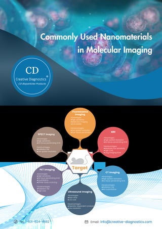
  • 1. Commonly Used Nanomaterials in Molecular Imaging CD Email: info@creative-diagnostics.com Tel: 1-631-624-4882 Target Fluorescence Imaging Advantages: ●High sensitivity ●Multicolor imaging ●Activatable Disadvantages: ●Low spatial resolution ●Poor tissue penetration MRI Advantages: ●High spatial resolution ●No tissue penetrating limit Disadvantages: ●Relatively low sensitivity ●High Cost ●Long imaging time CT Imaging Advantages: ●High spatial resolution ●No tissue penetrating limit Disadvantages: ●Radiation risk ●Not quantitative Ultrasound Imaging Advantages: ●Real-time ●Low cost Disadvantages: ●Operator dependent analysis ●Low resolution PET Imaging Advantages: ●High sensitivity ●No issue penetrating limit ●Quantitative ●Whole-body scanning Disadvantages: ●Radiation risk ●High cost SPECT Imaging Advantages: ●High sensitivity ●NO tissue penetrating limit Disadvantages: ●Radiation risk ●Low spatial resolution
  • 2. Nanomaterials (e.g. nanoparticles) display special physical and biological behavior and unique interactions with biomolecules. They possess large surface area and inherent functionalities, making structural modifications feasible to change their pharmacokinetics, prolong their vascular circulation life-time, improve their extravasation capacity, ensure an enhanced biodistribution in vivo and sustainably control the delivering efficacy for drug cargoes. In addition, nanoparticles conjugated with specific targeting ligands show a high targeted binding capability to diseased regions. Nanomaterials have been applied as efficient carriers for targeted drug delivery and therapeutic agents as well as for gene transportation. Molecular imaging is an attractive and fast-growing research field, in which several nanomaterials have been used as imaging agents (Table 1). As an emerging interdisciplinary research field, molecular imaging combines chemistry, biology, pharmacology, and medicine to monitor in vitro and in vivo biomedical or physiological processes at molecular and cellular levels, which provides valuable information for treatment strategies for various diseases. Table 1: Characteristics of several representative nanomaterials and their biomedical applications. Email: info@creative-diagnostics.com Tel: 1-631-624-4882 Type of Nanoparticle Synthetic Protocol Size Range Possible Surface Modifications Imaging Modality Applicable Gold nanoparticle Biological reduction, colloidal synthesis, vapor precipitation Several to hundreds of nm Lipids, polymeric shell, targeting ligands or biomolecules CT, optical imaging Silica nanoparticle Chemical polymerization, microemulsion, sol-gel Tens to hundreds of nm Charge, polymer, targeting ligands or biomolecules MRI, optical imaging Carbon nanotube Arc discharge, laser ablation, vapor precipitation Tens of nm Polymeric shell, targeting ligands or biomolecules MRI, optical, radionuclide imaging Quantum dot Colloidal synthesis, self- assembly, viral assembly Several to tens of nm Lipids, polymer, targeting ligands or biomolecules Optical imaging Iron oxide Coprecipitation, decomposition, microemulsion, sol-gel, thermal Several to tens of nm Charge, dextran, lipids, polymer, targeting ligands or biomolecules MRI Dendrimer Organic chemistry techniques Several nm varies from different “generation” Charge, polymer, targeting ligands or biomolecules MRI, optical imaging Liposome Emulsion, polymerization Tens to hundreds of nm Charge, polymer, targeting ligands or biomolecules, viral protein coating, MRI, optical, radionuclide imaging Microbubble Emulsion, layer-by-layer fabrication, polymerization Tens to thousands of nm Polymeric shell, targeting ligands or biomolecules Ultrasound imaging Micelle Microemulsion Tens of nm Charge, polymer, targeting ligands or biomolecules MRI, optical, radionuclide imaging Adenovirus Replication in host nucleus Tens to hundreds of nm Charge, polymer, targeting ligands or biomolecules MRI, optical imaging
  • 3. ◆ Five aspects need to be considered when developing an imaging agent with nanoparticles: 1) The toxicity of nanoparticles for living subjects and humans; 2) Any possible metabolites after vascular circulation or cell uptake; 3) The biocompatibility and biodegradability to avoid harmful accumulations in organs, tissues, and blood; 4) The availability for chemical modifications of nanoparticles; 5) The in vitro and in vivo comprehensive assessments of synthesized nanoparticles before practical applications for living subjects or humans. ◆ The common methodologies for design and functionalization strategies of nanoparticles are summarized below: 1) Select and fabricate nanoparticles core Based on imaging purposes and a specific imaging modality, the core of the nanoparticle is selected and the synthesis method of fabrication of the structure of imaging probes is determined. 2) Synthesize shell structure Compared with the core of nanoparticles, the shell structure usually possesses more complicated functions including preventing the core from the external microenvironment and improving the core stability and physical property. 3) Modify surface To maintain the nanoparticles' stability, surface coatings with stabilizers or emulsions may be necessary if the outer interface of the shell is too sensitive when exposed to bio-medium. • Fluorescence imaging Fluorescence imaging is one of the major techniques in optical imaging to analyze the propagation of nonionizing radiation, light photons through a medium such as tissue. It is the visualization of molecular processes or structures via fluorescent dyes or proteins. Fluorescence imaging can be applied in a wide range of experiments such as the location and dynamics of gene expression, protein expression, and molecular interactions in cells and tissues. Email: info@creative-diagnostics.com Tel: 1-631-624-4882 Design and Functionalization of Nanoparticles in Molecular Imaging Nanomaterials Used in Different Molecular Imaging
  • 4. • MRI MRI, magnetic resonance imaging, is one of the most widely used and powerful tools for noninvasive clinical diagnosis. It possesses a high degree of soft-tissue contrast, spatial resolution, and depth of penetration. There are several advantages of using nanoparticles as imaging probes compared with conventional imaging agents: 1) the concentration of the imaging agent is controllable in each nanoparticle during the synthesis process; 2) the tunability of the nanoparticles' surface is beneficial to target a specific location in the body and prolong the circulation time of the agent in the blood; 3) nanoparticles can be used as multifunctional molecular imaging agents due to their two or more properties. Table 2. Selected examples of nanomaterials used in fluorescence imaging. Table 3. Selected examples of nanomaterials used in MRI. Email: info@creative-diagnostics.com Tel: 1-631-624-4882 AuNP: gold nanoparticle; QD: quantum dot; UCNP: upconverting nanoparticle. NP: nanoparticle; USPIO: ultra-small superparamagnetic iron oxide; MNP: magnetic nanoparticle. NP Type Size (nm) Applications Imaging Alpha(nu)beta(3)-Gd (paramagnetic particle) 273 Imaging angiogenesis T1 Liposomal gadolinium 125 Imaging placenta as blood-pool contrast T1 RBC encapsulated iron particles 60 Blood-pool contrast with longer lifetime T1, T2 PEGMnCaP NPs 60 PH-activatable contrast in cancer T1, T2 USPIO-PEI 100 Determining nanoparticle vehicle unpackaging for gene T2 P-selectin-MNP(iron oxide)-PBP 50 Imaging post-stroke neuroinflammation T2 NP Type Imaging Agent Size (nm) Applications Cy5.5-substrate/AuNP Cy5.5 20 Detecting protease activity Cy5.5-DEVD-DOPAK/AuNP Cy5.5 37.8 Testing caspase-3 to identify apoptosis activity in cells QD710-Cy7-PEGylated lipids Cy7 20 Monitoring NP accumulation and dissociation kinetics in tumor QD710-Dendron/RGD (InP/ZnS core/shell QDs) Quantum dots 12 Targeted imaging tumor cells Perylenediimide-containing polysiloxane core and silica shell Perylenediimide 18, 70 Detecting nanotoxicity in alive cells AB3-UCNP(NaYF4:Yb/Tm/Er)- RB/KE108 UCNP 14 Monitoring cellular uptake of nanoparticles and combined with therapy
  • 5. • CT imaging Computed tomography (CT) is an X-ray based, a whole-body imaging technique and is widely used in medicine. Although iodinated small molecules or barium suspensions are clinically approved contrast agents for CT, developing nanoparticle-based CT contrast agents have attracted more attention due to the growing population of renally impaired patients and those who are hypersensitive to iodinated contrast. Nanoparticle-based CT contrast agents possess several advantages over small molecule ones, including long blood-pool residence times, and the potential for cell tracking and targeted imaging applications. • Ultrasound imaging Ultrasound (US) imaging is a diagnostic medical procedure that uses high-frequency sound waves to view inside the body. As real-time captured imaging, ultrasound images can not only show the movement of the body's internal organs but also present the blood flowing through the blood vessels. This technique does not require the use of ionizing radiation, nor the injection of nephrotoxic contrast agents. Since there is a great progress in the discovery of various disease-specific biomarkers and in the development of nanoparticle fabrication, nanoparticle-based ultrasound contrast agents have made a big development. Those contrast agents could extravasate through the leaky vasculature of a tumor into the interstitial space with less echogenic than microbubbles. Table 4. Selected examples of nanomaterials used in CT imaging. Table 5. Selected examples of nanomaterials used in ultrasound imaging. Email: info@creative-diagnostics.com Tel: 1-631-624-4882 AuNP: gold nanoparticle. AuNP: gold nanoparticle. NP Type Size (nm) Applications Classification Silica coated NP into perfluorobutane microbubble Near 3000 Ultrasound imaging agents with potential therapeutic applications Gas Exosome-like silica NP 30–150 Stem cell imaging agent Solid Gas-NP 290 PH related contrast agents in tumor Gas Porphyrin nanodroplet 185 Tumor imaging contrast agent Gas FA-PEG-CS and perfluorooctyl bromide nanocore 229.5 Molecular tumor imaging agents Liquid NP Type Size (nm) Applications PSMA-specific aptamer conjugated AuNP 29.4 Imaging prostate cancer cells Liposomal iodine 400 Imaging macrophage-rich atherosclerotic plaques Tantalum oxide <6 Producing greater imaging capability than iodine AuNP 20 Incorporating RBC to image blood flow AuNP 27–176 AuNP with CT contrast capability
  • 6. • PET/SPECT imaging As a powerful and widely used nuclear medicine technology, PET (positron emission tomography) possesses high tissue penetration and high sensitivity and is ideal for real-time quantitative imaging analysis. Similar to PET, SPECT (single-photon emission computed tomography) is also widely used nuclear medicine technology which can detect abnormal biochemical function before changes in anatomy. Both of them are suffered from high costs and radioactive exposure. Nanoparticles in PET/SPECT are generally used for tumor detection. Tumor imaging can occur through specific binding to receptors or via the EPR effect as well as being acquired through active and passive accumulation in target lesions. Each imaging modality has its own unique strengths. Multimodality imaging, combining two or more imaging modalities, can provide more comprehensive structural, functional and molecular information, which offers the prospect of improved diagnostics and therapeutic monitoring abilities. Nanoparticles have been used for the development of dual-modal or multimodal probes at an incredibly fast rate. For example, as dual-modal imaging probes, nanoparticles administrate a single contrast agent for different types of imaging modalities and possess signal consistency at the target region. Creative Diagnostics has presented a summary of commonly used nanomaterials in molecular imaging. If you desire any raw materials mentioned above, please visit our website to see more. Table 6. Selected examples of nanomaterials used in PET/SPECT imaging. Email: info@creative-diagnostics.com Tel: 1-631-624-4882 MSN: mesoporous silica nanoparticle; TNP: tri-reporter nanoparticle; AuNP: gold nanoparticle. NP Type Size (nm) Applications Imaging Modality 18 F-labeled DBCO-PEGylated MSN 100–150 Imaging tumor PET 64 Cu labeled CANF-comb nanoparticle 16–22 Imaging natriuretic peptide clearance receptor in prostate cancer PET 64 Cu-TNP 20 Imaging macrophages in inflammatory atherosclerosis PET 125 I silver nanoparticle 12 Monitoring distribution of nanoparticles SPECT 125 I labeled cRGD-PEG-AuNP 31 Detecting cancer cells and imaging tumor sites SPECT
  • 7. References: 1. Polyak, A., & Ross, T. L. (2018). Nanoparticles for SPECT and PET imaging: towards personalized medicine and theranostics. Current medicinal chemistry, 25(34), 4328-4353. 2. Pratiwi, F. W., Kuo, C. W., Chen, B. C., & Chen, P. (2019). Recent advances in the use of fluorescent nanoparticles for bioimaging. Nanomedicine, 14(13), 1759-1769. 3. Cormode, D. P., Naha, P. C., & Fayad, Z. A. (2014). Nanoparticle contrast agents for computed tomography: a focus on micelles. Contrast media & molecular imaging, 9(1), 37-52. 4. Mallidi, S., Wang, B., Mehrmohammadi, M., Qu, M., Chen, Y. S., Joshi, P., ... & Sokolov, K. (2009, September). Ultrasound-based imaging of nanoparticles: From molecular and cellular imaging to therapy guidance. In 2009 IEEE International Ultrasonics Symposium (pp. 27-36). IEEE. 5. Lee, D. E., Koo, H., Sun, I. C., Ryu, J. H., Kim, K., & Kwon, I. C. (2012). Multifunctional nanoparticles for multimodal imaging and theragnosis. Chemical Society Reviews, 41(7), 2656-2672. 6. Estelrich, J., Sánchez-Martín, M. J., & Busquets, M. A. (2015). Nanoparticles in magnetic resonance imaging: from simple to dual contrast agents. International journal of nanomedicine, 10, 1727. 7. Burke, B. P., Cawthorne, C., & Archibald, S. J. (2017). Multimodal nanoparticle imaging agents: design and applications. Philosophical Transactions of the Royal Society A: Mathematical, Physical and Engineering Sciences, 375(2107), 20170261. 8. Liu, Z., Kiessling, F., & Gätjens, J. (2010). Advanced nanomaterials in multimodal imaging: design, functionalization, and biomedical applications. Journal of Nanomaterials, 2010, 51. 9. Han, X., Xu, K., Taratula, O., & Farsad, K. (2019). Applications of nanoparticles in biomedical imaging. Nanoscale, 11(3), 799-819. 5 Tel: 1-631-624-4882 Email: info@creative-diagnostics.com Fax: 1-631-938-8221 Address: 45-1 Ramsey Road, Shirley, NY 11967, USA For more information, view our website: www.cd-bioparticles.com