
Recommended
More Related Content
What's hot
What's hot (20)
Similar to Intron and types
Similar to Intron and types (20)
Recently uploaded
Recently uploaded (20)
Intron and types
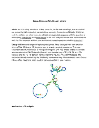
- 1. Group I introns :A(i). Group I introns Introns are noncoding sections of an RNA transcript, or the DNA encoding it, that are spliced out before the RNA molecule is translated into a protein. The sections of DNA (or RNA) that code for proteins are called exons. An intron is any nucleotide sequence within a gene that is removed by RNA splicing during maturation of the final RNA product.The term intron refers to both the DNA sequence within a gene and the corresponding sequence in RNA transcripts Group I introns are large self-splicing ribozymes. They catalyze their own excision from mRNA, tRNA and rRNA precursors in a wide range of organisms. The core secondary structure consists of nine paired regions (P1-P9). These fold to essentially two domains - the P4-P6 domain (formed from the stacking of P5, P4, P6 and P6a helices) and the P3-P9 domain (formed from the P8, P3, P7 and P9 helices). The secondary structure mark-up for this family represents only this conserved core. Group I introns often have long open reading frames inserted in loop regions. Mechanism of Catalysis
- 2. Splicing of group I introns is processed by two sequential ester-transfer reactions. The exogenous guanosine or guanosine nucleotide (exoG) first docks onto the active G- binding site loc ated in P7, and its 3'-OH is aligned to attack the phosphodiester bond at the 5' splice site located in P1, resulting in a free 3'-OH group at the upstream exon and the exoG being attached to the 5' end of the intron. Then the terminal G (omega G) of the intron swaps the exoG and occupies the G-binding site to organize the second ester-transfer reaction: the 3'-OH group of the upstream exon in P1 is aligned to attack the 3' splice site in P10, leading to the ligation of the adjacent upstream and downstream exons and release of the catalytic intron. Two-metal-ion mechanism seen in protein polymerases and phosphatases was proposed to be used by group I and group II introns to process the phosphoryl transfer reactions. B(i). Group 2 introns Group II introns are a large class of self-catalytic ribozymes and mobile genetic elements found within the genes of all three domains of life. Ribozyme activity (e.g., self-splicing) can occur under high-salt conditions in vitro. However, assistance from proteins is required for in vivo splicing. In contrast to group I introns, intron excision occurs in the absence of GTP and involves the formation of a lariat, with an A-residue branchpoint strongly resembling that found in lariats formed during splicing of nuclear pre-mRNA. It is hypothesized that pre-mRNA splicing (see spliceosome) may have evolved from group II introns, due to the similar catalytic mechanism as well as the structural similarity of the Domain V substructure to the U6/U2 extended snRNA. Finally, their ability to site-specifically mobilize to new DNA sites has been exploited as a tool for biotechnology.
- 3. Structure and catalytic site The Domain V substructure that is shared between Group II introns and U6 spliceosomal RNA. The secondary structure of group II introns is characterized by six typical stem-loop structures, also called domains I to VI or D1 to D6. The domains radiate from a central core that brings the 5' and 3' splice junctions into close proximity. The proximal helix structures of the six domains are connected by a few nucleotides in the central region (linker or joiner sequences). Due to its enormous size, the domain 1 was divided further into subdomains a, b, c, and d. Sequence differences of group II introns that led to a further division into subgroups IIA and IIB were identified. Group II introns also form very complicated RNA Tertiary Structure. • Group II introns have a very few conserved nucleotides, and the nucleotides important for the catalytic function are spread over the complete intron structure. • The few strictly conserved primary sequences are the consensus at the 5' and 3' splicing site (...↓GUGYG&... and ...AY↓...), some of the nucleotides of the central
- 4. core (joiner sequences), a relatively high number of nucleotides of D5 and some short-sequence stretches of D1. • The unpaired adenosine in D6 marked by an asterisk (7 or 8 nt away from the 3' splicing site, respectively) is also conserved and plays a central role in the splicing process. • During splicing of Group II introns, all reactants are preorganized before the initiation of splicing (A. De Lencastre et al. ;2005, ). • The catalytically essential regions of D5 and J2/3, and epsilon−epsilon' are in close proximity before the first step of splicing occurs. • Apart from the bulge and AGC triad regions of D5, the J2/3 linker region, the epsilon−epsilon' nucleotides and the coordination loop in D1 are crucial for the architecture and function of the active-site. Group II catalytic intron • Group II catalytic introns are found in rRNA, tRNA, and mRNA of organelles (chloroplasts and mitochondria) in fungi, plants, and protists, and also in mRNA in bacteria. • The length of Type II introns can be up to 3 kb. • They are large self-splicing ribozymes and have 6 structural domains (usually designated dI to dVI). • A subset of group II introns also encode essential splicing proteins in intronic ORFs. • Splicing occurs in almost identical fashion to nuclear pre-mRNA splicing with two transesterification steps. • The 2' hydroxyl of a bulged adenosine in domain VI attacks the 5' splice site, followed by nucleophilic attack on the 3' splice site by the 3' OH of the upstream exon. • Many proteins and intron-intron and intron-exon interactions important for splice site positioning for splicing in vivo. Group II introns sub-classified into groups: IIA and IIB, which differ in: i) splice site consensus sequence ii) and the distance of the bulged adenosine in domain VI (the prospective branch point forming the lariat) from the 3' splice site.
- 5. Structure of group II intron (This model and alignment represents only domains V and VI)