PSYPACT- Practicing Over State Lines May 2024.pptx
Arhthritis
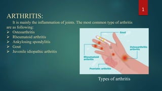
1. ARTHRITIS:
It is mainly the inflammation of joints. The most common type of arthritis
are as following:
Osteoarthritis
Rheumatoid arthritis
Ankylosing spondylitis
Gout
Juvenile idiopathic arthritis
1
Types of arthritis
2. 1. Osteoarthritis:
Osteoarthritis (OA), also called degenerative joint disease.
CAUSE:
It occurs when the protective cartilage that cushions the ends of your
bones wears down over time.
SYMPTOMS:
i. Pain
ii. Stiffness
iii. Tenderness
iv. Loss of flexibility
v. Grating sensation
vi. Bone spurs
vii. Swelling.
2
3. SITE OF OCCURANCE:
Osteoarthritis usually occur in weight bearing joints, other than this it
commonly affects joints in hands, knees, hips and spine.
3
OSTEOARTHRITIS OF THE SPINEOSTEOARTHRITIS OF THE HIP
4. DIAGNOSIS:
I. Imaging tests → X-rays. Magnetic resonance imaging (MRI)
II. Lab tests → Joint fluid analysis.
TREATMENT:
Therapy includes management of pain, NSAIDs to reduce inflammation,
intra-articular corticosteroids, activity modification, and, for severe cases,
arthroplasty.
SURGICAL AND OTHER PROCEDURES:
I. Lubrication injections
II. Cortisone injections
III. Realigning bones
IV. Joint replacement.
4
SURGERY OF HIP
KNEE OSTEOTOMY
5. 2.Rheumatoid Arthritis :
Rheumatoid arthritis (RA) is a chronic inflammatory disorder of
autoimmune.
CAUSE:
Rheumatoid arthritis occurs when your immune system
attacks the synovium resulting inflammation which can
eventually destroy the cartilage and bone within the joint. The
tendons and ligaments that hold the joint together weaken and
stretch. Gradually, the joint loses its shape and alignment.
SYMPTOMS:
i. Tender, warm, swollen joints
ii. Joint stiffness that is usually worse in the mornings and
after inactivity
iii. Fatigue, fever and loss of appetite
5
RHEUMATOID ARTHRITIS
6. SITE OF OCCURANCE:
i. Smaller joints (especially of hands and feet fingers).
ii. Other than this wrists, ankles, elbows, and knees are most commonly affected.
DIAGNOSIS:
i. Blood tests → ESR, CRP and anti-CCP.
ii. Imaging tests → X-rays and MRI.
TREATMENT:
The treatment for RA consists of corticosteroids, other immune suppresants such as
methotrexate, and most notably, TNF antagonists.
SURGICAL AND OTHER PROCEDURES:
i. Synovectomy
ii. Tendon repair
iii. Joint fusion
iv. Total joint replacement
6
SYNOVECTOMY OF KNEE
7. 3.Ankylosing spondylitis:
Ankylosing spondylitis is an inflammatory disease that,
over time, can cause some of the small bones in your spine
(vertebrae) to fuse.
CAUSE:
Ankylosing spondylitis has no known specific cause.
People who have a gene called HLA-B27 are at a great risk.
SITE OF OCCURANCE:
It usually occur in joint between the base of your spine and
your pelvis, lower back, tendons and ligaments that attach
to spine
7
Figure showing healthy and
ankylosing spondylitis
8. SYMPTOMS:
i. Pain and stiffness in your lower back and hips
ii. Neck pain and fatigue
DIAGNOSIS:
Imaging tests → X-rays, MRI, Dual energy CT scan.
TREATMENT:
NSAIDs, (TNF) blocker
8
Spinal cord showing stages of ankylosing
spondylitis
9. 4.Gout:
Gout is marked by transient attacks of acute arthritis initiated by
urate crystals deposited within and around joints.
Gout and pseudogout result from inflammatory responses
triggered by precipitation of urate or calcium
pyrophosphate, respectively.
CAUSE:
Gout occurs when urate crystals accumulate in your joint, causing the
inflammation and intense pain of a gout attack. Urate crystals can form
when you have high levels of uric acid in your blood.
SITE OF OCCURANCE:
Gout usually affects the large joint of your big toe,.
9
GOUT
10. SYMPTOMS:
i. Intense joint pain
ii. Lingering discomfort
iii. Inflammation and redness
iv. Limited range of motion
DIAGNOSIS:
i. Imaging tests → X-rays, MRI, Dual energy CT
scan.
ii. Lab tests → Joint fluid analysis
TREATMENT:
NSAIDs, Pain reliever, Corticosteroids
10
Figure 1 showing X-ray of normal foot
and figure 2 showing gout condition
11. 5.Juvenile Idiopathic Arthritis:
JIT is the most common type of arthritis in children under the
age of 16.
CAUSE:
The pathogenesis is unknown but similar to adult RA.
Symptoms:
i. Pain
ii. Swelling
iii. Stiffness
iv. Fever
v. swollen lymph nodes and rash
DIAGNOSIS:
i. Blood tests → ESR, C-reactive protein, Anti-nuclear
antibody, Rheumatoid factor, CCP
ii. Imaging tests → X-rays, MRI
TREATMENT:
NSAIDs, DMARDs, Biologic agents, Corticosteroids.
11
Figure showing a healthy and affected
joint of a kid
12. 12
0.00%
10.00%
20.00%
30.00%
40.00%
50.00%
60.00%
18-44 YEARS 45-64 YEARS ABOVE 60 YEARS
Prevalance rate of arthrits in
age group
Prevalence rate of arthritis
according to age group
WOMEN
58%
MEN
42%
Prevalance rate of
arthritis in gender
WOMEN MEN
Prevalence rate of arthritis
according to gender
PREVALANCE RATE OF ARTHRITIS:
13. 13PREVALANCE RATE OF OSTEOARTHRITIS:
9.60%
18.00%
0.00%
5.00%
10.00%
15.00%
20.00%
MEN WOMEN
Prevalance rate of osteoarthritis according
to gender
PREVALANCE RATE OF RHEUMATOID ARTHRITIS:
The prevalence varies between 0.3% and 1% and is more common in women and in
developed countries.
Within 10 years on onset, at least 50% of patients in developed countries are unable to
hold down a full-time job.
41 OUT OF 100,000.
14. 14PREVALENCE RATE ANKYLOSING SPONDYLITIS:
0.00%
10.00%
20.00%
30.00%
40.00%
AS
prevalance
per 10,000
23.80%
16.70%
31.90%
10.20%
7.40%
PREVALANCE RATE OF ANKYLOSING
SPONDYLITIS
Europe Asia North America Latin America Africa
PREVALENCE RATE GOUT:
0%
1%
2%
3%
4%
5%
Gout per 1000 person - year
PREVALANCE RATE OF GOUT
Prevalance Incidence
PREVALENCE OF JUVENILE IDIOPATHIC ARTHRITIS:
About 7/1000,00 newly diagnosed children with JIA per year.
Prevalence about 1/ 1000 children.