
Recommended
Recommended
More Related Content
What's hot
What's hot (20)
Similar to Cultural and Morphological Characteristics of Pyrenochaeta terrestris Isolates
Similar to Cultural and Morphological Characteristics of Pyrenochaeta terrestris Isolates (20)
More from QUESTJOURNAL
More from QUESTJOURNAL (20)
Recently uploaded
Recently uploaded (20)
Cultural and Morphological Characteristics of Pyrenochaeta terrestris Isolates
- 1. Quest Journals Journal of Research in Agriculture and Animal Science Volume 4 ~ Issue 8 (2017) pp: 10-14 ISSN(Online) : 2321-9459 www.questjournals.org *Corresponding Author: Mawahib Ahmed Elsiddig1 10 | Page Department, Plant Protection, College Of Agricultural Studies Sudan University Of Science And Technology, Shambat,Khartoum State, Sudan Research Paper Cultural and Morphological Characteristics of Pyrenochaeta terrestris Isolates Mawahib Ahmed Elsiddig*1 Siddig Mohamed El Hassan2 1 Department, Plant Protection, College Of Agricultural Studies Sudan University Of Science And Technology, Shambat, Khartoum State, Sudan 2 Department Of Crop Protections, Faculty Of Agriculture, University Of Khartoum, Shambat, Sudan mawahibahmed@hotmail.com Received 13 Jan, 2017; Accepted 16 Mar, 2017 © The author(s) 2017. Published with open access at www.questjournals.org ABSTRACT: The study on the different isolates of the fungus Pyrenochaeta terrestris that cause pink root rot disease on onion Allium cepa L., collected from different areas in Sudan, was carried out to elucidate the morphological, cultural and physiological variations of the pathogen. The PDA medium was found suitable for the fungus growth, while the Watson's medium was suitable for isolation. The white Kassala (WK) isolate outgrew the rest of isolates significantly, followed by the red Hudeiba (RHu) isolate, which scored more than two fold the rates of other isolates. The darker pigment of the culture seemed to be maintained only when the fungus utilized sucrose as the carbon source and/or sodium nitrate as the nitrogen source. The fungus Pyrenoehaeta terrestris was found to be more suited to slightly acidic to alkaline pH, although it could grow in a pH range of >4.0-8.0. The colour shade of the culture was found to be affected by the H ion concentration of the medium. Keywords: Cultural, morphological characteristics Pyrenochaeta terrestris I. INTRODUCTION The onion(Allium cepa L.) ,demand for it is worldwide and is fairly constant throughout the year is probably a native of Asia, perhaps from Palestine to India and Egypt. It has been in cultivation and used as a food from earliest period of history. It is mentioned in the Bible as one of the things for which the Israelies longed in the wilderness (Thompson and Kelly, 1957; Tindall, 1983).In the Sudan, it is mainly grown in Khartoum, Kassala, Gezira, White Nile, Darfour and Northern State. The total area put under onion production is increasing steadily in the last few decades. The average yield between the years 1981 and 1994 was 7,164 kg per hectare. The total production in 1994 was estimated at 81,000 MT. The pink root rot disease is incited by a soil inhabiting fungus (Pyrenochaeta terrestris (Hansen) Gorenz, Walker and Larson) which attacks onion plants from the seedling to maturity stages. The first observation of the pink root rot disease in the Sudan was made by Prof. ElHilo at Hudeiba Research Station in early 1960's and in 1982 the causal organism was identified on the basis of cultural characteristics on a specified medium using symptomatic onion roots (Yassin et al, 1982). However, the disease is a serious constraint to onion production in some of the main producing areas of the country such as River Nile, Khartoum, Gezira and Kassala State (El Amin, 1999) as well as in certain parts of Darfur in Western Sudan (Dr. S. M. El Hassan, Personal communication). The present study is therefore intended to: Elucidate the cultural, and morphological among characteristics of Pyerenochaeta terrestris isolates. II. Materials And Methods The original Pyrenochaeta terrestris isolates were provided by Dr. Siddig M. El Hassan, Dept. of Crop Protection Fac. of Agric. Univ. of Khartoum as pure culture of the fungus stored in potato dextrose agar (PDA) in McCartney bottles in the refrigerator. These fungus isolates were originally isolated from onion samples showing typical pink root disease symptoms collected from Kassala (Kassala State), Hilalyiah (Gezira State), Wad-Elbasal and Islanj (Khartoum State), and Hudeiba (River Nile State). The six isolates were first re-vitalized by sub culturing in PDA medium
- 2. Cultural and Morphological Characteristics of Pyrenochaeta terrestris Isolates *Corresponding Author: Mawahib Ahmed Elsiddig1 11 | Page under aseptic conditions and incubated for 7 days at 28° C. Plants Seeds of cultivars Saggai (red), Kamlin (yellow), and ElHilo (white) were used and sown in October, 1999 after being sterilized by NaOC1 in plastic trays containing sterilized sand/clay soil mixture (1:2) and kept in the nursery of Faculty of Agric. Univ. of Khartoum. The seedlings were watered when needed. The seedlings were transplanted in December 1999 to plastic bags (10 seedlings/bag). The soil mixture was inoculated with P. terrestris prior to seedlings transplanting. The onion seedlings were then maintained in the screen house. After 1.5 months from transplanting in inoculated soil, the fungus was re-isolated from infected seedlings. The respective new isolates were named after the area and colour of onion cultivar into which it was passed. These were: - Hudieba red (RHu), Hilalyiah red (RHi, Kassala red (RK), Kassala yellow (YK), Islanj yellow (YIs), Hudieba yellow (YHu), Wad - Elbasal white (WW) and Kassala white (WK). The effect of different media In this experiment different media were used to determine the best composition for growth of P. terrestris isolates. These media included: Potato dextrose agar (PDA) Watson's medium, and modified Watson's medium (Watson's minus NaNO3, and Watson's minus MgSO4). A number of sterilized Petri-dishes were poured with the different media and tetracycline anti-biotic was added at a rate of 0.1 g/I to suppress bacterial contamination. These plates were inoculated, respectively, with the eight fungus isolates (RHu, RHi, RK, YK, YIs, YHu, WW and WK) using 9 mm discs cut froth the edge of the 7-day old culture. The inoculated Petri-dishes, were incubated at 27° C in 4 replicates. Two perpendicular diameters were drawn on the back of each Petri-dish to intersect at the centre of the culture. The rate of fungal growth was measured daily along the two diameters drawn previously and the mean colony diameter, was calculated for each isolate. The colour, texture, shape and pattern of culture growth were observed and recorded for each fungus isolate in the different media tested. The effect of hydrogen ion concentration (pH) Two buffers were used in this study: phosphate buffer (NaH2PO4 and Na2HPO4) and acetate buffer (Acetic acid and Na acetate). The first one was used for the high pH levels, whereas the other one was used for the low pH levels. A number of flasks were prepared with 25 ml of potato dextrose broth (PDB) (Appendix 4) per each flask. Similar amount of buffers were added to each flask. Five levels of pH were used 4, 5, 6, 7 and 8. The pH of all media was checked before inoculation of the pathogen by Beckman pH meter. After adding buffer solution, the flasks were sterilized, cooled, and then, inoculated with 9 mm discs cut from the edge of 7- days old culture of each isolate under test, respectively. Three replicates in a randomized complete design were used. The inoculated flasks were inoculated at 27°C for 10 days, after which the mycelial mat was filtered through a cheese cloth and the mycelium was dried in an oven at 70°C/48 h. Finally, the dry weight of the mycelium of each isolate was obtained. III. Results The effect of different media The pink root fungus was one of the slow growing fungi. The aerial mycelium of the fungus was septate, gutulate compact, delicate, velvety, grey in colour and developed a range of pigmentation from pink to purple in the culture medium. In comparison, potato dextrose agar, Watson's medium and modified Watson's mediums were all found to be suitable for the fungus growth, while Watson's medium was the best for isolation. The pigment varied from pink to slight pink or purple but was usually a shade of red. In Watson's medium and Watson's minus NaNO3 medium the fungus isolates recorded significantly the lowest radius of colony growth while PDA supported significantly the greatest growth. The data also disclosed significant differences in the growth rates of different P. terrestris isolates under test. The isolate `WK' scored significantly the greatest mean colony diameter followed by `RHu' isolate while 'RK' and `RHi gave the lowest mean colony diameter. The interaction of the fungus and the culture media was also found to be significant while 'WK and `RHu' isolate producing the fastest growth in PDA and `RK displaying the slowest growth in W-MgSO4 (Table 1; Figs. 1 and 2).
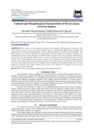
- 3. Cultural and Morphological Characteristics of Pyrenochaeta terrestris Isolates *Corresponding Author: Mawahib Ahmed Elsiddig1 12 | Page Table 1. Effect of different media on colony growth of Pyrenochaeta terrestris isolates measured 5 days post- inoculation Fungus isolates* Media** PDA W W-NaNO3 W-MgSO4 Mean RHu 19.875a 8.625d 9.505d 12.875b 12.720b RK 5.315g-1 5.020h-1 4.115kl 2.188u 4.159e RHi 5.320g-1 3.925i 4.165kl 3.9131 4.331e YHu 6.875ef 6.450e-i 4.950i-1 6.775e-g 6.262c YK 5.150hykl 4.325kl 5.595f-k 6.590e-h 5.415d YIs 5.12h-1 6.887ef 5.525f-k 6.145e-j 5.917cd WW 6.600eh 5.650f-k 4.850j-k 7.275e 6.094cd WK 20.950a 12.400bc 11.425c 13.125b 14.475a Mean 9.400a 6.660c 3.266c 7.361b * Fungus isolates: RHu = Hudeiba isolate passed into Saggai (Red) RK = Kassala isolate passed into Saggai (Red) RHi = Hilalyih isolate passed into Saggai (Red) Yls = Islanj isolate passed into Kamlin (yellow) YHu= Hudeiba isolate passed into Kamlin (yellow) YK = Kassala isolate passed into Kamlin (yellow) WW= Wad Elbasal isolate passed into El Hilo (white) WK = Kassala isolate passed into El Hilo (white) ** Media: PDA= Potato dextrose agar. W = Watson's medium. W-NaNO3 = Watson's minus NaNO3. W-MgS 04 =Watson's minus MgSO4. Figures represent the mean colony radii (mm). Means followed by the same letter(s) are not significantly different (P=0.05). Fig. 1.Effect of medium on growth rate of Pyrenocheata terrestris isolates (5-day-old culture) From left to right Upper: The growth and pigmentation of RK , RHi and RHu%) isolates on Watson's minus NaNO3 Below: Same isolates on Watson's minus MgSO4
- 4. Cultural and Morphological Characteristics of Pyrenochaeta terrestris Isolates *Corresponding Author: Mawahib Ahmed Elsiddig1 13 | Page Fig. 2. Effect of medium on growth rate of (RK and Yls) isolates (5-d ay-old culture) from left to right Upper: The isolates on Watson's medium Below: The same isolates on FDA medium The effect of hydrogen ion concentration (pH) on mycelial dry weight of P. terrestris isolates The results in Table 2 reveal that the different fungus isolates grew considerably at pH levels between 5.0 and 8.0, but the growth was significantly minimal at pH 4.0. The best average mycelial growth was obtained at pH 6.0 followed by that at pH 8.0 and 7.0, respectively; however, the differences were significant. The mean mycelial dry weights under the pH range tested (4.0 – 8.0) of these fungus isolates were found to be significantly different. The greatest average mycelial dry weight was produced by the isolate `RHu) followed by `RK, `YIs' and 'WK', respectively, while the lowest weights were shown by YK and `RHi isolates. The colour of these isolates ranged from pink at pH 8.0 to dark pink at pH 5.0 (Fig. 4). There were no significant differences in mycelial weights between isolates at pH 4.0. Table 2. Effect of H-ion concentration on mycelial dry weight (g) of Pyrenochaeta terrestris isolates Fungus isolates* pH- level 8.0 7.0 6.0 5.0 4.0 Mean RHu 0.403a 0.230a-d 0.353abc 0.273cde 0.010I 0.272a RK 0.317a-d 0.240d-g 0.393ab 0.037j-I 00.017I 0.201b RHi 0.123i-k 0.110i-I 0.160g-i 0.093kI 0.010I 0.099e YHu 0.163f-I 0.230d-h 0.267c-f 0.027kI 0.010I 0.139d YK 0.123i-k 0.077i-I 0.170e-I 0.093i-I 0.017I 0.096e YIs 0.247d-g 0.230a-d 0.357abc 0.038j-I 0.010I 0.194bc WW 0.210red 0.133hij 0.267c-f 0.263c-g 0.010I 0.193bc WK 0.233d-h 0.177e-i 0.303a-d 0.030j-I 0.020kI 0.153cd Mean 0.237b 0.201c 0.284a 0.107d 0.013e * Fungus isolates RHu Hudeiba isolate passed into Saggai (Red) RK Kassala isolate passed into Saggai (Red) RHi Hilalyih isolate passed into Saggai (Red) YIs Islanj isolate passed into Kamlin (yellow) YHu Hudeiba isolate passed into Kamlin (yellow) YK Kassala isolate passed into Kamlin (yellow) WW Wad Elbasal isolate passed into El Hilo (white) WK Kassala isolate passed into El Hilo (white) Means followed by the same letter(s) are not significantly different (P=0 05).
- 5. Cultural and Morphological Characteristics of Pyrenochaeta terrestris Isolates *Corresponding Author: Mawahib Ahmed Elsiddig1 14 | Page IV. DISCUSSION The present investigation was conducted to study the different isolates of Pyrenochaeta terrestris (Hansen) Gorenz, Walker and Larson from the main onion producing areas in the Sudan and elucidate their variability in cultural, morphological and physiological characteristics. The difficulty faced during isolation of the pink root organism from infected onion roots might be due to the presence of co-inhabiting fungi, which usually impeded the isolation. This was also suggested by previous workers (Davis and Henderson, 1937; Kreutzer, 1941; Hess and Vaughan, 1962). The variation in P. terrestris isolated from .the different parts of the surveyed areas (eight isolates in this thesis) is an indicative of a high variability of the pathogen and this a further attribute of ecological fitness of the fungus. The isolates of the fungus under study were found to differ in cultural properties. The results that potato dextrose agar medium (PDA) sustained a better fungus growth, while Watson's medium was best for isolation indicated the correlation between nutritional status of the medium and a specific step in fungus isolation and identification. Hence, a rich medium such as PDA is best for growth but usually encourages contaminants and rapid growth of co-inhabiting fungi, while poor medium (e.g. Watsons medium) may inhibit or delay growth of contaminations giving the causal organism a chance to outgrow them. This interpretation is in agreement with those presented by previous workers (Hess and Vaughan, 1962; El Amin, 1999). The data on fungus colony diameter as affected by culture media illustrated the fast growth of the isolate `WK, which was isolated from Kassala and passed through the onion cultivar `ElHilo'. The significant differences in the rate of growth between this isolate and the other ones including the original Kassala isolate, which was passed into Saggai' (red) and `Kamlin' (yellow) indicated a clear genetic variability between these isolates. The data also indicated that the `RHu' isolate although significantly less faster in growth than 'WK', yet it scored the second best growth rate, which was more than two-fold the rates of the remaining isolates. The darker pigment of P. terrestris in culture seemed to be maintained only when the fungus utilized sucrose as the carbon source and/or sodium nitrate as the nitrogen source. This was found to be in accord with the conclusions of Horton (1964) and Gunasekaran and Weber (1981). The results that the isolates of P. terrestris grew well at all hydrogen ion concentrations within the range 4.0-8.0 illustrate that the fungus can adapt well to a wide range of soil types, although it is obvious that the fungus is more suited to slightly acidic to alkaline soil reaction. The peak at pH 6.0 is significantly greater than the beak at pH 8.0 and the curve decline steeply with the increase in the acidity. These data agree well with those of Kreutzer (1939) and Gorenz et al. (1949). The variability in the fungus was partly expressed in the colour variation illustrated by the different isolates at various pH levels. The colour shades due to the pH change in the medium could be explained on the basis of availability of certain elements such as the C and N sources and their ratios. The significant differences in the dry mycelium weight displayed by the different fungus isolates indicate clearly the variability in P. terrestris as affected by the medium pH. The growth of `RHu' isolate was significantly greater than the rest of the isolates under test followed by `RK', `YIs' and 'WW'. However, the 'WK' isolates, which displayed the best growth performance using different growth media failed to maintain it at a pH value other below or above 6.0. This indicates the reduced virulence of "WK" isolate in non- ideal conditions of infection. Future lines - The perfection of a procedure for simple and fast isolation of the pathogen from infected onion roots. Finding a selective medium and/or ability to induce formation of pycnidia on infected roots would facilitate and simplify isolation of the pathogen. REFERENCES [1]. Davis, G, N., and Henderson, W. J. (1937). The interrelation of the pathogenicityof a phoma and fusarium on onion. Phytopathology 27: 763 – 772. [2]. El Amin, I. H. (1999). Investigations on onion pink root disease in Sudan. Ph. D. thesis, University of Khartoum, Sudan. [3]. Gorenz, A. M., Larson, R. H., and walker, J. C. (1949). Factors affecting p a t h o g e n i c i t y o f p i n k r o o t f u n g u s o f o n i o n s . J o u r n a l o f Agricultural Research 78: 1 – 18. [4]. Gunasekaran, M., and Weber, D. J. (1981). Influence of physiochemical fact ors on growth and pi gment synth esi s b y Pyrenocha eta terrestris. Mycologia 73: 844 – 852. [5]. Hess, W. M., and Vaughan, E. K. (1962). Pathogenicity of Pyrenochaeta terrestris and Fusarium sp. from onion roots. Phytopathology 52: 735 (Abstr). [6]. Horton, J. C. (1964). Extracellular pectolytic and cellulytic enzymes of Pyrenochaeta terrestris. Horton, J. C. and keen, N. T. Glucose inhibition of cellulase synthesis by P. terrestris. [7]. Kreutzer, W.A. (1939). Host- parasite relationships in pink root of Allium cepa. L. I.The pigment of Phoma terrestris. Phytopathology 29: 629-632. [8]. Kreutzer, W. A. (1941). Host parasite relationship in pink root of Allium cepa and other hosts. Phytopathology 31: 907 – 915. [9]. Thompson, H. C. and Kelly, W. C. (1957). Vegetable crops. Text book MCGRAW. Hill book. P 348-355. [10]. Tindall, H.D. (1983). Vegetable in the tropics. 1st edition. Lonodn and Basingtoke. P. 20-23. [11]. Yassin, A. M., Babiker, A. G. T., and Ahmed, M. K. (1982). First report of pink root of onion in Sudan (Pyrenochcteta terrestns) Plant Dis. 66: 741.