Prevalence, occurrence and biochemical characterization of Xanthomonas campestris pv. vesicatoria in District Swat, Pakistan and its management through host resistance | JBES

Xanthomonas campestris pv. vesicatoria the causal organism of bacterial spot in tomato results in heavy losses both in the form of quality and. In this study a survey was carried out to report the incidence of bacterial spot disease of tomato in district Swat. We reported maximum disease incidence in tehsil Kabal (71.66%), followed by Charbagh (61.66%) and Barikot (58.33%). For resistant screening a total of 13 tomato germplasms were screened against the disease. The foliar severity ranged from 3.33% to 73.33%, while severity for fruits was ranged from 18.33% to 30.66%. In case of phenotypic data the highest numbers of fruits obtained were 34, plant height 79.5cm and fruit weight was 470 grams/ten tomatoes. While the lowest average numbers of fruits were 6.67, plant height 45.7cm and fruit weight recorded was 215.67 grams/ten tomatoes. Line 1288 showed highest level of resistance followed by Red-stone. However, line 9708 showed highest susceptibility when exposed to artificial inoculation. Our study showed that bacterial spot is a major issue in some part of Pakistan and germplasm screening are linked to increased host resistance and could offer an important contribution to future integrated bacterial spot management programs.

Recommended
Recommended
More Related Content
What's hot
What's hot (19)
Similar to Prevalence, occurrence and biochemical characterization of Xanthomonas campestris pv. vesicatoria in District Swat, Pakistan and its management through host resistance | JBES
Similar to Prevalence, occurrence and biochemical characterization of Xanthomonas campestris pv. vesicatoria in District Swat, Pakistan and its management through host resistance | JBES (20)
More from INNS PUBNET
More from INNS PUBNET (15)
Recently uploaded
Recently uploaded (20)
Prevalence, occurrence and biochemical characterization of Xanthomonas campestris pv. vesicatoria in District Swat, Pakistan and its management through host resistance | JBES
- 1. J. Bio. & Env. Sci. 2020 130 | Khan et al. RESEARCH PAPER OPEN ACCESS Prevalence, occurrence and biochemical characterization of Xanthomonas campestris pv. vesicatoria in District Swat, Pakistan and its management through host resistance Aftab Ali Khan1 , Abdul Rafi1 , Asma Akbar*2 , Zahoor Ahmad3 , Azra Nadeem1 1 The University of Agriculture, Peshawar, Pakistan 2 Crop Diseases Research Institute, National Agriculture Research Centre, Islamabad, Pakistan 3 Adaptive Research Program, Quetta, Baluchistan, Pakistan Article published on August 30, 2020 Key words: Tomato, Bacterial spot, Xanthomonas campestris pv. vesicatoria, Host resistance Abstract Xanthomonas campestris pv. vesicatoria the causal organism of bacterial spot in tomato results in heavy losses both in the form of quality and. In this study a survey was carried out to report the incidence of bacterial spot disease of tomato in district Swat. We reported maximum disease incidence in tehsil Kabal (71.66%), followed by Charbagh (61.66%) and Barikot (58.33%). For resistant screening a total of 13 tomato germplasms were screened against the disease. The foliar severity ranged from 3.33% to 73.33%, while severity for fruits was ranged from 18.33% to 30.66%. In case of phenotypic data the highest numbers of fruits obtained were 34, plant height 79.5cm and fruit weight was 470 grams/ten tomatoes. While the lowest average numbers of fruits were 6.67, plant height 45.7cm and fruit weight recorded was 215.67 grams/ten tomatoes. Line 1288 showed highest level of resistance followed by Red-stone. However, line 9708 showed highest susceptibility when exposed to artificial inoculation. Our study showed that bacterial spot is a major issue in some part of Pakistan and germplasm screening are linked to increased host resistance and could offer an important contribution to future integrated bacterial spot management programs. *Corresponding Author: Asma Akbar asmaakbar139@gmail.com Journal of Biodiversity and Environmental Sciences (JBES) ISSN: 2220-6663 (Print) 2222-3045 (Online) Vol. 17, No. 2, p. 130-143, 2020 http://www.innspub.net
- 2. J. Bio. & Env. Sci. 2020 131 | Khan et al. Introduction Tomato (Lycopersicon esculentum Mill) is an important vegetable and fruit crop (Akbar et al., 2018) after potato both in consumption and production worldwide (Lemma et al., 1992). It is consumed as a raw or as an ingredient in a number of dishes and drinks (Anonymous, 2009). Tomato contains essential amino acids, dietary fibers, minerals, vitamins and sugars (Akbar et al., 2018). The metabolites of tomato have reducing effect on acidity of urine when compared to orange juice (Saywell et al., 1933). Furthermore, lycopene contents in tomato act as an antioxidant and play great role in prevention of cancer heart diseases and many other health problems (Miller et al., 2002) (Akbar et al., 2018). In terms of production China is in the list of top tomato producing country and accounts for one quarter of tomato acreage of the world. Furthermore Asia and Africa is shares approximately 65% to the world tomato production (FAO, 2008). Under divers agro-ecological conditions of Pakistan tomatoes are cultivated over 52.8 thousand hectares with the annual production of about 529.9 thousand tons Pakistan (Agriculture statistics of Pakistan, 2011-12). The tomato production of Baluchistan is more (45%) as compared to other provinces like KP which 35% followed by Punjab 14% and Sindh 10%. (Anonymous, 2007). In Khyber Pakhtunkhwa, tomato occupied an area of 16.5 thousand hectares with the average yield of 9.8 tons/ha (Federal Bureau of Statistics, 2012). Off season tomatoes are grown in the frost-free area of the Katha, Sughral in Punjab province and Dargai area in KP (Burney, 1995). In Pakistan tomato yield is very low when compared to other leading tomato growing countries like USA, India and is much less than world average production which is 36 ton/ha. Low tomato production is attributed to several insect’s attack and diseases. Different fungal, bacterial, and viral and nematodes diseases occur in tomato. Among those bacterial diseases, tomato spot is one of the destructive disease. Bacterial spot caused by Xanthomonas vesicatoria causes a heavy loss than any other bacterial disease. In 1921 bacterial spot was reported by Doidge in USA and South Africa for the first time and later reported on capsicum for the first time in Florida in 1923 by Gardner (Jones et al., 2004). Xanthomonas vesicatoria is having rod shape, motile aerobic and gram negative and having single or polar flagellum (Thieme et al., 2005). On Nutrients Agar colonies are circular, wet, The pathogen can affect all above ground parts of tomato including stem, leaf, fruit, etc. Lesions on leaves are small (<3mm in diameter) brown and circular with water-soaked margins. Lesion on fruits begin as raised blisters. Older lesions are brown, scab like and sunken in the middle. Keeping in view the importance of this disease in our province particularly in tomato growing areas of district swat, the study was planned with the following objectives. 1. To determine the incidence of bacterial spot caused by Xanthomonas vesicatoria in tomato growing areas of District Swat. 2. To isolate and identify the pathogen through biochemical tests. 3. To screen tomato germplasm for resistance to bacterial spot. Material and methods Assessment of disease incidence and samples collection Survey was conducted in tomato growing areas of district Swat for bacterial spot of tomato incidence. The district was divided into three different areas and in each tehsil three locations were surveyed and two fields in each location were selected at a well apart distance i.e., 5 to 10Km (Table 1). To find out disease incidence 10 plants from 2 to 3 spots in each field were randomly selected and their average is obtained. The disease incidence was determined by using the following formula. Disease incidence (%) = No of diseased plants Total no. of plants observed x 100 To isolate pure culture of the causal bacterium diseased samples were collected at the same locations. Samples were kept in bags, properly
- 3. J. Bio. & Env. Sci. 2020 132 | Khan et al. labeled and brought to plant pathology laboratory at Agriculture Research Institute (ARI) Swat and stored at 4oC for further processing. Table 1. Tomato growing areas of district Swat visited for disease incidence and samples collection. SL Locality Tehsil 1 Jawand Kabal 2 Nasrat 3 Sogaley 4 Ghari Barikot 5 Parrhi 6 Dadahara 7 Charbagh Charbagh 8 Gulibagh 9 Alabad Experiment In-vitro Isolation and identification of pathogen The technique developed by Chiejina. (2008) which is used to isolate bacterium. 2mm thin sections were cut from infected fruits and leaves and sterilized in 70% ethyl alcohol. After sterilization, the pieces were washed three times with sterilized distilled water and blotted dried on paper towels aseptically. Then 4-5 sterilized pieces were placed on Nutrient agar (NA) plates. Plates were kept at 30oc in incubator for 24 to 48 hours upside down. To obtain pure culture a smear of well grown colony was steaked on NA medium and incubated for 24 to 48hours. Then the isolates obtained from samples were subjected for identification using gram staining reaction (Schaad., 1988), Hypersensitive response test (James et al., 1996), (Sahin et al., 1996) and through use of various biochemical tests by (Holt et al., 1994) such as KOH test (Ryu, 1940; Suslow et al., 1982), Catalase test (He et al., 1993), Oxidase test (Schaad, 1988) and Starch hydrolysis test (Sands, 1990). Experiment in-vivo for screening germplasms against Bacterial spot Nursery preparation and transplantation for screening tomato germplasms For this purpose, diseased free seeds of different available tomato germplasms (13) were sown initially in trays. Trays were maintained in screen house for four weeks till germination of seeds. After four weeks, transplantation was done in the field at ARI (Agriculture Research Institute) Mingora Swat. Six plants per plot were planted, the distance from plant to plant was 45cm and the rows distance was 90cm in a randomized complete block design. The management practices for fertilization, insect and foliar fungal diseases were done according to standard recommendations (Ivors et al., 2013). Copper was sprayed on control plants to control bacterial pathogens. Table 2. Tomato germplasms and its source, evaluated for resistance against bacterial spot. SL Germplasms Source 1 AVTO 9802 ARI Swat 2 1405 -- 3 1409 -- 4 1455 -- 5 AVTO 1003 -- 6 AVTO 1130 -- 7 9708 -- 8 1429 -- 9 1288 -- 10 U-RO -- 11 Rio-Grande Market 12 Red stone -- 13 Roma -- Inoculum preparation and inoculation A 3-days old culture of Xanthomonas vesicatoria was flooded with SDW (sterilized distilled water). The colonies were scrapped by using a glass slide. The colonies concentration in suspension was adjusted to OD 600 and abs 0.3 with spectrophotometer. Plants were inoculated with suspension in the field by using a sprayer after 30 days of transplantation. Inoculation process was done in morning time to increase the chances of infection. Bacterial spot symptoms developed in about 14 to 21 days on leaf surface. Data on the following Parameters was recorded and statistically analyzed using statistic 8.1 software. The determined were subjected to ANOVA at the least significant difference Test (at 5% significance level) (steel et al., 1997). Disease severity After four weeks of inoculation Plants were calculated for foliar bacterial disease severity according to the following scale, adopted by Suárez-Estrella1 et al., 2014.
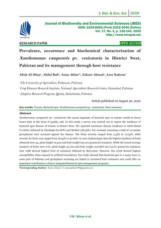
- 4. J. Bio. & Env. Sci. 2020 133 | Khan et al. Table 3. Disease Severity Scale used for grading germplasm resistance. Disease rating Symptoms Category 0 Symptomless Plants Resistant 1 Mildly infected plants (<50% of spotted leaves) Moderately Resistant 2 Highly infected plants (>50% of spotted leaves but plants not dead) Susceptible 3 Dead plants Highly Susceptible (Suárez-Estrella1 et al., 2014). Number of fruits per plant Number of fruits per plant was counted after each picking. Mean number of fruits per plant were calculated after the last picking and were analyzed statistically. Plant height The growth of a plant is highly reflected by plant height which contributes to the final yield. Statistical results of data pertaining to the plant height (cm) were measured at 4, 6 and 8 weeks after planting. Weight of fruits The disease affects the weight of the fruits as well, so data about the weight of fruits at maturity was calculated through digital balance. Statistical analyses Analyses of variance of RCBD was performed using Statistical Software statistix 8.1. Least Squares of Means were calculated, and lines were separated using LSD at P = 0.05 levels (Steel et al., 1997). Disease severity on leaves and fruits were calculated on ten severely infected leaves and fruits of each plot. Results Incidence of bacterial spot in tomato growing areas of Swat Survey was done for incidence of bacterial spot disease in different growing areas of Swat. A total of nine locations were visited in the district. Highest bacterial spot incidence was found for tehsil Kabal which was 71.6%, followed by tehsil Charbagh 61.6% and in tehsil Barikot 58.3% disease incidence was recorded (Table 4). Table 4. Incidence of Bacterial Spot disease in Tomato growing areas of District Swat. SL Areas Incidence (%) 1 Kabal 71.6 A 2 Charbagh 61.6 B 3 Barikot 58.3 B 4 LSD Value 3.60 Experiments in vitro Isolation and purification Small pieces of diseased leaves/fruits samples, placed on Nutrient Agar plates and re-streaking was done. Bacterial colonies were obtained at 30ºC after 48 hours of incubation. (Fig.1). The visual observation was made visually to identify the pathogen (Xanthomonas vesicatoria) through colonies morphology. The color of colonies was light yellow to yellow. The colonies shape was found to be mucoid and convex. A total of 9 isolates were obtained which were used further (Table 5). Table 5. Morphology of bacterial colonies obtained after 24 hours grown on NA medium. SL Locality Colony/24 hour Mucoid, convex, Yellow colony 1 Jawand yes + 2 Nasrat yes + 3 Sogaley yes _ 4 Garrhi yes + 5 Pharri yes + 6 Dadahara yes + 7 Charbagh yes _ 8 Gulibagh yes + 9 Alabad yes + += Xanthomonas vesicatoria (Xv) -= bacterial culture other than Xv Hypersensitive response test HR-inducing ability of the isolates was tested on tobacco plant. Complete collapse of tissue followed by necrosis after 24 hours was found as positive (Fig. 2) (Table 6). Out of nine isolates, seven showed H.R response. However, isolate representing Tehsil Kabal showed sever HR response that was used for inoculation in In-vivo experiments.
- 5. J. Bio. & Env. Sci. 2020 134 | Khan et al. A F E D C B Fig 1. Isolation of pathogen from leaves and fruits samples and inoculation on NA medium. A & B (Isolation and inoculation), C& D (Bacterial culture), E (Bacterial colonies), F (Gram staining). Table 6. Hypersensitive Response of bacterial isolates on tobacco plants 24 hours after inoculation. SL Isolates Hypersensitive Response 1 I. k1. + 2 I. k2 + 3 I. k3 + 4 I. c1 + 5 I. c2 + 6 I. c3 _ 7 I. b1 + 8 I. b2 _ 9 I. b3 + The symbol “+” = express positive result that the bacterium is pathogenic while the symbol “_” denotes a negative result. Fig 2. Hypersensitive Response on Tobacco plants 24 hours after inocultaion with representative bacterial isolates. B(bacteria) W(water). Biochemical characterization of pathogen The biochemical characterization results have been shown (Table 7). The bacterial stained mounts were observed under light microscope using oil immersion in gram staining. Xanthomonas vesicatoria isolates were found pinkish in color and termed as gram negative. KOH test Thread like slime was formed from viscous suspension when picked up by the toothpick during KOH test (Fig. 3a), indicating that the bacterium was Gram-negative. Catalase test The bacterium produced bubbles by dropping Hydrogen peroxide on smear of culture indicating the bacteria as catalase positive (Fig. 3b). Oxidase test The Xanthomas vesicatoria was characterized as oxidase negative that gave no color after 60 seconds (Fig. 3d). Starch hydrolysis test After incubation of seven days at 30ºC a clear zone around the colonies was formed on starch medium when Lugol’s Iodine was applied. This test indicated that the bacterium hydrolyzed the starch which was present in the nutrient agar medium (Fig. 3c)
- 6. J. Bio. & Env. Sci. 2020 135 | Khan et al. Table 7. Bio-chemical characterization of bacterial isolates. SL Isolates Gram reaction KOHCatalaseOxidase Starch Hydrolysis 1 Isolate.k1 _ + + _ + 2 Isolate.k2 _ + + _ + 3 Isolate.k3 _ + + _ + 4 Isolate.c1 _ + + _ + 5 Isolate.c2 _ + + _ + 6 Isolate.c3 _ + + _ + 7 Isolate.b1 _ + + _ + 8 Isolate.b2 _ + + + + 9 Isolate.b3 _ + + _ + The symbol “+” = denotes a positive result while the symbol “_” denotes a negative result. Fig. 3. Identification of the bacterium through different bio-chemical tests i.e KOH Test (a), Catalase Test (b), Starch Hydrolysis (c), Oxidase Test (d). Experiment in vivo Disease severity There was high Bacterial Spot disease pressure in the field. Disease symptoms were seen on all parts of a plant (Fig. 4). ANOVA of the field study demonstrated that there were significant differences among tomato lines for foliar (P=0.00) (Table 8) and fruit disease scores (P=0.00) (Table 9). Mean data exhibited that the foliar disease severity among the germplasms ranged from 3.333 to 73.333 and the mean data for fruit disease severity ranged from 18.333 to 30.667. LSD of the severity of tomato germplasms in the field for fruit was 2.0728 (Table 9) and LS means of BS disease score on leaves was 4.6685 (Table 8). Tomato lines 1288 showed highest level of resistance among the germplasms and followed by Redstone, Rio- Grande respectively, while in contrast the line 9708 was found highly susceptible to BS disease followed by AVTO-9802, AVTO-1130 etc. respectively. Table 8. Disease severity of Leaves recorded on tomato lines. SL Germplasms Severity (Foliar) Conclusion 1 9708 73.333 A Susceptible 2 AVTO-9802 63.333 B Susceptible 3 AVTO-1130 53.333 C Susceptible 4 1455 43.333 D Moderately resistant 5 1409 36.667 DE Moderately resistant 6 1405 33.333 EF Moderately resistant 7 AVTO-1003 33.333 EF Moderately resistant 8 U-RO 26.667 FG Moderately resistant 9 Roma 23.333 GH Moderately resistant 10 1429 16.667 HI Moderately resistant 11 Rio-Grande 13.333 I Moderately resistant 12 Redstone 10.000 IJ Moderately resistant 13 1288 3.333 J Moderately resistant LSD Values 4.6685 Table 9. Disease severity of Fruits recorded on tomato lines. SL Germplasms Severity (Fruits) 1 9708 30.667 A 2 AVTO-9802 30.000 AB 3 AVTO-1130 29.667 AB 4 1455 29.000 ABC 5 1409 27.667 ABC 6 1405 27.000 ABC 7 AVTO-1003 26.667 ABC 8 U-RO 26.000 BC 9 Roma 26.000 B C 10 1429 25.333 C 11 Rio-Grande 20.667 D 12 Redstone 19.667 D 13 1288 18.333 D LSD Values 2.0728 Fig 4. Tomato leaves and fruits showing typical bacterial spot symptoms. Number of fruits per plant The germplasm displayed highly significant difference (p=0.001) for number of fruits per plant (Table 10). Mean data showed that number of fruits among the tomato germplasms ranged from 34 to 6.67. Maximum fruits were collected from line 1288 (34), followed by Redstone, (28.3), Rio-Grande (22.6).
- 7. J. Bio. & Env. Sci. 2020 136 | Khan et al. While minimum number of fruits were picked from line 9708 (6.6), followed by AVTO-9802 (8.6), AVTO- 1130 (9.6), 1455 (10.3) respectively. Table 10. No of Fruits of 13 germplasms of tomato. SL Germplasms No of Fruits/plant Control 1 1288 34.000 A 35 A 2 Redstone 28.333 AB 31 B 3 Rio-Grande 22.667 BC 35 A 4 1429 19.667 BCD 25 DE 5 Roma 18.333 CDE 29 BC 6 U-RO 16.333 CDEF 26 CD 7 AVTO-1003 15.333 CDEFG 22 EF 8 1405 13.333 DEFG 21 FG 9 1409 12.667 DEFG 20 FGH 10 1455 10.333 EFG 18 GH 11 AVTO-1130 9.667 EFG 17 H 12 AVTO-9802 8.667 FG 18 GH 13 9708 6.667 G 10 I LSD Values 4.5031 1.6845 Plant height Significant differences (0.00) among the germplasms were observed for plant height (Table 11). Plant height ranged from 45.7cm to 79.5cm. Minimum plant height was recorded for line 9708 (45.7cm), followed by AVTO-9802 (56.7cm), AVTO-1130 (60.9cm), 1455 (63.5cm) respectively. However, maximum plant height (79.5cm) was recorded for Redstone followed by line 1288 (78.7cm), and Rio-Grande (76.2cm). Table 11. Plant height (cm) of 13 germplasms of tomato. SL Treatment Plant height Control 1 Redstone. 1 79.587 A 80 B 2 1288. 2 78.740 A 80 B 3 1429. 3 76.200 AB 77 BC 4 Rio-Grande. 4 76.200 AB 80 B 5 Roma. 5 74.507 ABC 76 BC 6 U-RO. 6 72.813 BCD 85 A 7 AVTO-1003. 7 71.967 BCD 72.46 CDE 8 1405. 8 70.273 CD 75 CD 9 1409. 9 68.580 DE 73 CDE 10 1455. 10 63.500 EF 71 DE 11 AVTO-1130. 11 60.960 FG 70 E 12 AVTO-9802. 12 56.727 G 73 CDE 13 9708. 13 45.720 H 65 F LSD Values 2.5598 2.2632 Weight of fruits Mean squares revealed highly significant difference (0.00) among germplasms for fruits weight (Table 12). Weight of fruits ranged from 215.6 to 470.0 in which line 9708 displayed low weighted fruits followed by AVTO-9802, AVTO-1130,1455 respectively, while in contrast, maximum fruits weight was found for line 1288 followed by Redstone, Rio-Grande. Table 12. Weight (g) of Fruits of 13 germplasms of tomato. SL Treatment Fruit weight Control 1 1288 470.00 A 470 A 2 Redstone 460.00 A 476 A 3 Rio-Grande 405.00 B 402 B 4 U-RO 383.33 BC 380 CD 5 1429 381.67 BC 392 BC 6 1409 375.00 BCD 370 D 7 1405 372.00 CD 375 D 8 Roma 348.33 DE 340 E 9 AVTO-1003 316.6 EF 321 F 10 1455 305.0 F 306 FG 11 AVTO-1130 285.00 F 296 G 12 AVTO-9802 225.0 G 255 H 13 9708 215.67 G 237 I LSD Values 15.501 8.1072 Discussion Survey was determined to find out disease incidence (%) in tomato growing areas of district Swat. Incidence percentage of bacterial spot was 71.6%, 61.6% and 58.3% for Tehsil Kabal, Charbagh and Barikot. Incidence of disease in Kabal was on top among the three regions of the district. This could be because of inoculum buildup due to successive tomato cropping in environmental conditions, mostly moisture and temperature favored bacterial spot disease as compared to Charbagh and Barikot. Various biochemical tests like Gram reaction, Starch hydrolysis, Oxidase, Catalase and KOH tests characterized the Xanthomonas vesicatoria as gram negative, catalase positive and oxidase negative bacteria. Some of the isolates were not positive according to biochemical tests so may be those were the pathogens of bacterial speck or other bacterial diseases. In oxidase test the isolate (ib2) showed as oxidase positive which means that was pseudomonas spp the cause of bacterial speck. Our results correlate with the work of Mubeen et al. (2015), Vernière et al. (1998) and Suslow et al. (1982) who used Gram staining, Starch hydrolysis, Tween 80 hydrolysis, Gelatin Liquefaction, KOH test, Kovacs’ Oxidase and Fluorescent Pigmentation tests for identification of
- 8. J. Bio. & Env. Sci. 2020 137 | Khan et al. Xanthomonas axonopodis campestris pv. citri. Vernière et al. (1998) also used several biochemical tests to identify and differentiate pathotypes of citrus canker bacteria Xanthomonas axonopodis citri. Bacterial spot symptoms were seen on leaves and fruits. The appearance of the fruit symptoms generally appears when the disease pressure is high. When hair of fruit fall from the fruit, there is an injury or opening left for the bacteria to enter through the fruit surface and cause raised black lesions. Significant differences among the germplasm were observed for disease resistance. Most of the lines were moderately resistant to susceptible according to the scale reported by Suarez et al. (2014). Ten lines, in which lines 1288, 1429, AVTO-1003, 1405, 1409 and 1455 and in commercial varieties Red-stone, Rio- Grande, Roma and U-RO were moderately resistant. These lines were resistant as compared to the rest of lines and variety because they may have resistance genes to bacterial spot while another three lines were found susceptible including lines 9708, AVTO-9802 and AVTO-1130. The symptoms were found more as compared to fruits because the plants were inoculated at the early stage and fruits were not produced and other reason for more symptoms on leaves could be the stomata through which the bacteria easily enter as compared to fruits. These results are in uniformity with the findings of Bhattarai et al. (2014) who screened 63 varieties for resistance to Bacterial spot, where eleven genotypes were found resistant. Similar results were obtained by Wang (1992) where different genotypes were tested both in greenhouse and field. Our results are also in similarity of Alballat et al. (2016), five out of 35 hybrids were found resistant. Phenotypic traits were measured i-e, plant height, number of fruits, and weight of fruits. Significant differences were found for all the traits among the germplasm because the disease effects the plant height as well when compared with control. For plant height the best result was recorded (79.5cm) for commercial variety Red-stone followed by 1288, Rio- Grande, 1429, Roma, U-RO, AVTO-1003, 1405, 1409, and 1455. (78.7, 76.2, 76.2,74.5,72.8,71.9,70.2,68.5 and 63.5). While the minimum plant height was recorded 45.7cm for 9708 followed by AVTO-9802, AVTO-1130 and 1455. (56.7cm, 60.9cm, 63.5cm). More number of fruits were found for line 1288 which was 34 followed by Red-stone, Rio-Grande, 1429, Roma, U-RO, and 1405, while less number of fruits was recorded for line 9708 followed by AVTO-9802, 1455, and 1409. Fruit weight was found more for (470g) line 1288 while the minimum result for fruit weight was shown by 9708 (215.6). The results of these phenotypic traits are in similarity with the findings of Bhattarai et al. (2014), in which the traits measured were growth type, height, leaf type, leaf color, fruit shape, fruit size. The findings of this study demonstrated that screening is an effective step in the management of tomato bacterial spot. It’s also reported that host genetic resistance is the most effective strategy in managing bacterial -speck and -spot diseases Jones et al. (1986) and Hulbert et al. (2001). Survey was conducted in tomato growing areas of swat for bacterial spot incidence. Severe incidence was found in Tehsil Kabal (71.6%), Tehsil Charbagh (61.6%), and Tehsil Barikot (58.3%). Samples were collected from same tomato fields of District Swat, visited for bacterial spot incidence. The experiment was carried out at Agriculture Research Institute Mingora Swat. The collected samples were brought to laboratory for further isolation and identification. Physiological and biochemical tests were done for the identification of the bacterium. The bacterium was confirmed as Xanthomonas vesicatoria through different biological tests, such as gram reaction, KOH, catalase, oxidase and starch hydrolysis test. While in vivo study, 13 germplasms were planted in a RCB design with 3 replications in the field. Nursery was raised in greenhouse and then transplanted into field after 27 days of sowing. For inoculation purpose, A 3-days old culture of Xanthomonas vesicatoria was flooded with SDW (sterilized distilled water). The colonies were scrapped by using a glass slide. The colonies concentration in suspension was adjusted to
- 9. J. Bio. & Env. Sci. 2020 138 | Khan et al. 108 CFU/ml, OD 600 with spectrophotometer. Plants were inoculated with suspension in the field after 30 days of transplantation. Bacterial spot symptoms developed in about 14 to 21 days on leaf surface. Data were recorded according to the parameters. The variability for all the traits was studied. The germplasms revealed high significant difference (p=0.00) for disease severity, incidence, plant height, fruit weight and number of fruits per plant. Average of varieties were moderately resistant according to scale for foliar severity but among 13 varieties the minimum disease severity was revealed (3.3%) by 1288 while the maximum disease severity was shown (73.3%) by 9708. For fruit severity, the germplasm 9708 showed severity of 30.6% while the severity of 1288 was 18.3%. The best result for number of fruits (34), fruit weight (470 gram) and plant height (78.4cm) after Redstone was displayed by 1288. Thus, it is concluded that the breeding line 1288 showed best result among all the germplasms evaluated and should be used in future to prevent significant losses of bacterial spot disease. Conclusions In the light of present research work, it is concluded that the Bacterial spot disease is widespread in district Swat and severely affected tomato crop. In screening for disease resistance, the line 1288 exhibited more resistant among all the germplasms evaluated. 1288 was tallest, had heavy fruits and having more number of fruits per plant. The second resistant was commercial variety, Redstone and the then Rio-Grande. Further, most of the lines were moderately resistant to susceptible. References Abbasi PA, Soltani N, Cuppels DA, Lazarovits G. 2002. Reduction of bacterial spot disease severity on tomato and pepper plants with foliar applications of ammonium lignosulfonate and potassium phosphate. Plant Dis 86, 1232-1236.FV Agrios GN. 2005. Plant Pathology, 5th. Ed. Academic Press, Inc. New York pp. 523-526. Akbar A, Hussain S, Ullah K, Fahim M, Ali GS. 2018. Detection, virulence and genetic diversity of Fusarium species infecting tomato in Northern Pakistan. PLoS One 13, e0203613. Alballat IA, Panthee DR. 2016. Assessment of tomato genotypes for resistance to bacterial spot disease. Researchgate. https://www.researchgate.net /publication/309745361 Anonymous. 2007. Agriculture Statistics of Pakistan, Govt. of Pakistan, Ministry of Food and Agriculture, Food and Agriculture Division. (Economic wing). Islamabad 12-13. Anonymous. 2009. Cherry tomato nutritional information, USDA National Nutritional Database for Standard Reference (www.lose-weight- withus.com/ cherry tomato-nutrition. html). Anonymous. 2012. Pakistan Bureau of Agriculture Statistics pp 84. Astua-Monge G, Minsavage GV, Stall RE, Vallejos CE, Davis MJ, Jones JB. 2000. Xv4-vrxv4: a new gene-for-gene interaction identified between Xanthomonas campestris pv. vesicatoria race T3 and the wild tomato relative Lycopersicon pennellii. Mol.Plant-Microbe Interact 13(12), 1346-1355. Atherton and Rudich. 1986. The tomato crop: A scientific basis for improvement, Chapman and Hall, New York. Bacterial wilt of tomato and pepper. Crop Disease Research Institute. National Agriculture Research Center, Islamabad 185(1), 285-299. Balogh B, Jones JB, Momol MT, Olson SM, Obradovic A, King P, Jackson LE. 2002. Efficacy of bacteriophage formulations for control of bacterial spot on tomato. Phytopathology 92, S6. Balogh B, Jones JB, Momol MT, Olson SM, Obradovic A, King P, Jackson LE. 2002. Efficacy of bacteriophage formulations for control of bacterial spot on tomato. Phytopathology 92, S6.
- 10. J. Bio. & Env. Sci. 2020 139 | Khan et al. Bashan Y, Okon Y. 1986. Internal and external infection of fruits and seeds of peppers by Xanthomonas campestris pv. vesicatoria. Canadian Journal of Botany 64, 2865-2871. Bashan Y, Azaizeh M, Diab S, Yunis H, Okon Y. 1985. Crop loss of pepper plants artificially infected with Xanthomonas campestris pv. vesicatoria in relation to symptom expression. Crop Protection 4, 77-84. Bashan Y, Diab S, Okon Y. 1982a. Survival of Xanthomonas campestris pv. vesicatoria in pepper seeds and roots, in symptomless and dry leaves in non- host plants and in the soil. Plant and Soil 68, 161-170. Bashan Y, Okon Y, Henis Y. 1982 b. Long-term survival of Pseudomonas syringae pv. tomato and Xanthomonas campestris pv. vesicatoria in tomato and pepper seeds. Phytopathology 72, 1143-1144. Beecher GR. 1998. Nutrient Content of Tomatoes and Tomato Products. Proceedings of the Society for Experimental Biology and Medicine 218, 98-100. Bhattarai K, Frank JL, Williamson DJ, Panthee RD. 2005. Screening Tomato lines for Bacterial Spot resistance in North Carolina Department of Horticultural Science, North Carolina State University, 455 Research Drive, Mills River, NC, 28759, USA. Burney K. 1995. South Asian Vegetable Research Network. Final Report (1993-1995). CABI and EPPO. Data sheets on Quarantine pests Xanthomonas vesicatoria. EPPO quarantine pest. https://www.eppo.int/ QUARANTINE/bacteria/ Xanthomonas_ vesicatoria/ XANTVE_ds.pdf. Campbell CL, Madden LV. 1990. Introduction to Plant disease epidemiology. John Wiley and Sons, New York, USA. Chiejina NV. 2008. Microflora of some salad vegetables. Bio-Res 392-395. Conn EE, Stumph PK. 1970. Outlines of Biochemistry.3rd ed. John Wiley and Sons, New York 508pp. Conover RA, Gerhold NR. 1981. Mixture of copper and maneb or mancozeb for control of bacterial spot of tomato and their compatibility for control of fungus diseases. Proc. Fla. State HorticScience 94, 154-156. Diab S, Bashan Y, Okon Y. 1982b. Studies on infection with Xanthomonas campestris pv. vesicatoria, causal agent of bacterial scab of pepper in Israel. Phytoparasitica 10, 183-191. Diab S, Bashan Y, Okon Y, Henis Y. 1982a. Effects of relative humidity on bacterial scab caused by Xanthomonas campestris pv.vesicatoria on pepper. Phytopathology 72, 1257-1260. Doidge EM. 1921. A tomato canker. Annals of Applied Biology 7, 407 -430. Dougherty DE. 1979. Yield reduction in tomato caused by bacterial spot and disease control with copper sprays. Proceedings of the Florida State Horticultural Society 91, 291-293. EPPO quarantine pest. 1996. Data sheets on Quarantine pests-Xanthomonas campestris vesicatoria. Prepared by CABI and EPPO for the EU under contract 90/39900. Fahy PC, Persley GJ, Eds. 1983: Plant Bacterial Diseases. A Diagnostic Guide. Academic press. Sydney, New York. pp. 1 - 393. FAO STAT. 2009. Food and Agriculture Organization Corporate Statistical Database. FAO. 2009. Food Security and Agricultural Mitigation in Developing Countries: Options for Capturing Synergies. Rome, Italy. www. fao.org/docrep/ 012/i1318e/ i1318e00. FAO. 2012. Available from: http://en.Wikipedia. Org/wiki.
- 11. J. Bio. & Env. Sci. 2020 140 | Khan et al. Flaherty JE, Jones JB, Harbaugh BK, Somodi GC, Jackson LE. 2000. Control of bacterial spot on tomato in the greenhouse and field with h-mutant bacteriophages. Hort Science 35, 882-884. Gardner MW, Kendrick JB.1923. Bacterial spot of tomato and pepper. Phytopathology 13, 307-315. Gardner MW, Kendrick JB. 1921. Bacterial spot of tomato. Journal of Agricultural Research 21, 123-156. Goode MJ, Sasser M. 1980. Prevention - the key to controlling bacterial speck and bacterial speck of tomato. Plant Disease 64, 831-834. Hanson P, Chen JT, Kuo CG, Morris R, Opena RT. 2001. Tomato Production. http:// www. avrdc. org//tomato/production/0LC4climate.html[24 August He LY, Sequeria L, Kelman A. 1993. Characteristics of strains of pseudomonas solanacerum from china. Plant Dis. 67, 1357-1361. Holt JG, Krieg HR Sneath PHA, Staley TT, Williams ST. 1994.Bergey’s Manual for determinative bacteriology, 9th edition, Academic Press, London pp.125-324. Hulbert SH, Webb CA, Smith SM, Sun Q. 2001. Resistance gene complexes: evolution and utilization. Ann. Rev. Phytopathol 39, 285312. Jones JB, Bouzar H, Somodi G, Stall R, Pernezny K, El-Morsy G, Scott J. 1998. Evidence for the preemptive nature of tomato race 3 of Xanthomonas campestris pv. vesicatoria in Florida. Phytopathology 88(1), 33-38. Jones JB, Obradovic A, Balogh B, Momol MT, Jackson LE. 2002. Control of bacterial spot on tomato with bacteriophages. Phytopathology 92, S108. Jones JB. 1991. Bacterial Spot. In: Compendium of Tomato Diseases, eds: JB Jones et al., APS press. p. 27. Jones JB, Lacy GH, Bouzar H, Minsavage GV, Stall RE, Schaad NW. 2005. Bacterial spot worldwide distribution, importance and review. Acta Horticulture (695), 27 33. Jones JB, Lacy GH, Bouzar H, Minsavage1 GV, Stall RE, Schaad NW. 2004. Bacterial Spot - Worldwide Distribution, Importance and Review. University of Florida, Plant Pathology Department, P.O. Box 110680, Gainesville, FL 32611, USA. Jones JB, Lacy GH, Bouzar H, Stall RE, Schaad NW. 2004. Reclassification of the xanthomonads associated with bacterial spot disease of tomato and pepper. Systematic and Applied Microbiology 27, 755-762. Jones JB, Bouzar H, Stall RE, Almira EC, Roberts PD, Bowen BW, Sudberry J, Strickler PM, Chun J. 2000. Systematic analysis of xanthomonads (Xanthomonas spp.) associated with pepper and tomato lesions. Int. J. Syst. Evol. Microbiol. 50 Pt 3, 1211-1219. Jones RA, David WB, Timoothy M, Alan C. 1996. Analysis of the role of the pseudomonas syringae. Syringae HrpZ harpin in elicitation of the hypersensitive response in tobacco using functionally non-polar hrpZ deletion mutations, truncated HrpZ fragments, and hrmA mutations. Department of plant pathology, Cornell University, Ithaca, New York 14853, USA. Molecular microbiology 715-728. Jones JB, Scott JW. 1986. Hypersensitive response in tomato to Xanthomonas campestris pv. vesicatoria. Plant Dis 70, 337-339. Kaushik CS, Ritchie DF. 1996. Race shift in Xanthomonas campestris pv. vesicatoria within a season in field-grown pepper. Phytopathology 86, 952-958. Kavitha R, Umesha S. 2006. Prevalence of bacterial spot in tomato fields of Karnataka and effect of biological seed treatment on disease incidence. Sciencedirect 991-99.
- 12. J. Bio. & Env. Sci. 2020 141 | Khan et al. Koller W. 1998. Chemical approaches to managing plant pathogens. In: Handbook of Integrated Pest Management, ed. J. R Ruberson, Dekker. Kucharek T. 1994. Bacterial spot of tomato and pepper. Plant Pathology Fact Sheet PP-3, 2. Lemma D, Yayeh Z, Herath E. 1992. Agronomic Studies in Tomato and Capsicum. In: Herath and Lemma (Eds.). Horticulture Research and Development in Ethiopia: Proceedings of the Second National Horticultural Workshops of Ethiopia. 1-3 December. Addis Ababa, Ethiopia pp 153-163. Lerner B. 2001. Tomatoes. Purdue University Cooperative Extension Service. Marco GM, Stall RE. 1983. Control of bacterial spot of pepper initiated by strains of Xanthomonas campestris pv. vesicatoria that differ in sensitivity to copper. Plant Disease 67, 779-781. McInnes T, Gitaitis BRD, Carter SM, Jaworski CA, Phatak SC. 1988. Airborne dispersal of bacteria in tomato and pepper transplant fields. Plant Disease 72, 575-579. Miller EC, Hadley CW, Schwartz SJ, Erdman JW, Boileau TWM, Clinton SK. 2002. Lycopene, tomato products, and prostate cancer prevention. Pure and Applied Chemistry 74,1435–1441. Mubeen M, Arshad HMI, Iftikhar Y, Irfanullah M, Bilqees I. 2015. Bio-chemical characterization of Xanthomonas axonopodis pv. citri: a gram-negative bacterium causing citrus canker. International journal of science and nature I.J.S.N., VOL 6(2), 151-154. Mustafa M, Aysan Y, Cinar O. 2005. Prevalence and and incidence of bacterial spot disease caused by Xanthomonas campestris pv. Vesicatoria on pepper in eastern Mediterranean region of Turkey. Pakistan journal of biological sciences 8(12), 1656-1658 Naika S, Van J, Goffau D, Hilmi M, Dam BV. 2005. Cultivation of tomato. Production, processing and marketing. In: B. Van Dam (ed.), Digigrafi, Wageningen, The Netherlands. Obradovic A, Jones JB, Momol MT, Olson SM, King P, Balogh B. 2002. Management of tomato bacterial spot in the field by foliar applications of bacteriophages and SAR inducers. Phytopathology 92, S60 Obradovic A, Jones JB, Momol MT, Balogh B, Olson SM. 2004. Management of tomato bacterial spot in the field by foliar applications of bacteriophages and SAR inducers. Plant Disease 88, 736-740. Peggy TF. 2013. The history of tomatoes as food. Home cooking. Retrieved 2013-08-07. Peralta IE, Spooner DM. 2007. History, origin and early cultivation of tomato (Solanaceae) In: M. K. Razdan, A. K. Mattoo, editors. Genetic improvement of solanaceous crops. Vol. 2. Enfield, NH: Science Publishers pp. 1-27. Tomato. Pohronezny KL, Moss MA, Danker W, Schenk J. 1990. Dispersal and management of Xanthomonas campestris pv. vesicatoria during thinning of direct seeded tomato. Plant Disease 74, 800-805. Ritchie DF. 2000. Bacterial spot of pepper and tomato. The Plant Health Instructor. DOI: 10.1094 /PHI-I-2000-1027-01. Ryu E. 1940. A simple method of differentiation between Gram positive and Gram-negative organisms without staining. Kitasato Archives of Experimental Medicine 17, 58-63. Sahin F, Miller SA. 1996. Characterization of ohio strains of Xanthomonas campestris pv. vesicatoria, causal agent bacterial spot of pepper. Department of plant pathology, The Ohio state University, OARDC. The American pytopathological society. Publication no D-1996-04. Salomon D, Daniel D, Sreeramulu S, Sessa G. 2011. Expression of Xanthomonas campestris pv. vesicatoria Type III Effectors in Yeast Affects Cell Growth and Viability. The American Phytopathological Society, MPMI 24(3), 305-314.
- 13. J. Bio. & Env. Sci. 2020 142 | Khan et al. Sands DC. 1990. Phusiological criteria- Determinative test. In: klement Z., Rudolph K., Sands D.C (eds.). Methods in pytobacteriol Akademiaikiado, Budapest, Hungary pp.137-143. Saywell LG, Lane EW. 1933. Comparative effect of tomato and orange juice on urinary acidity. J. Nutrition 6, 263-270. Scott J, Francis D, Miller S, Somodi G, Jones J. 2003. Tomato bacterial spot resistance derived from PI 114490; Inheritance of resistance to race T2 and relationship across three pathogen races. J. Am. Soc. Hort. Sci. 128(5), 698-703. Scott J, Jones J, Somodi G, Stall R. 1995. Screening tomato accessions for resistance to Xanthomonas campestris pv. vesicatoria, Race T3. HortScience 30(3), 579581. Scott JW, Stall RE, Jones JB, Somodi GC. 1996. A single gene controls the hypersensitive response of Hawaii 7981 to race 3 (T3) of the bacterial spot pathogen 46, 23. Shaad NW. 1988. Initial identification of common genra. In: laboratory Guide for identification of plant pathogenic Bacteria. N.W. schaad (ed.), American pytopathol. Society, St. paul., MN, USA pp.1-15. Sjjam K, Chang CJ, Gitatis RD. 1991. An agar medium for isolation and identification of Xanthomonas campestris pv. vesicatoria from seed. Pytopathology 81, 831-834. Smith IM, Dunez J, Philips DH, Lelliott RA, Archer SA. 1988. European Handbook of Plant diseases. Blackwell Scientific Publications. Uk pp. 1-198. Somodi G, Jones J, Scott J, Wang J, Stall R. 1996. Relationship between the hypersensitive reaction and field resistance to tomato race 1 of Xanthomonas campestris pv. vesicatoria. Plant Dis 80(10), 1151-1154 Song WL, Zhou Yang C, Cao Zhang L, Liu X. 2004. Tomato Fusarium wilt and its chemical control strategies in hydroponic system. Crop protection 23, 243-247. Steel RGD, Torrie JH, Pickey DA. 1997. Principles and procedure of statistics. A Biometric Approach 3rd ED. McGraw Hill Book Co. Inc. New York 480. Sticher L, Mauch Mani B, Métraux JP. 1997. Systemic acquired resistance. Annual Reviw Phytopathology 35, 235-270. Suárez-Estrella F, Ros M, Vargas-García MC, López MJ, Moreno J. 2014. Control of Xanthomonas campestris pv. vesicatoria using agroindustrial waste-based compost. Journal of Plant Pathology 96(2), 243-248 Suslow TW, Schroth N, Isha M. 1982. Application of a rapid method for gram differentiation of plant pathogenic bacteria without staining. Pytopathology 72, 927-918. Tahir ZC, Sarfaraz A. 2008. An Assessment of Tomato Production Practices in Danna Katchely, Azad Jammu Kashmir. Pak. j. life soc. sci 6(2), 96-102 Thayer PL, Stall RE. 1961. A survey of Xanthomonas vesicatoria resistance to streptomycin. Proc. Fla. State Hortic. Soc 75, 163-165. Thieme and Frank. 2005. Insights into Genome Plasticity and Pathogenicity of the Plant Pathogenic Bacterium Xanthomonas campestris pv. vesicatoria Revealed by the Complete. Tim M, Jones J, Olson S, Obradovic A, Balogh B, King P. 2002. Integrated management of bacterial spot on tomato in Florida. University of Florida pp192. Uguru MI. 1996. Crop Production techniques. Fulladan Publication Company, Nsukka, Nigeria.
- 14. J. Bio. & Env. Sci. 2020 143 | Khan et al. Vauterin L, Hoste B, Kersters K, Swings J. 1995. Reclassification of Xanthomonas. International Journal of Systematic Bacteriology 45, 472-489. Verniere C, Hartung JS Pruvost OP Civerolo EL Alvarez AM, Maestri P, Luisetti J. 1998. Characterization of phenotypically distinct strains of Xanthomonas axonopodis pv. citri from Southwest Asia. Euro. J. Plan. Patho 104, 477-487. Wang J. 1992. Resistance to Xanthomonas campestris pv. vesicatoria in tomato. PhD Diss. Univ. Florida, Gainsville. Wang J, Jones J, Scott J, Stall R. 1994. Several Genes in Lycopersicon esculentum Control Hypersensitivity to Xanthomonas campestris pv. vesicatoria. Phytopathology 84(7), 702-706. Whalen M, Wang J, Carland F, Heiskell M, Dahlbeck D, Minsavage G, Jones J, Scott J, Stall R, Staskawicz B. 1993. Avirulence Gene AvrRxv from Xanthomonas campestris pv. vesicatoria Specifies Resistance on Tomato Line Hawaii-7998. Mol. Plant- Microbe Interact 6(5), 616-627. Willson M, Campbell HL, Jones JB, Suslow TV, Cuppels DA. 1997. Biological control of bacterial speck of tomato. Phytopathology 86, 49.