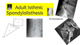
SlideShare a Scribd company logo
1 of 55
Adult Isthmic
Spondylolisthesis
Dr.Ponnilavan
Spondylolisthesis
- the ventral (or anterior) displacement of
one vertebra relative to the subjacent
vertebra.
Spondylolysis - considered to occur prior to appearance of
spondylolisthesis, as the anterior column of the vertebra is no longer in
continuity with the posterior column.
• Over time, vertebral body then displaces ventrally once disc &
supporting soft tissues are no longer able to maintain the structural
integrity & anatomic alignment of the vertebrae.
Spondylolisthesis Classification- Meyerding
Etiologic classification system was described by
Wiltse, which includes
• .
Children < 6 yrs- incidence - lumbar spondylolysis - 4.4% & spondylolisthesis-2.6%
At adulthood- L. spondylolysis - 5.4% & spondylolisthesis - 4%.
- reported pars defects did not typically heal & slippage occurred throughout f/u
period
Fredrickson et al..
prospective, pop-based
study of 500
schoolchildren from
northern Pennsylvania in
1950s
Fredrickson also reported that 2/3rd of spondylolysis cases were
males, & > 90% occurred at L5–S1.
Spondylolisthesis-found in 74% of pts with b/l pars defects at L5–S1,
but not in Pts with unilateral defects or pars defects at other levels.
Females - lower incidence of defects but higher rate of slip
progression.
• A more recent study by Urrutia et al. found a similar incidence of isthmic
spondylolisthesis in a non-US adult population of 3.8% (range, 1.7–6.8%).
• Incidence appeared not to change into adulthood, & authors concluded
spondylolysis is more likely an acquired disorder.
Pathophysiology
• Isthmic spondylolisthesis is a defect in pars interarticularis, a critical
structural component of posterior element of the vertebra.
• The pars is the intersection of the lamina, inferior and superior
articular processes, and pedicle.
• Etiologies for spondylolysis are numerous but many believe that the
vast majority are due to stress fractures of the pars interarticularis.
• Biomechanical studies have shown this region is exposed to the
highest extension forces in the lumbar spine.
It has also been suggested that this region of bone is the weakest
structural component of the posterior neural arch.
Repetitive lumbar extension loading appears to result in a
localized stress reaction in the vulnerable bone of the pars
region, and if the stress is sustained or excessive & if the bone is
unable to heal, then a spondylolysis develops.
Rosenberg et al. support this hypothesis, reporting their observation
that spondylolysis does not occur in nonambulatory individuals.
Additional support for the relationship b/w extension stress &
spondylolysis is the observation that a high incidence of pars defects
are observed in athletes such as wrestlers, football linemen, &
gymnasts—who frequently undergo repetitive hyperextension loads.
M/c level for spondylolysis is L5 &
- Isthmic spondylolisthesis at L5–S1 (90%)
- Due to previous data suggesting that sagittally oriented facet joints
predispose individuals to degenerative spondylolisthesis, they
analyzed facet joint orientation in an isthmic spondylolisthesis group.
• The group with isthmic spondylolisthesis had significantly more
coronal orientation of the facets of L3–L4 and L4–L5, that is, above
the affected level.
• They concluded that facet joints with more coronal—less sagittal—
orientation allow for less dorsal-ventral translation of cranial motion
segments, which leads to greater extension stresses on the L5 pars
and results in spondylolysis.
• Ward and Latimer evaluated the intrafacet distances of lumbar
vertebrae in individuals with & without spondylolysis & found an
increase in cranial-to-caudal intrafacet distance in nonaffected
individuals, which they concluded allowed for overlap of lamina
during lordosis.
potentially resulting in impingement of the L4 inferior articular process on the pars of L5.
Biomechanics
Biomechanical environment at the lumbosacral
junction is a complex one that normally
functions as a harmonious linkage between the
trunk & pelvis.
During radiographic assessment of patients,
lumbar lordosis (LL), pelvic incidence (PI), sacral
slope (SS), and pelvic tilt (PT) are most
commonly measured.
Measurements
pelvic incidence
• correlates with severity of disease
pelvic incidence = pelvic tilt +
sacral slope
• pelvic tilt
• sacral slope
PI is considered a fixed anatomic measurement & does not change for
any given individual through adulthood.
LL, SS, and PT are measurements of the relative position of lumbosacral
spine
Sagittal spinopelvic parameters have been found to correlate significantly
with spondyloptosis as well as the severity of isthmic spondylolisthesis.
• Labelle et al. - 214 young adults
(ages 10–40 years) that PI is
significantly greater in
individuals with isthmic
spondylolisthesis and correlates
linearly with higher (Meyerding)
slip severities.
• They also showed that PI strongly correlates with the other
parameters:
• LL, SS, and PT they concluded that individuals with isthmic
spondylolisthesis stand with increased SS, PT, and LL.
• Moreover, these increased values—in particular, LL—are an
important factor in the amount of shear stress on the L5 lamina and
pars.
Diagnosis
• History
• Most individuals with spondylolysis are asymptomatic.
• While it is the most common cause of LBP in children, the same does
not hold true in the adult population.
• Andrade et al.recently reported a review of observational studies on
the association of spondylolysis and isthmic spondylolisthesis with
LBP.
• They reported that only 1 of 15 eligible studies found an association
and 11 did not.
• In fact, they found that LBP was significantly more prevalent in
individuals without spondylolysis/isthmic spondylolisthesis.
• Moller and Hedlund reported on 201 patients with isthmic
spondylolysis and found that their patients presented with
- back pain only in 27%,
- back pain and sciatica in 65%,
- and sciatica only in 8% of individuals.
• Back pain may be positional & may be worsened with standing and/or
lumbar extension maneuvers, while it may be relieved with forward
flexion or sitting.
• Lower extremity pain can be radicular in nature, as it is often caused
by impingement of the exiting L5 nerve root due to frequent
occurrence of associated foraminal stenosis at L5–S1.
• Pain in lower extremities can often be positional, similar to typical
LBP complaints.
Physical Examination
• There are no pathognomonic physical examination findings for
isthmic spondylolisthesis.
• Lumbar extension will often elicit LBP, lower extremity radicular
complaints, or both.
There can be a palpable or visible step-of in cases of highgrade slips.
The step-of occurs between the L4 and L5 spinous processes as the
posterior elements of L5 remain dorsal and in line with the S1 spinous
process
• Hamstring tightness is often described but difficult to assess
objectively.
• A positive straight-leg raise test will be present in approximately 50%
of patients.
• Sensory or motor abnormalities can be found due to associated
foraminal stenosis and exiting root compression.
Imaging
• Routine radiographic images can detect spondylolysis, especially if
spondylolisthesis is present, and slip severity can be measured on
lateral images.
• Additionally, standing images that include Flexion and extension
positioning should be evaluated.
• If spondylolysis is suspected but not visualized, oblique radiographic
images 45 degrees to the sagittal plane can be obtained that can
detect up to 96% of pars defects.
• Abnormalities in the “neck of the Scotty dog” is the hallmark
radiographic finding.
Complete defects are most common, but pars dysplasia & hypoplasia
can also be observed.
• Sagittal reformatted images best show the pars defect as being
distinct from facet joints and are the definitive finding in
spondylolysis.
• One limitation of CT is the lower sensitivity for soft tissue densities,
especially the internal anatomy of the neural foramen and the extent
of any associated nerve root compression.
- MRI is increasingly used as the primary imaging modality in patients
with LBP with or without radiculopathy.
• Sagittal T1-weighted images provide the greatest level of contrast
between hyperintense bone marrow and the signal void of bony
cortex at the pars defect.
• Single-photon emission computed tomography can be used in the
evaluation of suspected acute or impending spondylolysis.
• However, SPECT has been reported to have notable false-positive and
false-negative results in spondylolysis & thus should be used with
caution, although it may be of particular value in cases in which MRI
is contraindicated.
Differential Diagnosis
• The differential diagnoses for isthmic spondylolisthesis are those of its
clinical presentations: LBP and sciatica.
• First, spinal trauma, tumors & infections should be ruled out.
• Next, degenerative disc disease, spondylosis, spinal stenosis, or disc
herniation should be considered.
• Other causes include systemic diagnose-
- rheumatoid arthritis &
- spondyloarthropathies, and other
- nonspinal,
- musculoskeletal etiologies, such as sacroiliac joint arthrosis,
hip arthritis, & more.
Finally, abdominal/visceral considerations would include renal,
gastrointestinal, and vascular disorders.
Treatment
• Nonoperative Treatment
• Initial Rx for patients presenting with acute LBP should be
nonoperative.
• The mainstays are patient education, activity modification, and
medications - NSAIDS
• The addition of physical therapy and exercise can be considered when
early treatments fail and LBP becomes more long-standing.
• Other alternative pain management modalities—such as chiropractic
care, acupuncture, and massage—have been widely utilized, with
reported improvements in pain and function.
• Short-term use of narcotic analgesics should be considered with
caution.
• A minor neurologic deficit, such as radicular numbness or
paresthesias, but excluding severe motor weakness, can also be
managed nonoperatively but may benefit from corticosteroid
injection via injection therapies, such as Fluoroscopically guided
selective nerve root blocks.
Operative Treatment
• Indications-
• Severe, persistent back
• &/or lower extremity pain that is associated with functional
limitations or that significantly impacts quality of life—with or
without flexion–extension instability on radiographs, progressive
motor weakness, or cauda equina syndrome—are all generally
accepted indications for operative intervention.
• Patients should complete a rigorous course of nonoperative
treatment prior to considering surgery unless a signiicant neurologic
deficit exists.
Surgery
• Goals of operative intervention in isthmic spondylolisthesis are to
decompress neural elements & stabilize the affected motion segment.
• Uncommonly, decompression alone can be performed in certain
circumstances when fusion is not necessary.
• More typically, surgery involves stabilization traditionally performed
with posterior in situ fusion techniques with or without pedicle screw
instrumentation and sometimes without decompression.
• Supplemental anterior column support using interbody fusion
techniques approached posteriorly (posterior lumbar interbody
fusion [PLIF]) or anteriorly (anterior lumbar interbody fusion) has
been recently popularized and may currently represent the most
popular form of surgical treatment in the United States.
Decompression
• Decompression without fusion can be performed on individuals who
have only radicular symptoms & stable spondylolisthesis on dynamic
radiographs or a bony fusion seen on CT scans.
• Low-demand individuals or those with significant medical
comorbidities may be reasonable candidates.
• Gill laminectomy entails removal of the entire posterior arch and the
hypertrophied fibrocartilaginous tissue at the pars, as well as partial
facetectomies to decompress the nerve root.
• Long-term results of laminectomy alone have not been favorable,
leading most experts to believe that addition of fusion is required in
most cases to obtain good clinical outcomes.
• Direct Pars Repair
• Fusion
PEARLS
Isthmic spondylolisthesis are rarely symptomatic, but those who do
develop severe back pain, radiculopathy, or both can be effectively
treated without surgery.
Significant evidence exists that isthmic spondylolisthesis develops in
adolescence as a result of an extension stress injury.
Spondylolisthesis develops as the soft tissues around the vertebral
motion segment become incompetent.
Symptomatic individuals who fail nonoperative treatment is commonly
treated with a spinal fusion.
Ideal treatment technique has not been agreed upon and often involves
use of pedicle screw instrumentation and interbody techniques.
PITFALLS
1. Failure to identify spondylolysis on imaging studies is common; if
high suspicion exists, further imaging should be considered.
2. Because spinal fusion is the mainstay of treatment, individuals
with high risk of nonunion should be approached with caution.
3. Decompression alone should be reserved in individuals who are
low demand and poor operative candidates.
4. Nerve root involvement occurs at the level of the foramen and
should correlate with clinical symptoms at presentation.
5. Reduction of high-grade slips can increase risk of nerve injuries.
THANK U

More Related Content

What's hot

What's hot (20)

Vertical talus
Vertical talusVertical talus
Vertical talus
 
Developmental dysplasia of hip
Developmental dysplasia of hipDevelopmental dysplasia of hip
Developmental dysplasia of hip
 
Bilateral Transfemoral amputation and stubbies (Prostheses).pptx
Bilateral Transfemoral amputation and stubbies (Prostheses).pptxBilateral Transfemoral amputation and stubbies (Prostheses).pptx
Bilateral Transfemoral amputation and stubbies (Prostheses).pptx
 
Metatarsus adductus
Metatarsus adductusMetatarsus adductus
Metatarsus adductus
 
The distal radioulnar joint and tfcc
The distal radioulnar joint and tfccThe distal radioulnar joint and tfcc
The distal radioulnar joint and tfcc
 
Intoeing gait
Intoeing gaitIntoeing gait
Intoeing gait
 
Knee Arthrodesis
Knee ArthrodesisKnee Arthrodesis
Knee Arthrodesis
 
Pffd
PffdPffd
Pffd
 
HKAFO
HKAFOHKAFO
HKAFO
 
Hip biomechanics
Hip biomechanicsHip biomechanics
Hip biomechanics
 
Floor reaction orthosis
Floor reaction orthosisFloor reaction orthosis
Floor reaction orthosis
 
Patellofemoral instability
Patellofemoral instabilityPatellofemoral instability
Patellofemoral instability
 
Dislocation of patella
Dislocation of patellaDislocation of patella
Dislocation of patella
 
Spondylolisthesis ms
Spondylolisthesis msSpondylolisthesis ms
Spondylolisthesis ms
 
Lower cross syndrome
Lower cross syndromeLower cross syndrome
Lower cross syndrome
 
Kyphosis (curved thoracic spine)
Kyphosis (curved thoracic spine)Kyphosis (curved thoracic spine)
Kyphosis (curved thoracic spine)
 
Pediatric hip orthoses.pptx
Pediatric hip orthoses.pptxPediatric hip orthoses.pptx
Pediatric hip orthoses.pptx
 
Genu varum orthotic management
Genu varum orthotic managementGenu varum orthotic management
Genu varum orthotic management
 
Spondylolisthesis review
Spondylolisthesis reviewSpondylolisthesis review
Spondylolisthesis review
 
Hip joint biomecanics
Hip joint biomecanicsHip joint biomecanics
Hip joint biomecanics
 

Similar to Spondylolisthesis

Hip and spine syndrome (PMR)
Hip and spine syndrome (PMR)Hip and spine syndrome (PMR)
Hip and spine syndrome (PMR)mrinal joshi
 
Spondylolisthesis
Spondylolisthesis Spondylolisthesis
Spondylolisthesis Mahak Jain
 
Diffuse idiopathic skeletal hyperostosis
Diffuse idiopathic skeletal hyperostosisDiffuse idiopathic skeletal hyperostosis
Diffuse idiopathic skeletal hyperostosisAnkit Raiyani
 
Congenital anomalies of upper and lower limb
Congenital anomalies of upper and lower limbCongenital anomalies of upper and lower limb
Congenital anomalies of upper and lower limbMohammad azharuddin
 
SPONDYLOLISTHESIS_masoom.pptx
SPONDYLOLISTHESIS_masoom.pptxSPONDYLOLISTHESIS_masoom.pptx
SPONDYLOLISTHESIS_masoom.pptxmasoom parwez
 
HighGradeSpondylolisthesis.pptx
HighGradeSpondylolisthesis.pptxHighGradeSpondylolisthesis.pptx
HighGradeSpondylolisthesis.pptxTejasvi Agarwal
 
Pediatric knee copy
Pediatric knee   copyPediatric knee   copy
Pediatric knee copyluay hassan
 
Alan moelleken-md-santa-barbara-spondylolisthesis
Alan moelleken-md-santa-barbara-spondylolisthesisAlan moelleken-md-santa-barbara-spondylolisthesis
Alan moelleken-md-santa-barbara-spondylolisthesisAlan Moelleken
 
SCFE / slipped capital femoral epiphysis
SCFE / slipped capital femoral epiphysis SCFE / slipped capital femoral epiphysis
SCFE / slipped capital femoral epiphysis Surya Vijay Singh
 
Congenital kyphosis
Congenital kyphosisCongenital kyphosis
Congenital kyphosissaurabh rai
 
Differential diagnosis of hip
Differential diagnosis of hipDifferential diagnosis of hip
Differential diagnosis of hipRutuja Patharkar
 
Legg-Calve-Perthes Disease.pptx
Legg-Calve-Perthes Disease.pptxLegg-Calve-Perthes Disease.pptx
Legg-Calve-Perthes Disease.pptxNellyPhiri5
 
imaging in intrauterine skeletal dysplasia
imaging in intrauterine skeletal dysplasia imaging in intrauterine skeletal dysplasia
imaging in intrauterine skeletal dysplasia Shail Padmani
 

Similar to Spondylolisthesis (20)

Lumbar spinal stenosis
Lumbar spinal stenosisLumbar spinal stenosis
Lumbar spinal stenosis
 
Lumbar spinal stenosis
Lumbar spinal stenosisLumbar spinal stenosis
Lumbar spinal stenosis
 
Spondylolisthesis
SpondylolisthesisSpondylolisthesis
Spondylolisthesis
 
Spondylolithesis (1)
Spondylolithesis (1)Spondylolithesis (1)
Spondylolithesis (1)
 
Hip and spine syndrome (PMR)
Hip and spine syndrome (PMR)Hip and spine syndrome (PMR)
Hip and spine syndrome (PMR)
 
Spondylolisthesis
Spondylolisthesis Spondylolisthesis
Spondylolisthesis
 
Diffuse idiopathic skeletal hyperostosis
Diffuse idiopathic skeletal hyperostosisDiffuse idiopathic skeletal hyperostosis
Diffuse idiopathic skeletal hyperostosis
 
Congenital anomalies of upper and lower limb
Congenital anomalies of upper and lower limbCongenital anomalies of upper and lower limb
Congenital anomalies of upper and lower limb
 
Ankylosing spondylitis
Ankylosing spondylitisAnkylosing spondylitis
Ankylosing spondylitis
 
SPONDYLOLISTHESIS_masoom.pptx
SPONDYLOLISTHESIS_masoom.pptxSPONDYLOLISTHESIS_masoom.pptx
SPONDYLOLISTHESIS_masoom.pptx
 
HighGradeSpondylolisthesis.pptx
HighGradeSpondylolisthesis.pptxHighGradeSpondylolisthesis.pptx
HighGradeSpondylolisthesis.pptx
 
Pediatric knee copy
Pediatric knee   copyPediatric knee   copy
Pediatric knee copy
 
Spondylolisthesis
SpondylolisthesisSpondylolisthesis
Spondylolisthesis
 
Alan moelleken-md-santa-barbara-spondylolisthesis
Alan moelleken-md-santa-barbara-spondylolisthesisAlan moelleken-md-santa-barbara-spondylolisthesis
Alan moelleken-md-santa-barbara-spondylolisthesis
 
Spondylolisthesis
SpondylolisthesisSpondylolisthesis
Spondylolisthesis
 
SCFE / slipped capital femoral epiphysis
SCFE / slipped capital femoral epiphysis SCFE / slipped capital femoral epiphysis
SCFE / slipped capital femoral epiphysis
 
Congenital kyphosis
Congenital kyphosisCongenital kyphosis
Congenital kyphosis
 
Differential diagnosis of hip
Differential diagnosis of hipDifferential diagnosis of hip
Differential diagnosis of hip
 
Legg-Calve-Perthes Disease.pptx
Legg-Calve-Perthes Disease.pptxLegg-Calve-Perthes Disease.pptx
Legg-Calve-Perthes Disease.pptx
 
imaging in intrauterine skeletal dysplasia
imaging in intrauterine skeletal dysplasia imaging in intrauterine skeletal dysplasia
imaging in intrauterine skeletal dysplasia
 

More from Ponnilavan Ponz (20)

Cubitus varus and valgus
Cubitus varus and valgusCubitus varus and valgus
Cubitus varus and valgus
 
Rickets
RicketsRickets
Rickets
 
Meniscal injury
Meniscal injury Meniscal injury
Meniscal injury
 
Poliomyelitis
PoliomyelitisPoliomyelitis
Poliomyelitis
 
Anatomy of cervical spine
Anatomy of cervical spineAnatomy of cervical spine
Anatomy of cervical spine
 
Congenital pseudoarthrosis of tibia
Congenital pseudoarthrosis of tibiaCongenital pseudoarthrosis of tibia
Congenital pseudoarthrosis of tibia
 
screws and plate
screws and platescrews and plate
screws and plate
 
Distal femoral fresh osteochondral allografts
Distal femoral fresh osteochondral allograftsDistal femoral fresh osteochondral allografts
Distal femoral fresh osteochondral allografts
 
External fixation
External fixation External fixation
External fixation
 
Im nail
Im nailIm nail
Im nail
 
Krukenberg surgery
Krukenberg surgeryKrukenberg surgery
Krukenberg surgery
 
Patellofemoral disorders
Patellofemoral disordersPatellofemoral disorders
Patellofemoral disorders
 
Avn
AvnAvn
Avn
 
Osteochondroma
OsteochondromaOsteochondroma
Osteochondroma
 
Dupuytren
Dupuytren   Dupuytren
Dupuytren
 
Chopart amputation
Chopart amputationChopart amputation
Chopart amputation
 
Acl reconstruction
Acl reconstructionAcl reconstruction
Acl reconstruction
 
Bladder innervation
Bladder innervationBladder innervation
Bladder innervation
 
maduramycosis
maduramycosis   maduramycosis
maduramycosis
 
Adult acquired flat foot deformity
Adult acquired flat foot deformityAdult acquired flat foot deformity
Adult acquired flat foot deformity
 

Recently uploaded

18-04-UA_REPORT_MEDIALITERAСY_INDEX-DM_23-1-final-eng.pdf
18-04-UA_REPORT_MEDIALITERAСY_INDEX-DM_23-1-final-eng.pdf18-04-UA_REPORT_MEDIALITERAСY_INDEX-DM_23-1-final-eng.pdf
18-04-UA_REPORT_MEDIALITERAСY_INDEX-DM_23-1-final-eng.pdfssuser54595a
 
Software Engineering Methodologies (overview)
Software Engineering Methodologies (overview)Software Engineering Methodologies (overview)
Software Engineering Methodologies (overview)eniolaolutunde
 
Employee wellbeing at the workplace.pptx
Employee wellbeing at the workplace.pptxEmployee wellbeing at the workplace.pptx
Employee wellbeing at the workplace.pptxNirmalaLoungPoorunde1
 
A Critique of the Proposed National Education Policy Reform
A Critique of the Proposed National Education Policy ReformA Critique of the Proposed National Education Policy Reform
A Critique of the Proposed National Education Policy ReformChameera Dedduwage
 
Incoming and Outgoing Shipments in 1 STEP Using Odoo 17
Incoming and Outgoing Shipments in 1 STEP Using Odoo 17Incoming and Outgoing Shipments in 1 STEP Using Odoo 17
Incoming and Outgoing Shipments in 1 STEP Using Odoo 17Celine George
 
POINT- BIOCHEMISTRY SEM 2 ENZYMES UNIT 5.pptx
POINT- BIOCHEMISTRY SEM 2 ENZYMES UNIT 5.pptxPOINT- BIOCHEMISTRY SEM 2 ENZYMES UNIT 5.pptx
POINT- BIOCHEMISTRY SEM 2 ENZYMES UNIT 5.pptxSayali Powar
 
How to Make a Pirate ship Primary Education.pptx
How to Make a Pirate ship Primary Education.pptxHow to Make a Pirate ship Primary Education.pptx
How to Make a Pirate ship Primary Education.pptxmanuelaromero2013
 
The Most Excellent Way | 1 Corinthians 13
The Most Excellent Way | 1 Corinthians 13The Most Excellent Way | 1 Corinthians 13
The Most Excellent Way | 1 Corinthians 13Steve Thomason
 
Science lesson Moon for 4th quarter lesson
Science lesson Moon for 4th quarter lessonScience lesson Moon for 4th quarter lesson
Science lesson Moon for 4th quarter lessonJericReyAuditor
 
Science 7 - LAND and SEA BREEZE and its Characteristics
Science 7 - LAND and SEA BREEZE and its CharacteristicsScience 7 - LAND and SEA BREEZE and its Characteristics
Science 7 - LAND and SEA BREEZE and its CharacteristicsKarinaGenton
 
Introduction to AI in Higher Education_draft.pptx
Introduction to AI in Higher Education_draft.pptxIntroduction to AI in Higher Education_draft.pptx
Introduction to AI in Higher Education_draft.pptxpboyjonauth
 
BASLIQ CURRENT LOOKBOOK LOOKBOOK(1) (1).pdf
BASLIQ CURRENT LOOKBOOK  LOOKBOOK(1) (1).pdfBASLIQ CURRENT LOOKBOOK  LOOKBOOK(1) (1).pdf
BASLIQ CURRENT LOOKBOOK LOOKBOOK(1) (1).pdfSoniaTolstoy
 
Sanyam Choudhary Chemistry practical.pdf
Sanyam Choudhary Chemistry practical.pdfSanyam Choudhary Chemistry practical.pdf
Sanyam Choudhary Chemistry practical.pdfsanyamsingh5019
 
Paris 2024 Olympic Geographies - an activity
Paris 2024 Olympic Geographies - an activityParis 2024 Olympic Geographies - an activity
Paris 2024 Olympic Geographies - an activityGeoBlogs
 
Blooming Together_ Growing a Community Garden Worksheet.docx
Blooming Together_ Growing a Community Garden Worksheet.docxBlooming Together_ Growing a Community Garden Worksheet.docx
Blooming Together_ Growing a Community Garden Worksheet.docxUnboundStockton
 
Class 11 Legal Studies Ch-1 Concept of State .pdf
Class 11 Legal Studies Ch-1 Concept of State .pdfClass 11 Legal Studies Ch-1 Concept of State .pdf
Class 11 Legal Studies Ch-1 Concept of State .pdfakmcokerachita
 
Call Girls in Dwarka Mor Delhi Contact Us 9654467111
Call Girls in Dwarka Mor Delhi Contact Us 9654467111Call Girls in Dwarka Mor Delhi Contact Us 9654467111
Call Girls in Dwarka Mor Delhi Contact Us 9654467111Sapana Sha
 

Recently uploaded (20)

18-04-UA_REPORT_MEDIALITERAСY_INDEX-DM_23-1-final-eng.pdf
18-04-UA_REPORT_MEDIALITERAСY_INDEX-DM_23-1-final-eng.pdf18-04-UA_REPORT_MEDIALITERAСY_INDEX-DM_23-1-final-eng.pdf
18-04-UA_REPORT_MEDIALITERAСY_INDEX-DM_23-1-final-eng.pdf
 
Software Engineering Methodologies (overview)
Software Engineering Methodologies (overview)Software Engineering Methodologies (overview)
Software Engineering Methodologies (overview)
 
Employee wellbeing at the workplace.pptx
Employee wellbeing at the workplace.pptxEmployee wellbeing at the workplace.pptx
Employee wellbeing at the workplace.pptx
 
A Critique of the Proposed National Education Policy Reform
A Critique of the Proposed National Education Policy ReformA Critique of the Proposed National Education Policy Reform
A Critique of the Proposed National Education Policy Reform
 
Incoming and Outgoing Shipments in 1 STEP Using Odoo 17
Incoming and Outgoing Shipments in 1 STEP Using Odoo 17Incoming and Outgoing Shipments in 1 STEP Using Odoo 17
Incoming and Outgoing Shipments in 1 STEP Using Odoo 17
 
POINT- BIOCHEMISTRY SEM 2 ENZYMES UNIT 5.pptx
POINT- BIOCHEMISTRY SEM 2 ENZYMES UNIT 5.pptxPOINT- BIOCHEMISTRY SEM 2 ENZYMES UNIT 5.pptx
POINT- BIOCHEMISTRY SEM 2 ENZYMES UNIT 5.pptx
 
How to Make a Pirate ship Primary Education.pptx
How to Make a Pirate ship Primary Education.pptxHow to Make a Pirate ship Primary Education.pptx
How to Make a Pirate ship Primary Education.pptx
 
The Most Excellent Way | 1 Corinthians 13
The Most Excellent Way | 1 Corinthians 13The Most Excellent Way | 1 Corinthians 13
The Most Excellent Way | 1 Corinthians 13
 
9953330565 Low Rate Call Girls In Rohini Delhi NCR
9953330565 Low Rate Call Girls In Rohini  Delhi NCR9953330565 Low Rate Call Girls In Rohini  Delhi NCR
9953330565 Low Rate Call Girls In Rohini Delhi NCR
 
Model Call Girl in Tilak Nagar Delhi reach out to us at 🔝9953056974🔝
Model Call Girl in Tilak Nagar Delhi reach out to us at 🔝9953056974🔝Model Call Girl in Tilak Nagar Delhi reach out to us at 🔝9953056974🔝
Model Call Girl in Tilak Nagar Delhi reach out to us at 🔝9953056974🔝
 
Science lesson Moon for 4th quarter lesson
Science lesson Moon for 4th quarter lessonScience lesson Moon for 4th quarter lesson
Science lesson Moon for 4th quarter lesson
 
Science 7 - LAND and SEA BREEZE and its Characteristics
Science 7 - LAND and SEA BREEZE and its CharacteristicsScience 7 - LAND and SEA BREEZE and its Characteristics
Science 7 - LAND and SEA BREEZE and its Characteristics
 
Introduction to AI in Higher Education_draft.pptx
Introduction to AI in Higher Education_draft.pptxIntroduction to AI in Higher Education_draft.pptx
Introduction to AI in Higher Education_draft.pptx
 
BASLIQ CURRENT LOOKBOOK LOOKBOOK(1) (1).pdf
BASLIQ CURRENT LOOKBOOK  LOOKBOOK(1) (1).pdfBASLIQ CURRENT LOOKBOOK  LOOKBOOK(1) (1).pdf
BASLIQ CURRENT LOOKBOOK LOOKBOOK(1) (1).pdf
 
Sanyam Choudhary Chemistry practical.pdf
Sanyam Choudhary Chemistry practical.pdfSanyam Choudhary Chemistry practical.pdf
Sanyam Choudhary Chemistry practical.pdf
 
Paris 2024 Olympic Geographies - an activity
Paris 2024 Olympic Geographies - an activityParis 2024 Olympic Geographies - an activity
Paris 2024 Olympic Geographies - an activity
 
Blooming Together_ Growing a Community Garden Worksheet.docx
Blooming Together_ Growing a Community Garden Worksheet.docxBlooming Together_ Growing a Community Garden Worksheet.docx
Blooming Together_ Growing a Community Garden Worksheet.docx
 
Class 11 Legal Studies Ch-1 Concept of State .pdf
Class 11 Legal Studies Ch-1 Concept of State .pdfClass 11 Legal Studies Ch-1 Concept of State .pdf
Class 11 Legal Studies Ch-1 Concept of State .pdf
 
Call Girls in Dwarka Mor Delhi Contact Us 9654467111
Call Girls in Dwarka Mor Delhi Contact Us 9654467111Call Girls in Dwarka Mor Delhi Contact Us 9654467111
Call Girls in Dwarka Mor Delhi Contact Us 9654467111
 
Staff of Color (SOC) Retention Efforts DDSD
Staff of Color (SOC) Retention Efforts DDSDStaff of Color (SOC) Retention Efforts DDSD
Staff of Color (SOC) Retention Efforts DDSD
 

Spondylolisthesis

  • 2. Spondylolisthesis - the ventral (or anterior) displacement of one vertebra relative to the subjacent vertebra.
  • 3. Spondylolysis - considered to occur prior to appearance of spondylolisthesis, as the anterior column of the vertebra is no longer in continuity with the posterior column.
  • 4. • Over time, vertebral body then displaces ventrally once disc & supporting soft tissues are no longer able to maintain the structural integrity & anatomic alignment of the vertebrae.
  • 6. Etiologic classification system was described by Wiltse, which includes
  • 7.
  • 8.
  • 9. • . Children < 6 yrs- incidence - lumbar spondylolysis - 4.4% & spondylolisthesis-2.6% At adulthood- L. spondylolysis - 5.4% & spondylolisthesis - 4%. - reported pars defects did not typically heal & slippage occurred throughout f/u period Fredrickson et al.. prospective, pop-based study of 500 schoolchildren from northern Pennsylvania in 1950s
  • 10. Fredrickson also reported that 2/3rd of spondylolysis cases were males, & > 90% occurred at L5–S1. Spondylolisthesis-found in 74% of pts with b/l pars defects at L5–S1, but not in Pts with unilateral defects or pars defects at other levels. Females - lower incidence of defects but higher rate of slip progression.
  • 11. • A more recent study by Urrutia et al. found a similar incidence of isthmic spondylolisthesis in a non-US adult population of 3.8% (range, 1.7–6.8%). • Incidence appeared not to change into adulthood, & authors concluded spondylolysis is more likely an acquired disorder.
  • 12. Pathophysiology • Isthmic spondylolisthesis is a defect in pars interarticularis, a critical structural component of posterior element of the vertebra. • The pars is the intersection of the lamina, inferior and superior articular processes, and pedicle.
  • 13.
  • 14. • Etiologies for spondylolysis are numerous but many believe that the vast majority are due to stress fractures of the pars interarticularis. • Biomechanical studies have shown this region is exposed to the highest extension forces in the lumbar spine.
  • 15. It has also been suggested that this region of bone is the weakest structural component of the posterior neural arch. Repetitive lumbar extension loading appears to result in a localized stress reaction in the vulnerable bone of the pars region, and if the stress is sustained or excessive & if the bone is unable to heal, then a spondylolysis develops.
  • 16. Rosenberg et al. support this hypothesis, reporting their observation that spondylolysis does not occur in nonambulatory individuals. Additional support for the relationship b/w extension stress & spondylolysis is the observation that a high incidence of pars defects are observed in athletes such as wrestlers, football linemen, & gymnasts—who frequently undergo repetitive hyperextension loads.
  • 17. M/c level for spondylolysis is L5 & - Isthmic spondylolisthesis at L5–S1 (90%) - Due to previous data suggesting that sagittally oriented facet joints predispose individuals to degenerative spondylolisthesis, they analyzed facet joint orientation in an isthmic spondylolisthesis group.
  • 18. • The group with isthmic spondylolisthesis had significantly more coronal orientation of the facets of L3–L4 and L4–L5, that is, above the affected level. • They concluded that facet joints with more coronal—less sagittal— orientation allow for less dorsal-ventral translation of cranial motion segments, which leads to greater extension stresses on the L5 pars and results in spondylolysis.
  • 19. • Ward and Latimer evaluated the intrafacet distances of lumbar vertebrae in individuals with & without spondylolysis & found an increase in cranial-to-caudal intrafacet distance in nonaffected individuals, which they concluded allowed for overlap of lamina during lordosis.
  • 20. potentially resulting in impingement of the L4 inferior articular process on the pars of L5.
  • 22. Biomechanical environment at the lumbosacral junction is a complex one that normally functions as a harmonious linkage between the trunk & pelvis. During radiographic assessment of patients, lumbar lordosis (LL), pelvic incidence (PI), sacral slope (SS), and pelvic tilt (PT) are most commonly measured.
  • 23. Measurements pelvic incidence • correlates with severity of disease pelvic incidence = pelvic tilt + sacral slope
  • 26. PI is considered a fixed anatomic measurement & does not change for any given individual through adulthood. LL, SS, and PT are measurements of the relative position of lumbosacral spine Sagittal spinopelvic parameters have been found to correlate significantly with spondyloptosis as well as the severity of isthmic spondylolisthesis.
  • 27. • Labelle et al. - 214 young adults (ages 10–40 years) that PI is significantly greater in individuals with isthmic spondylolisthesis and correlates linearly with higher (Meyerding) slip severities.
  • 28. • They also showed that PI strongly correlates with the other parameters: • LL, SS, and PT they concluded that individuals with isthmic spondylolisthesis stand with increased SS, PT, and LL. • Moreover, these increased values—in particular, LL—are an important factor in the amount of shear stress on the L5 lamina and pars.
  • 29. Diagnosis • History • Most individuals with spondylolysis are asymptomatic. • While it is the most common cause of LBP in children, the same does not hold true in the adult population.
  • 30. • Andrade et al.recently reported a review of observational studies on the association of spondylolysis and isthmic spondylolisthesis with LBP. • They reported that only 1 of 15 eligible studies found an association and 11 did not. • In fact, they found that LBP was significantly more prevalent in individuals without spondylolysis/isthmic spondylolisthesis.
  • 31.
  • 32.
  • 33. • Moller and Hedlund reported on 201 patients with isthmic spondylolysis and found that their patients presented with - back pain only in 27%, - back pain and sciatica in 65%, - and sciatica only in 8% of individuals. • Back pain may be positional & may be worsened with standing and/or lumbar extension maneuvers, while it may be relieved with forward flexion or sitting.
  • 34. • Lower extremity pain can be radicular in nature, as it is often caused by impingement of the exiting L5 nerve root due to frequent occurrence of associated foraminal stenosis at L5–S1. • Pain in lower extremities can often be positional, similar to typical LBP complaints.
  • 35. Physical Examination • There are no pathognomonic physical examination findings for isthmic spondylolisthesis. • Lumbar extension will often elicit LBP, lower extremity radicular complaints, or both. There can be a palpable or visible step-of in cases of highgrade slips. The step-of occurs between the L4 and L5 spinous processes as the posterior elements of L5 remain dorsal and in line with the S1 spinous process
  • 36. • Hamstring tightness is often described but difficult to assess objectively. • A positive straight-leg raise test will be present in approximately 50% of patients. • Sensory or motor abnormalities can be found due to associated foraminal stenosis and exiting root compression.
  • 37. Imaging • Routine radiographic images can detect spondylolysis, especially if spondylolisthesis is present, and slip severity can be measured on lateral images. • Additionally, standing images that include Flexion and extension positioning should be evaluated.
  • 38. • If spondylolysis is suspected but not visualized, oblique radiographic images 45 degrees to the sagittal plane can be obtained that can detect up to 96% of pars defects. • Abnormalities in the “neck of the Scotty dog” is the hallmark radiographic finding.
  • 39. Complete defects are most common, but pars dysplasia & hypoplasia can also be observed. • Sagittal reformatted images best show the pars defect as being distinct from facet joints and are the definitive finding in spondylolysis. • One limitation of CT is the lower sensitivity for soft tissue densities, especially the internal anatomy of the neural foramen and the extent of any associated nerve root compression.
  • 40. - MRI is increasingly used as the primary imaging modality in patients with LBP with or without radiculopathy. • Sagittal T1-weighted images provide the greatest level of contrast between hyperintense bone marrow and the signal void of bony cortex at the pars defect.
  • 41. • Single-photon emission computed tomography can be used in the evaluation of suspected acute or impending spondylolysis. • However, SPECT has been reported to have notable false-positive and false-negative results in spondylolysis & thus should be used with caution, although it may be of particular value in cases in which MRI is contraindicated.
  • 42.
  • 43.
  • 44. Differential Diagnosis • The differential diagnoses for isthmic spondylolisthesis are those of its clinical presentations: LBP and sciatica. • First, spinal trauma, tumors & infections should be ruled out. • Next, degenerative disc disease, spondylosis, spinal stenosis, or disc herniation should be considered.
  • 45. • Other causes include systemic diagnose- - rheumatoid arthritis & - spondyloarthropathies, and other - nonspinal, - musculoskeletal etiologies, such as sacroiliac joint arthrosis, hip arthritis, & more. Finally, abdominal/visceral considerations would include renal, gastrointestinal, and vascular disorders.
  • 46. Treatment • Nonoperative Treatment • Initial Rx for patients presenting with acute LBP should be nonoperative. • The mainstays are patient education, activity modification, and medications - NSAIDS • The addition of physical therapy and exercise can be considered when early treatments fail and LBP becomes more long-standing. • Other alternative pain management modalities—such as chiropractic care, acupuncture, and massage—have been widely utilized, with reported improvements in pain and function.
  • 47. • Short-term use of narcotic analgesics should be considered with caution. • A minor neurologic deficit, such as radicular numbness or paresthesias, but excluding severe motor weakness, can also be managed nonoperatively but may benefit from corticosteroid injection via injection therapies, such as Fluoroscopically guided selective nerve root blocks.
  • 48. Operative Treatment • Indications- • Severe, persistent back • &/or lower extremity pain that is associated with functional limitations or that significantly impacts quality of life—with or without flexion–extension instability on radiographs, progressive motor weakness, or cauda equina syndrome—are all generally accepted indications for operative intervention. • Patients should complete a rigorous course of nonoperative treatment prior to considering surgery unless a signiicant neurologic deficit exists.
  • 49. Surgery • Goals of operative intervention in isthmic spondylolisthesis are to decompress neural elements & stabilize the affected motion segment. • Uncommonly, decompression alone can be performed in certain circumstances when fusion is not necessary. • More typically, surgery involves stabilization traditionally performed with posterior in situ fusion techniques with or without pedicle screw instrumentation and sometimes without decompression.
  • 50. • Supplemental anterior column support using interbody fusion techniques approached posteriorly (posterior lumbar interbody fusion [PLIF]) or anteriorly (anterior lumbar interbody fusion) has been recently popularized and may currently represent the most popular form of surgical treatment in the United States.
  • 51. Decompression • Decompression without fusion can be performed on individuals who have only radicular symptoms & stable spondylolisthesis on dynamic radiographs or a bony fusion seen on CT scans. • Low-demand individuals or those with significant medical comorbidities may be reasonable candidates. • Gill laminectomy entails removal of the entire posterior arch and the hypertrophied fibrocartilaginous tissue at the pars, as well as partial facetectomies to decompress the nerve root. • Long-term results of laminectomy alone have not been favorable, leading most experts to believe that addition of fusion is required in most cases to obtain good clinical outcomes.
  • 52. • Direct Pars Repair • Fusion
  • 53. PEARLS Isthmic spondylolisthesis are rarely symptomatic, but those who do develop severe back pain, radiculopathy, or both can be effectively treated without surgery. Significant evidence exists that isthmic spondylolisthesis develops in adolescence as a result of an extension stress injury. Spondylolisthesis develops as the soft tissues around the vertebral motion segment become incompetent. Symptomatic individuals who fail nonoperative treatment is commonly treated with a spinal fusion. Ideal treatment technique has not been agreed upon and often involves use of pedicle screw instrumentation and interbody techniques.
  • 54. PITFALLS 1. Failure to identify spondylolysis on imaging studies is common; if high suspicion exists, further imaging should be considered. 2. Because spinal fusion is the mainstay of treatment, individuals with high risk of nonunion should be approached with caution. 3. Decompression alone should be reserved in individuals who are low demand and poor operative candidates. 4. Nerve root involvement occurs at the level of the foramen and should correlate with clinical symptoms at presentation. 5. Reduction of high-grade slips can increase risk of nerve injuries.

Editor's Notes

  1. - Spondylolysis is a defect in the pars interarticularis due to congenital, traumatic, dysplastic, or neoplastic etiologies.
  2. It is measured as a percentage using the length of slip of the cranial vertebra compared to the length of superior endplate of the caudal vertebra. grade 5, greater than 100% or spondyloptosis.
  3. dysplastic, isthmic, degenerative, traumatic, and neoplastic types.
  4. -
  5. Axial computed tomographic image showing spondylolysis (open triangles). Note the undulating course of lucency, making it distinct from a facet joint.
  6. a line is drawn from the center of the S1 endplate to the center of the femoral head a second line is drawn perpendicular to a line drawn along the S1 endplate, intersecting the point in the center of the S1 endplate the angle between these two lines is the pelvic incidence
  7. sacral slope =  pelvic incidence - pelvic tilt a line is drawn from the center of the S1 endplate to the center of the femoral head a second vertical line (parallel with side margin of radiograph) line is drawn intersecting the center of the femoral head
  8. pelvic tilt = pelvic incidence - sacral slope a line is drawn parallel to the S1 endplate a second horizontal line (parallel to the inferior margin of the radiograph) is drawn