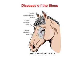
2. 1. Catarrh of the Maxillary Sinus
Definition:
It is a chronic catarrh or inflammation of the mucosa of the
maxillary sinus usually gives rise to the accumulation of
mucopurulent exudation. This condition is common in old
horses and is usually unilateral.
Causes:
(1) Traumatic.
(2) Extension from nasal catarrh or by extension diseases of
teeth and alveoli.
(3) In some infectious diseases (such as glanders and
malignant catarrhal fever).
3. •Symptoms:
(1)Unilateral nasal discharge, which at firstly is mucoid then
mucopurulant then purulent and foetid. It is more clear after
exercise and dropping of the head.
(2)The animal lowers the head, snorts and cough.
(3) White streaks is formed on the upper lip when the
affection continues for a long time.
(4) Conjunctivitis and lacrimation, due to extension of the
inflammation to the lacrimal ducts and sacs.
(5) Tenderness is usually present during pressure over the
sinus.
(6) Difficulty in respiration and swelling of sub-maxillary
lymph glands.
4. Diagnosis:
(1) From clinical symptoms.
(2) Exclude glanders by mallein test.
Treatment:
(1) Removal o f the affected tooth if
present.
(2) Trephining followed by repeated
irrigation o f the cavities by astringent
solutions and physiological saline.
5. 2. Catarrh of the Frontal Sinus
Definition:
It is a chronic inflammation in the mucous membrane of the frontal
sinus with the formation of mucopurulent masses of exudate.
Causes:
As catarrh of the maxillary sinus.
NB: Sinusitis in pet animals is usually caused by dental diseases. It
is usually involves the frontal and maxillary sinuses.
Symptoms:
(1) Unilateral fetid nasal discharge especially during snorting and
after cough.
(2) The frontal bone and base o f the horn are sensitive to pressure
and percussion.
(3) In cattle, the head is held to the side affected in unilateral
affection.
(4) Epileptic attacks may be present.
6.
7. Diagnosis:
(1) The affection is easily recognized by the tenderness to
pressure in the frontal region and base o f the horns.
(2) Nasal discharge is present.
(3) Area affected is warm to touch.
Treatment:
(1) Remove the initiating cause.
(2) Trephining of the frontal bones and irrigation by normal
saline.
(3) Antibiotic.
(4) Injection of enzymes.
In pet animals, local installation of enzymes (trypsin) helps
to liquefy the pus and tissue debris
8. Diseases o f the guttural pouch
Catarrh of the Guttural Pouch
Definition:
It is an acute or chronic inflammatory process in the pouch
with accumulation of masses of exudate.
Causes:
(1)Traumatic.
(2) Foreign bodies or food particles.
(3)May be secondary to pharyngitis due to the extension of
the inflammation from the upper parts of the nasal cavities.
(4) Glanders.
(5) Mycosis.
9.
10. Clinical findings:
(1) Pharangitis.
(2) Mucoid or purulent nasal
discharge.
(3) Slight enlargement of the sub-
maxillary glands
(4) Enlargement o f the parotid
regions.
11. Complications:
(1) Stenosis of the larynx.
(2) Dysphagia with regurgitation caused by narrowing o
f the pharyngeal cavity.
(3) Edema and swelling of the pharyngeal wall.
(4) Aspiration pneumonia may develop.
Treatment:
(1) Lower the head several times daily in order to
evacuate the exudate.
(2) Press on the guttural pouch area to help evacuation.
(3) Antibiotic.
NB: Irrigation is forbidden because o f the possibility of
aspiration pneumonia.
12. Tonsillitis (in pet animals)
The canine tonsils are elongated and fusiform
and are attached by a somewhat narrowed base.
The tonsils consist of aggregations of lymphoid
tissue. They play an important role in
preventing the entrance of microorganisms
into the general circulation because of the
phagocytic macrophages, which they contain.
Causes:
(1) Infection is usually caused by Streptococcus
hemolyticus.
(2) Chronic vomiting, regurgitation and bronchitis
result in secondary tonsillitis.
13. Symptoms:
(1)Cough.
(2)Fever.
(3) Inappetence.
(4) Dysphagia and salivation.
Diagnosis:
By inspection of the tonsils.
(1) Acutely inflamed tonsils appear bright
red, and inflammation of the surrounding
mucosa may be obvious.
(2) Punctuate hemorrhages may also be
seen.
(3) Localized abscesses may be visible
as white spots on the surface of the
tonsils.
14. Treatment:
(1) Antibiotic (as penicillin) or
broad-spectrum antibiotics.
(2) Analgesic drugs to relief pain.
•
NB: Tonsillectomy provides
permanent relief from clinical
signs.
15. Diseases o f larynx and trachea (next
lecture)
Laryngitis and trachitis
Definition:
It is an inflammation of the air
passages of the larynx, trachea
and sometimes bronchi. It is
characterized by cough, noisy
inspiration and respiratory troubles.
16. Causes:
(1) Sudden exposure to cold.
(2) Inhalation o f irritant gases and vapour or
dusty air.
(3) Bad usage of stomach tube or probage.
(4) Excessive drinking or blowing or barking.
(5) In course of some infectious agents such as
infectious equine bronchitis, strangles and
equine influenza virus infection, Equine viral
rhinopneumonitis, equine viral arteritis, calf
diphtheria, bovine rhino-trachitis, pharyngeal
abscess or retropharyngeal lymph node rupture.
17. Clinical signs:
(1) Cough is the classical sign, it is short, dry and harsh with
long interval in acute affections and can be easily induced by
pinching of the trachea or larynx then become moist long cough
with short interval (chronic).
(2) Inspiratory dyspnoea varies according to the degree of
obstruction.
(3) Fever in cases of infection.
(4) Nasal discharge and swelling of the nasal mucous
membranes if there is extension of inflammation.
(5) Palpation of the larynx reveals pain and cough.
(6) Swelling of the submaxillary lymph gland.
(7) Inspiratory dyspnoea in severe cases.
(8) Slight rise of body temperature, but it will be so high in
infectious diseases.
(9) Dysphagia, when the inflammation extended to the pharynx.
18. Course o f the
disease:
Only few days, but if
neglected, it may extend
to 2 weeks.
Diagnosis:
It depends on:
(1)History.
(2)Clinical symptoms.
19. Treatment:
(1) Remove or treat the real
cause.
(2) Non steroidal anti-
inflammatory drugs, such as
phenylbutazone used to decrease
fever and maintain the appetite
during the acute phase o f the
infection.
(3) Antibiotic for secondary
bacterial infection after culture and
sensitivity test or use broad-
spectrum antibacterial and or
trimethoprim sulpha, for 5-7 days.