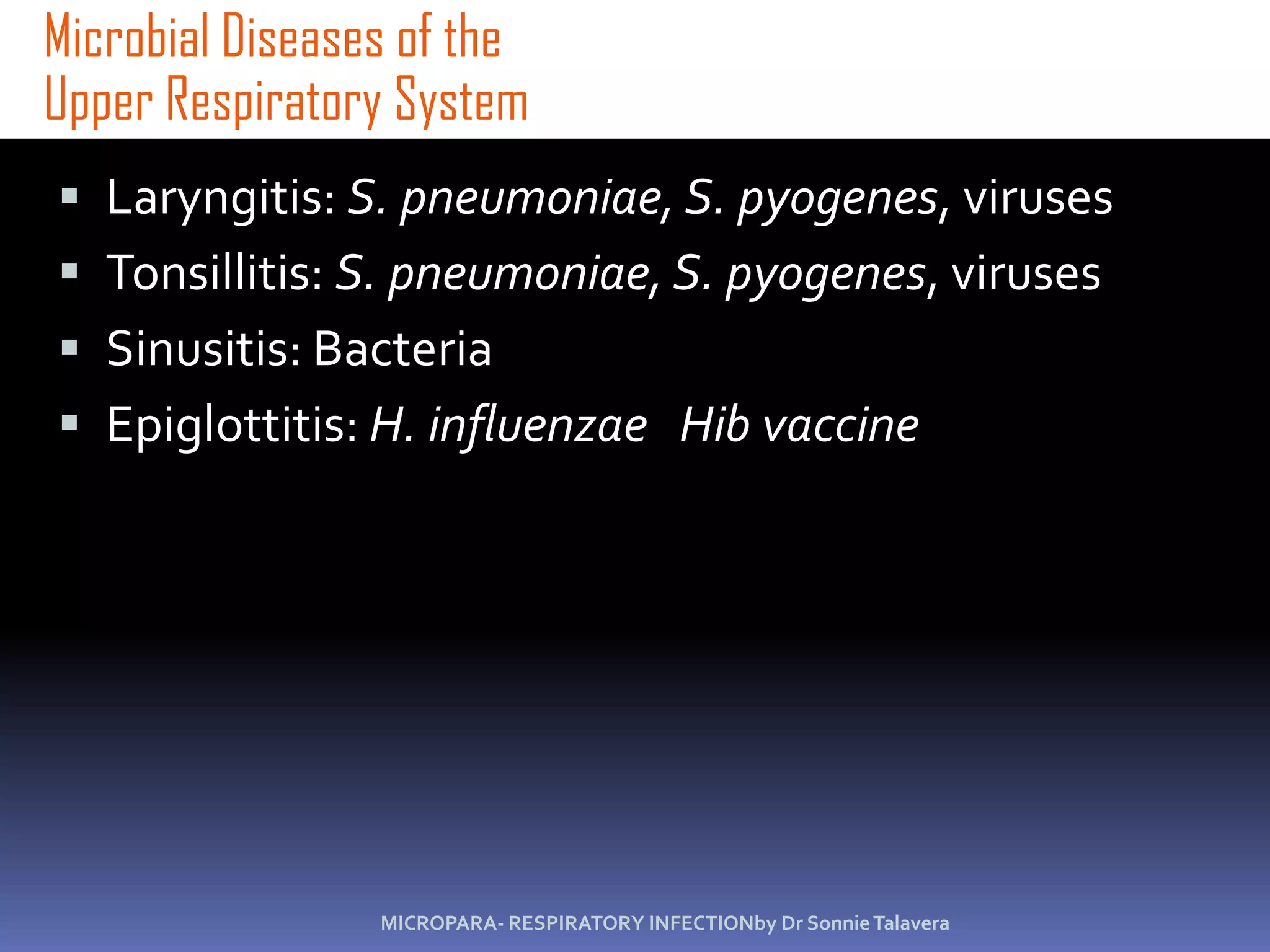
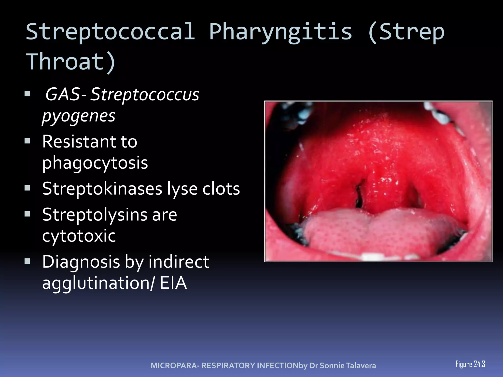
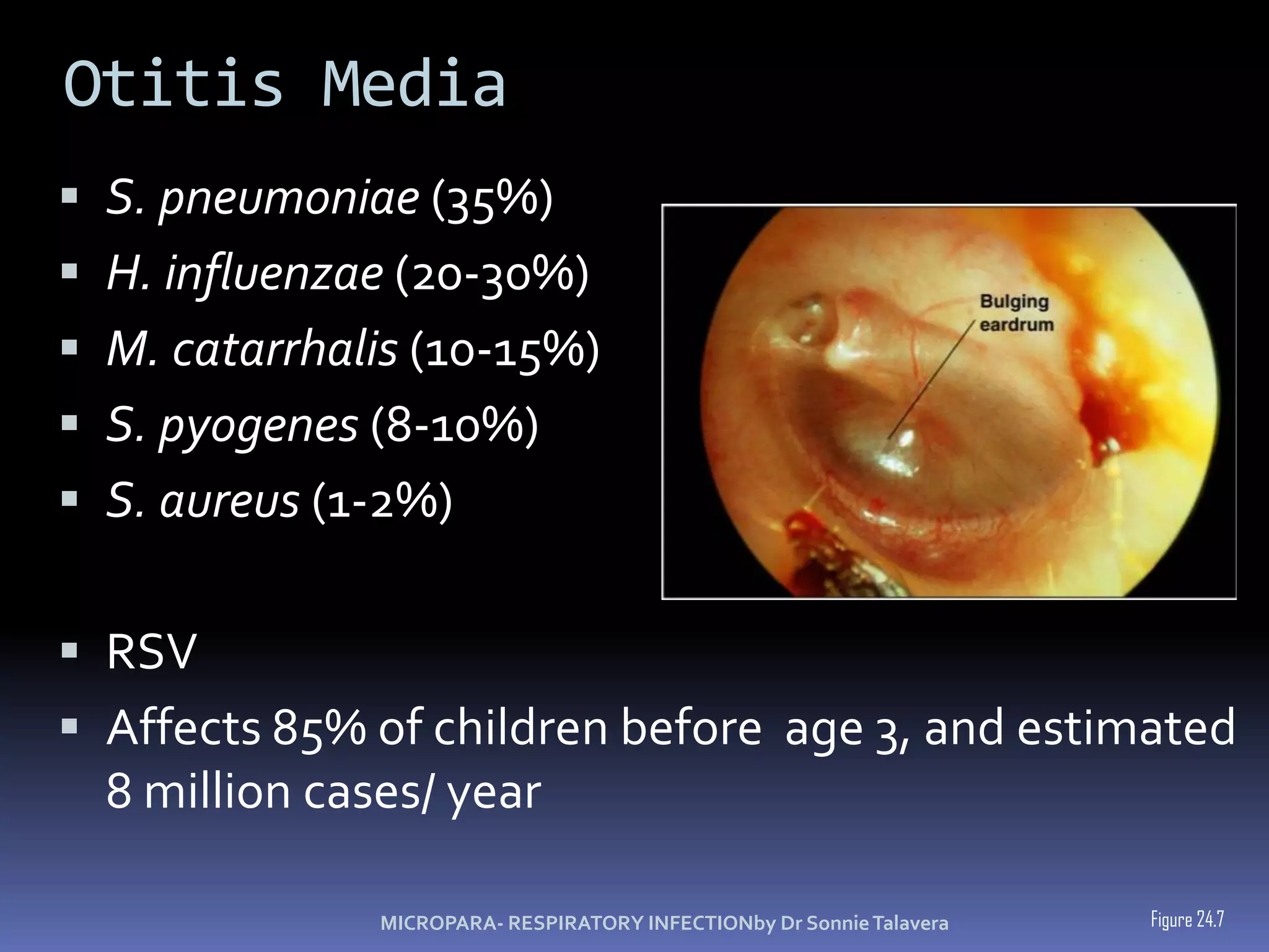
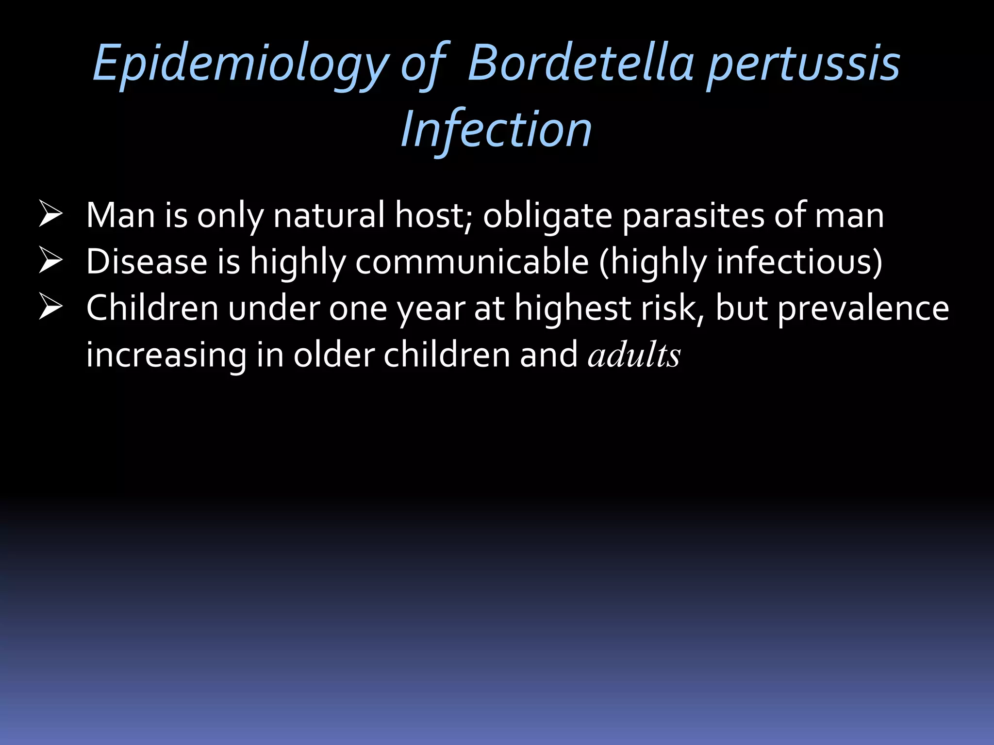
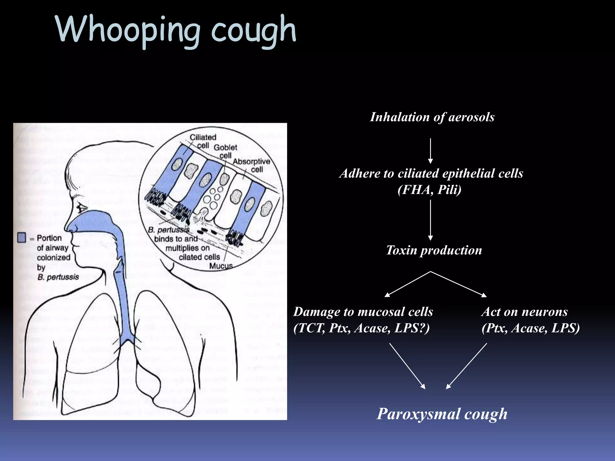
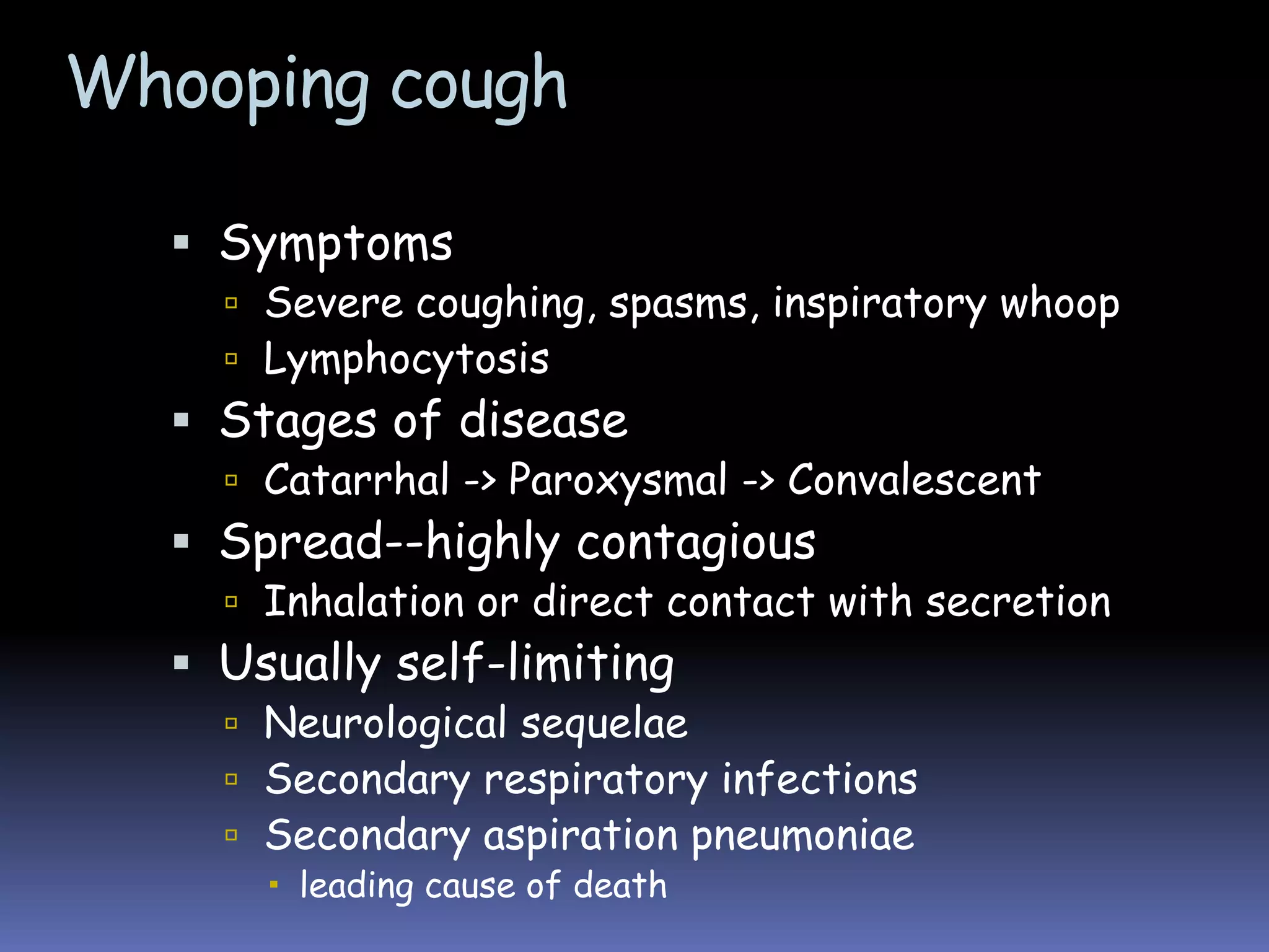
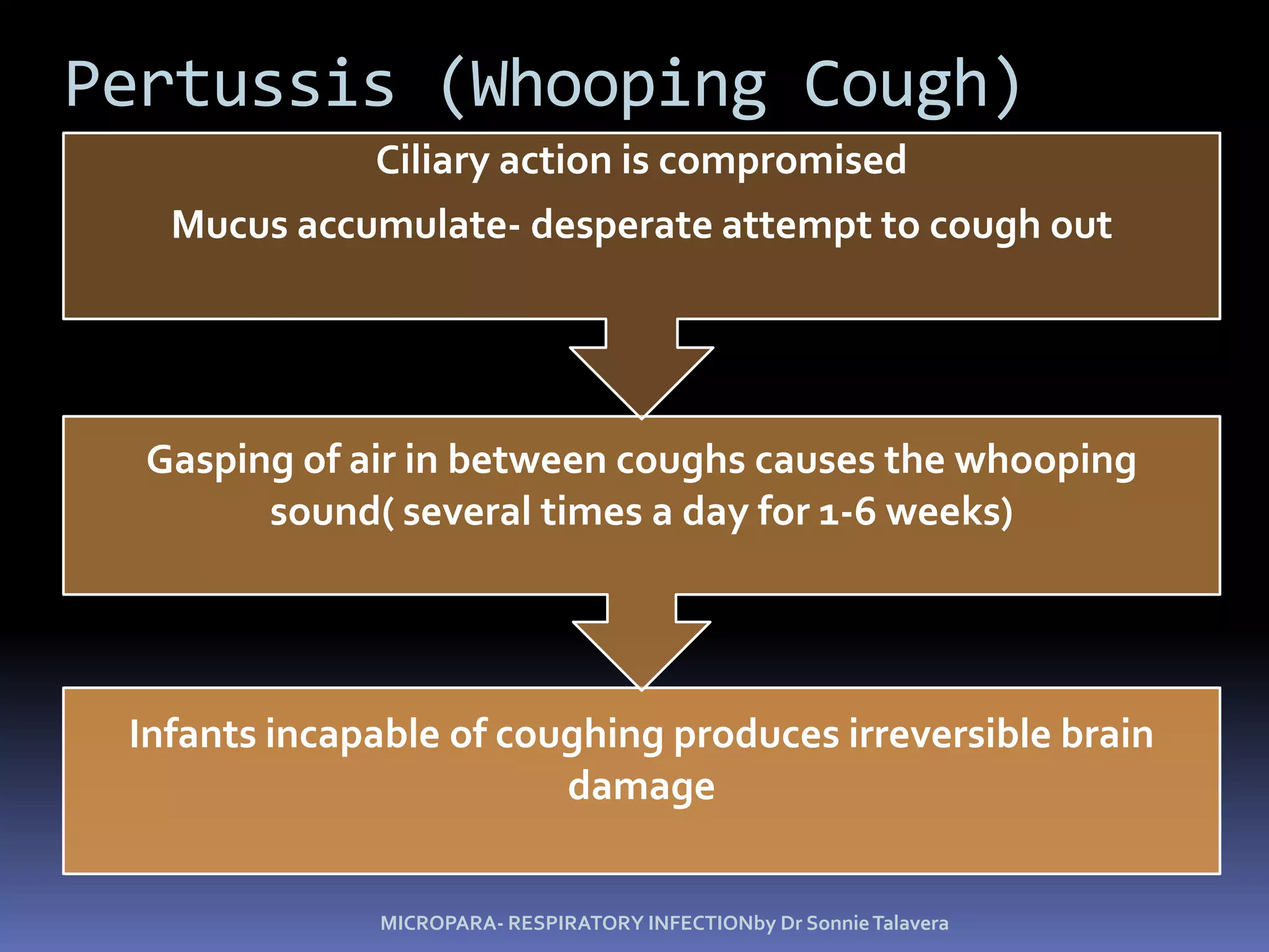
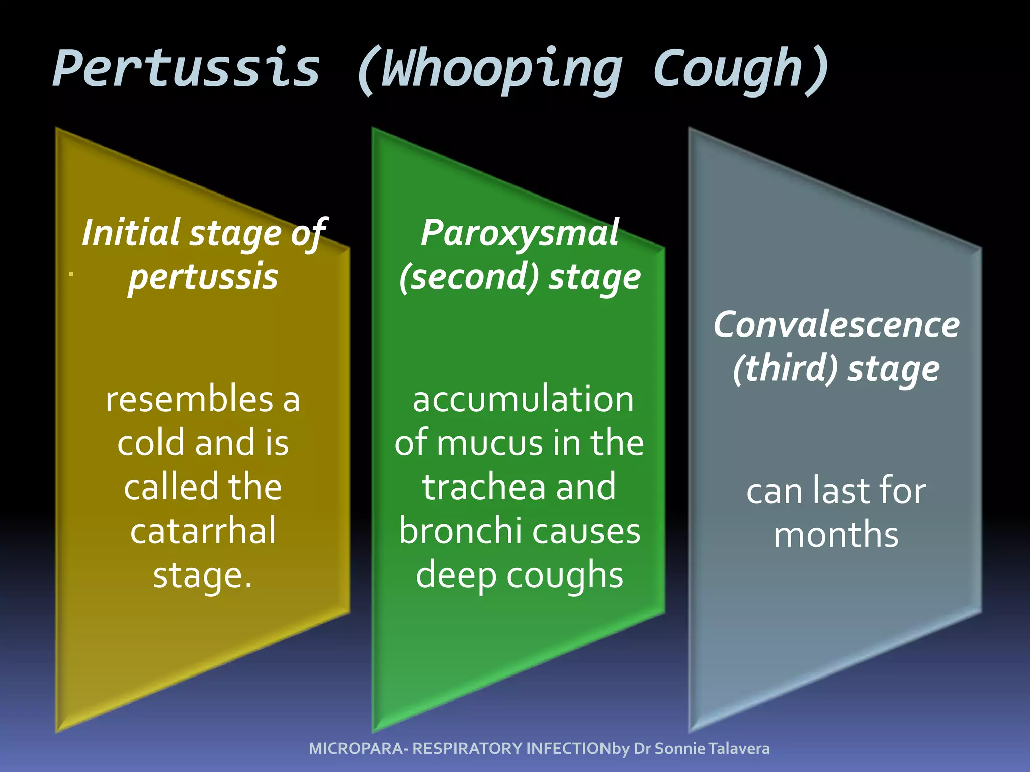
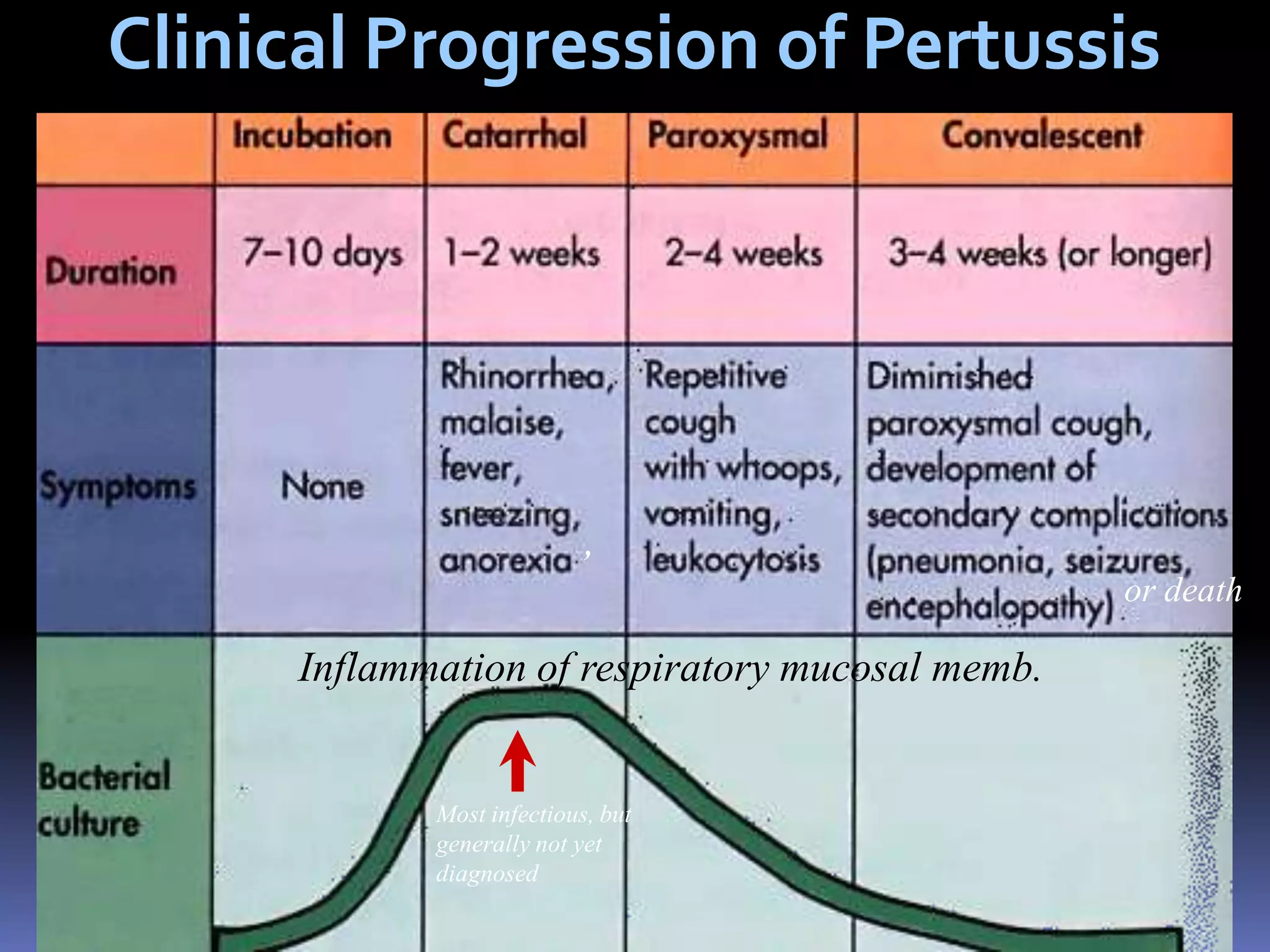
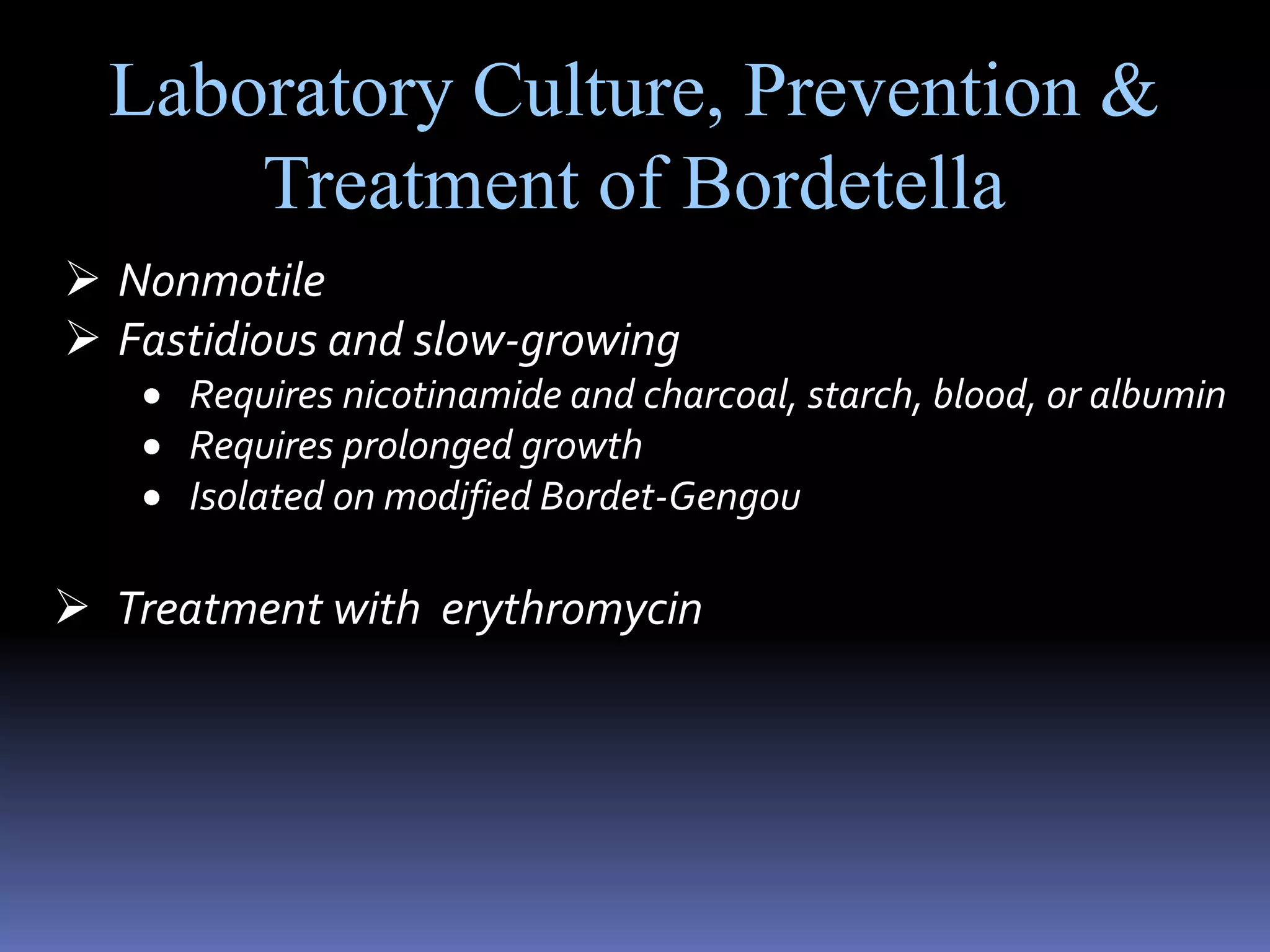
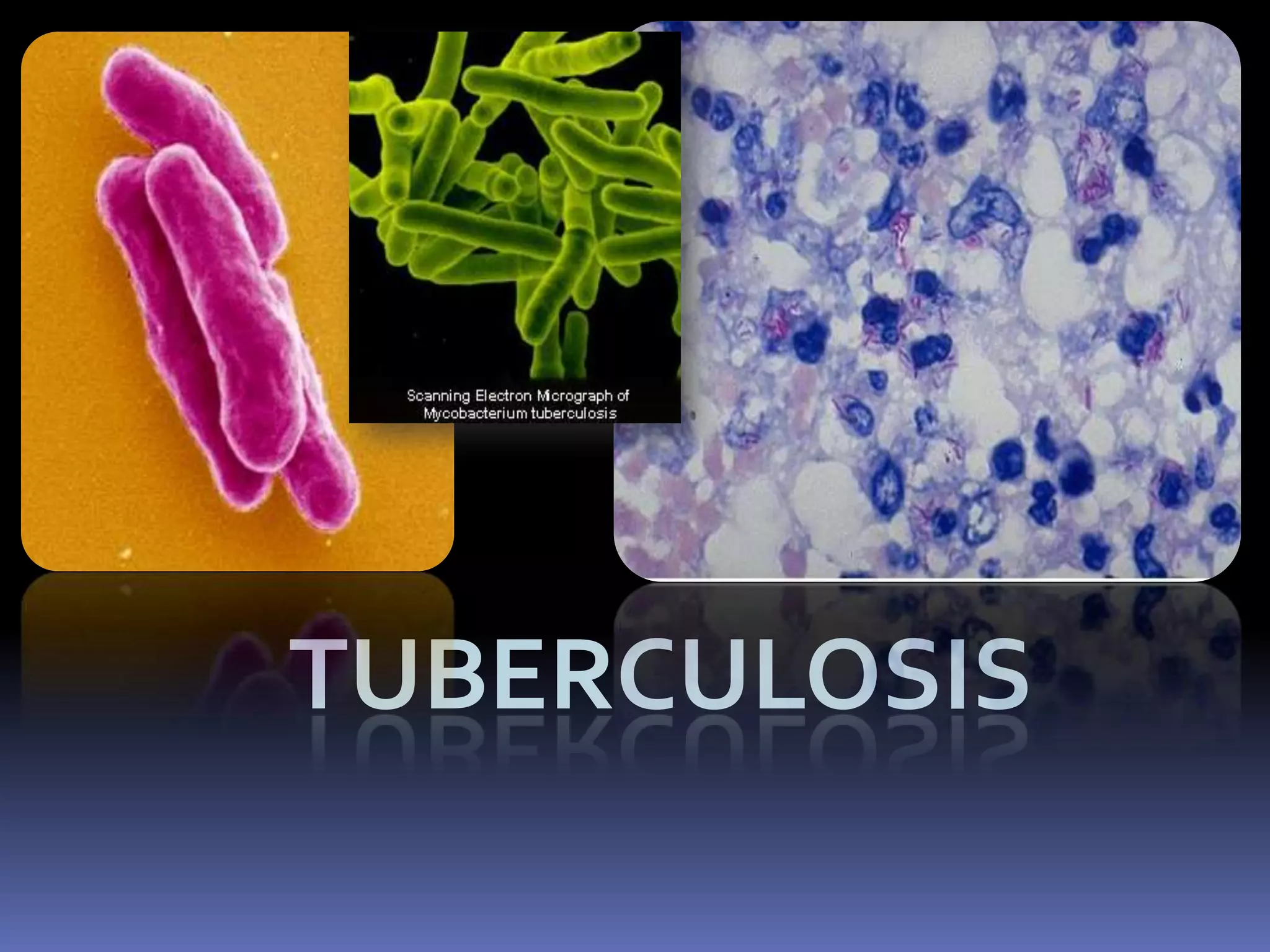
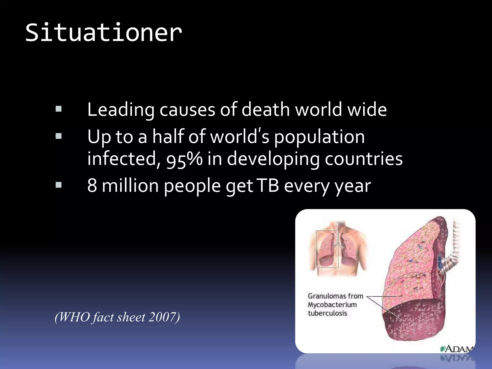
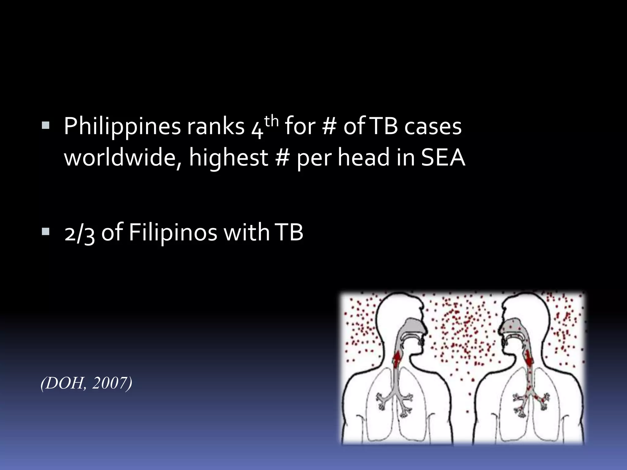
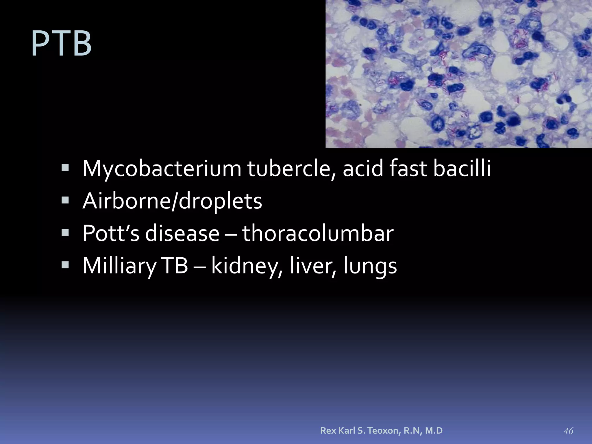
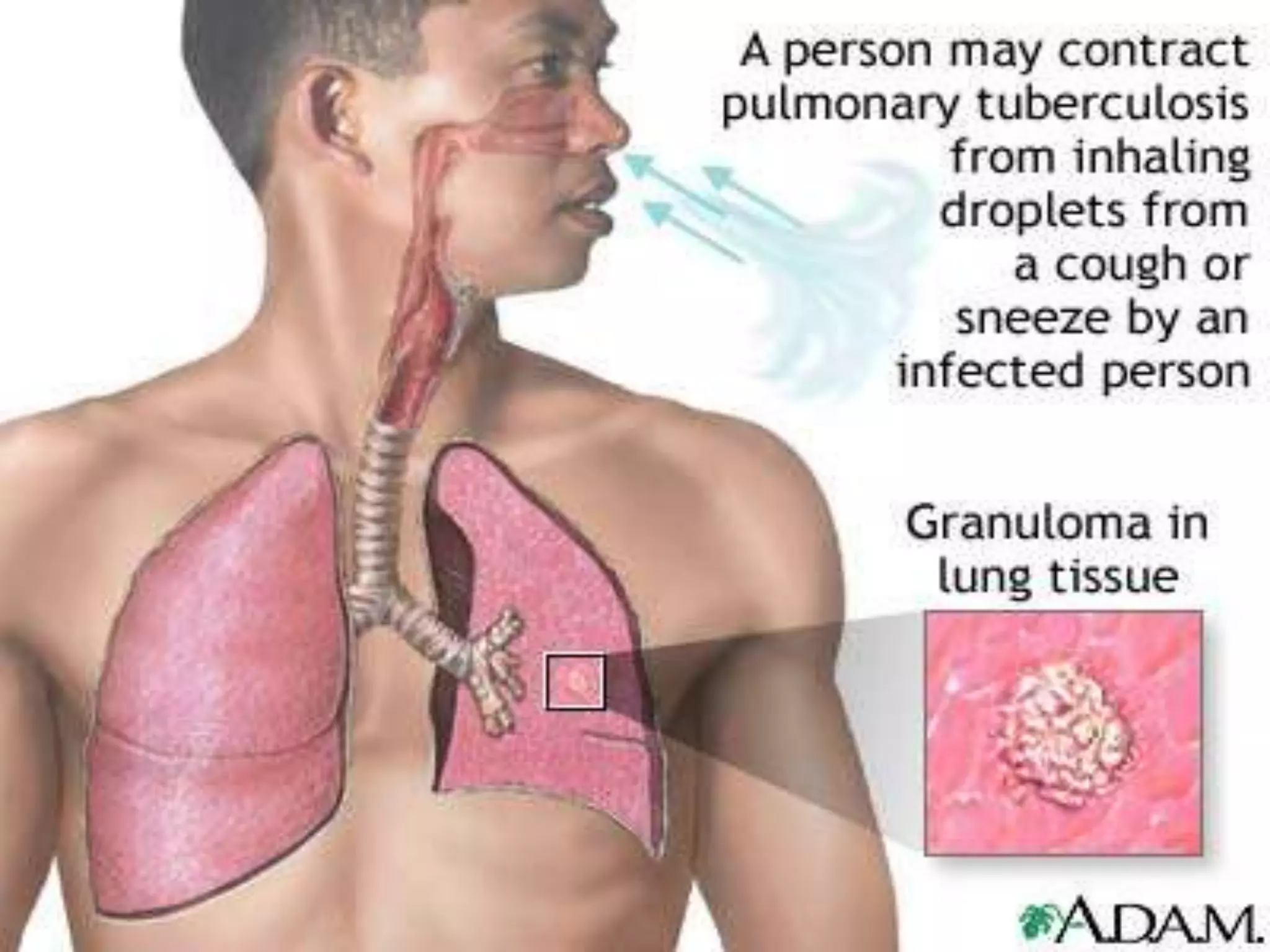
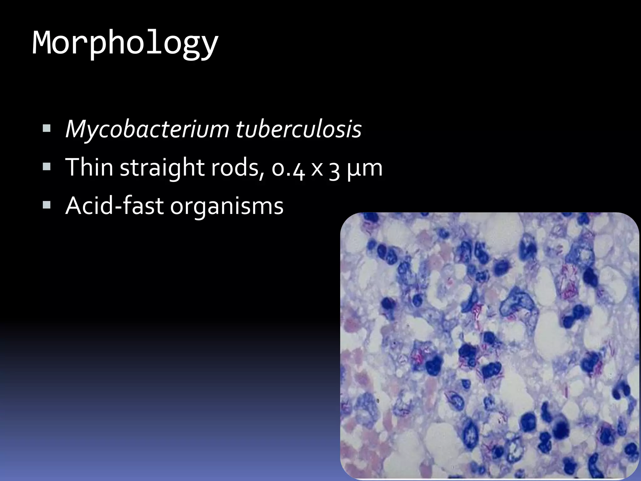
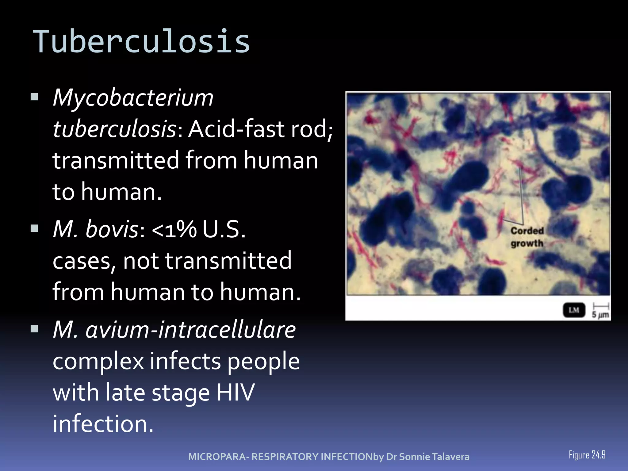
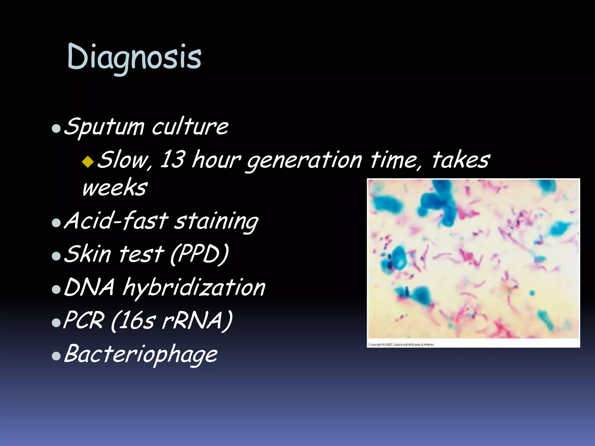
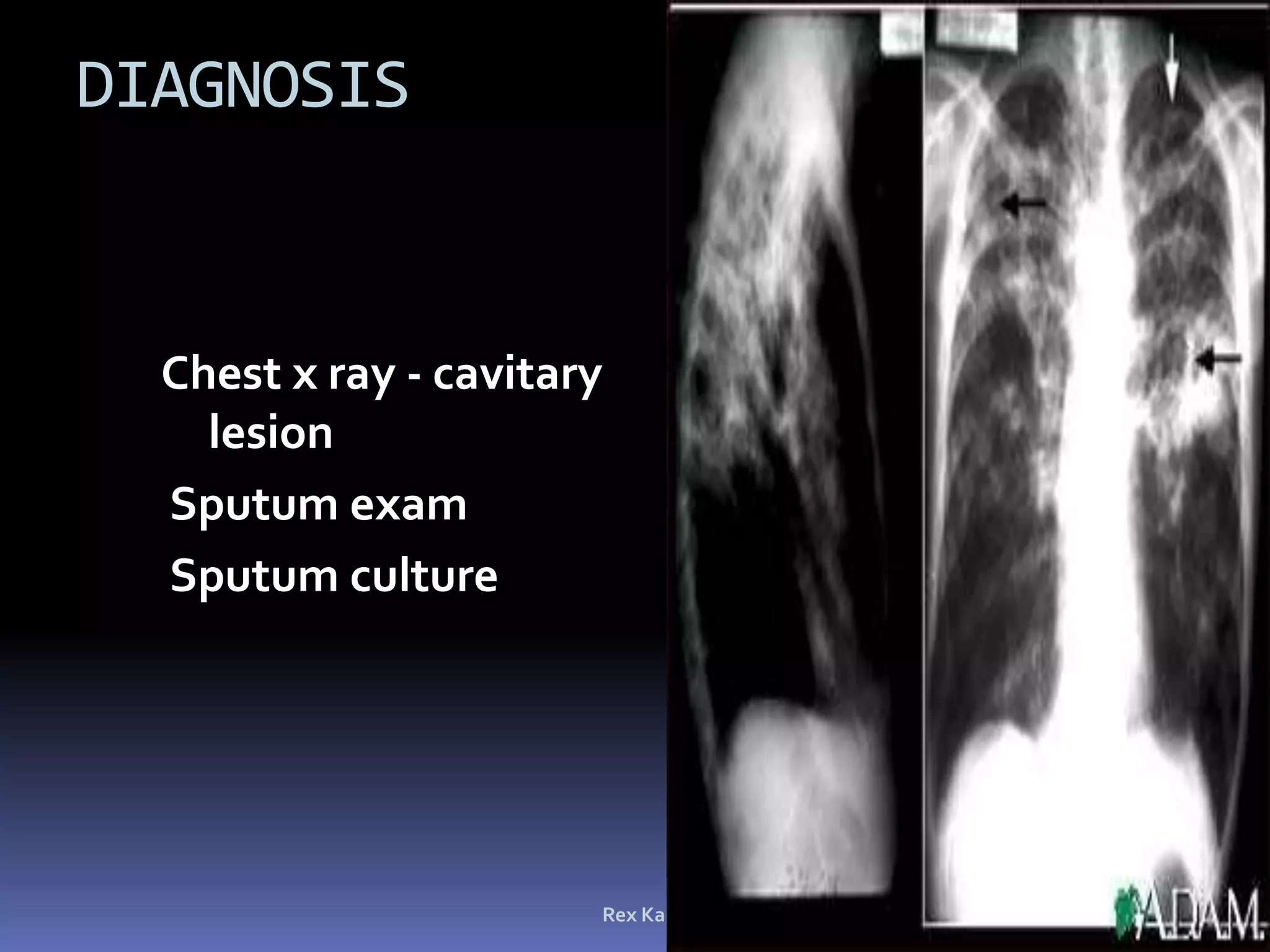
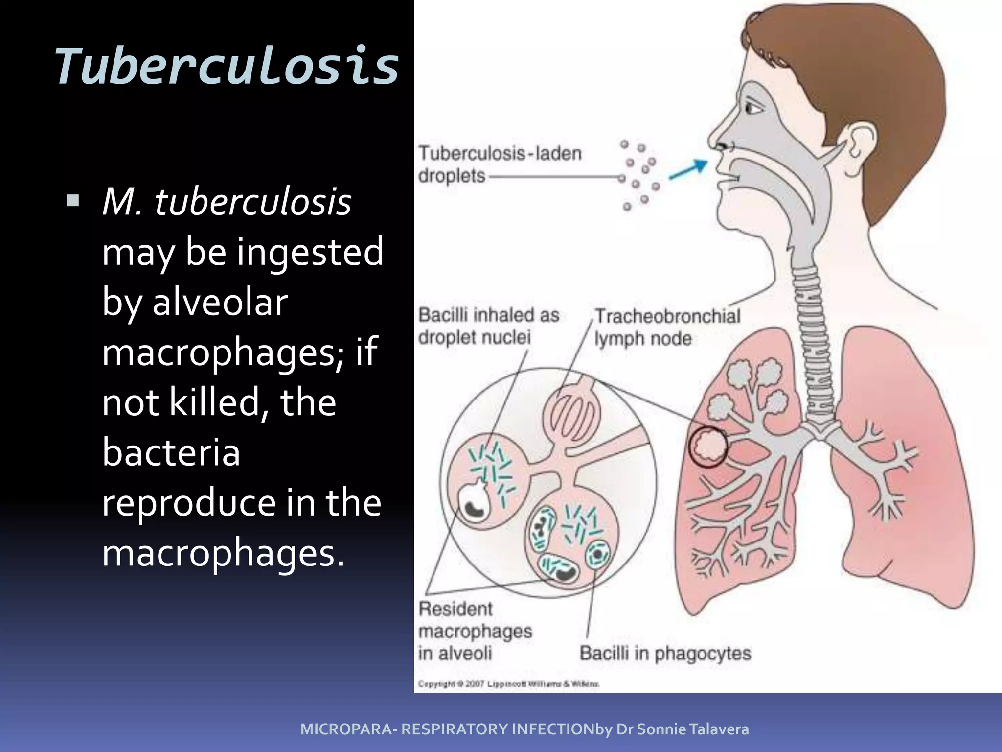
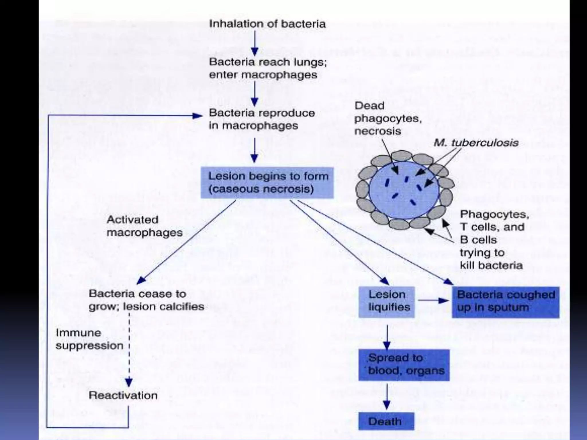
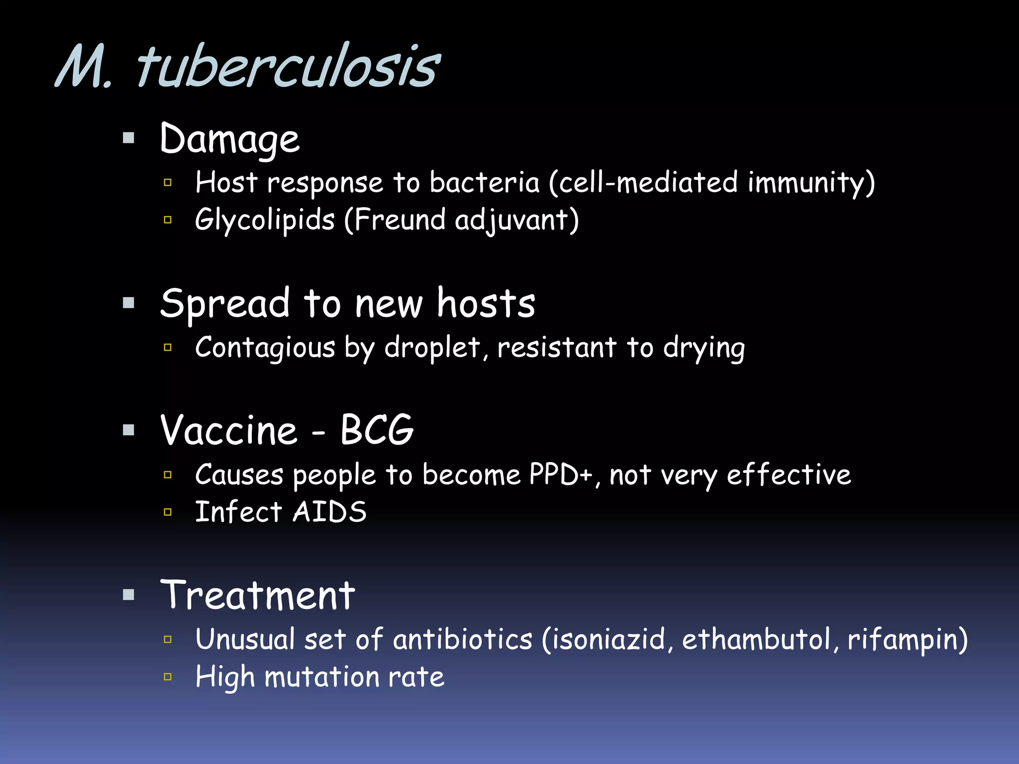
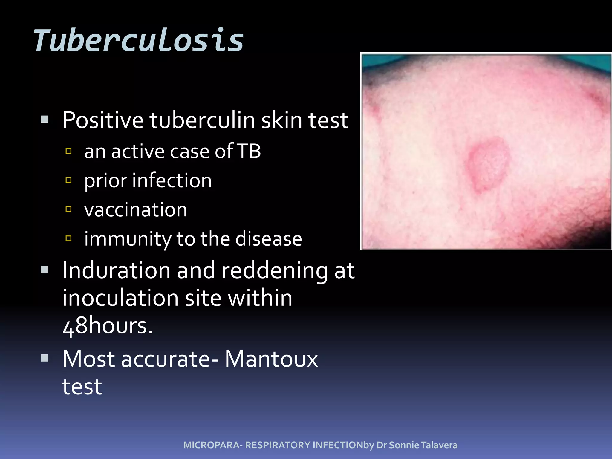
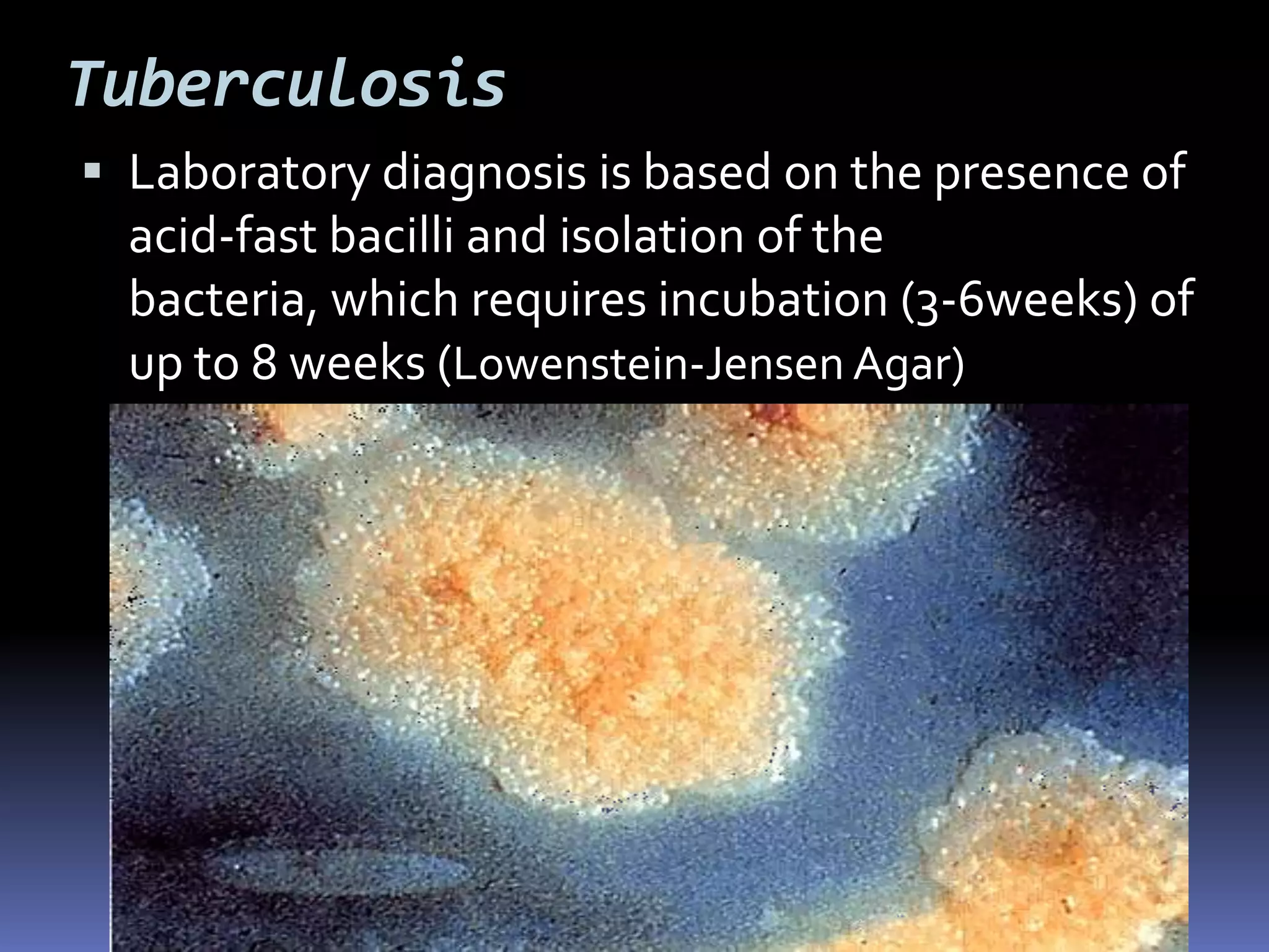
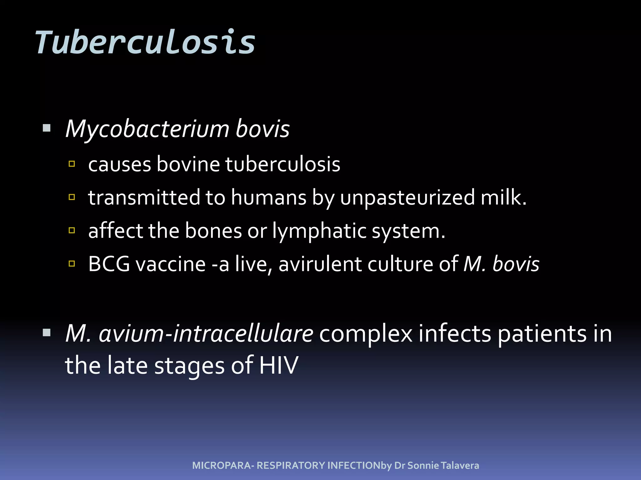
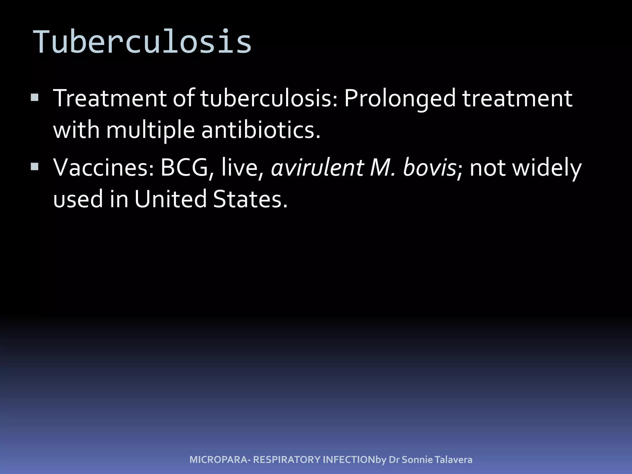
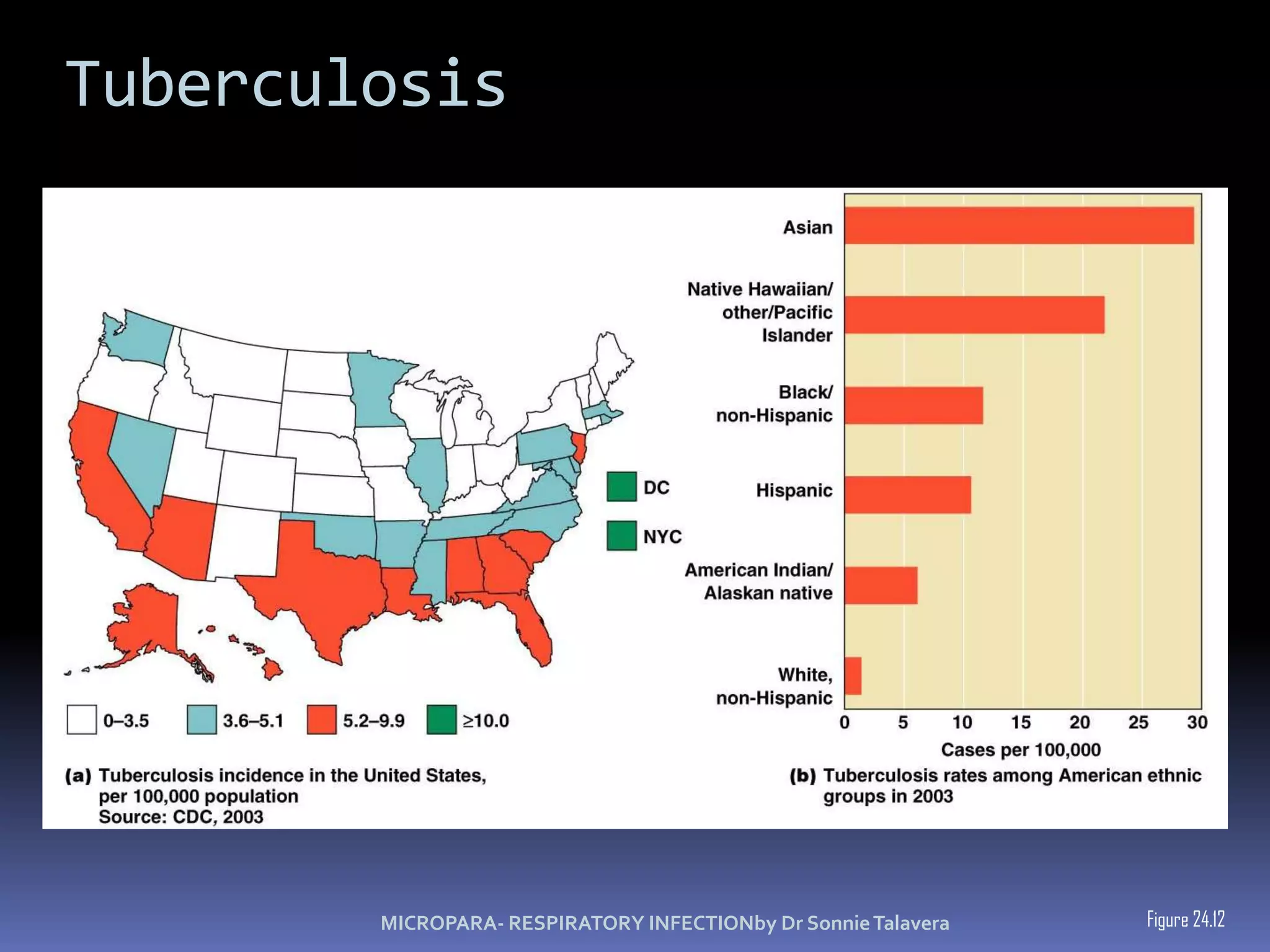
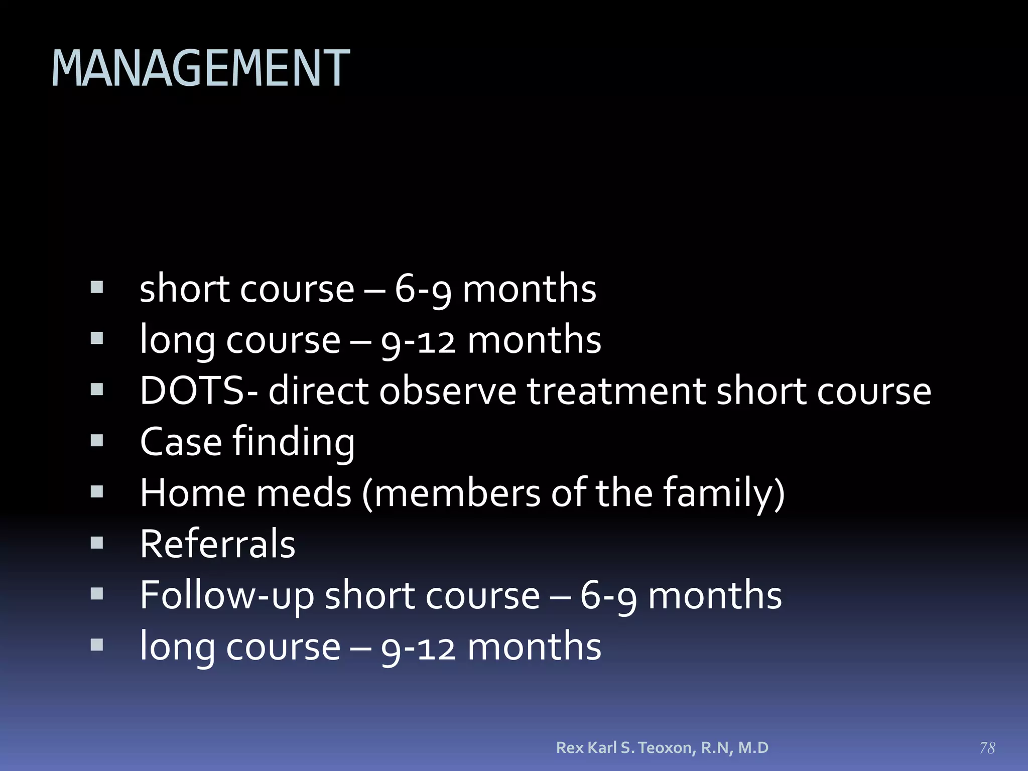
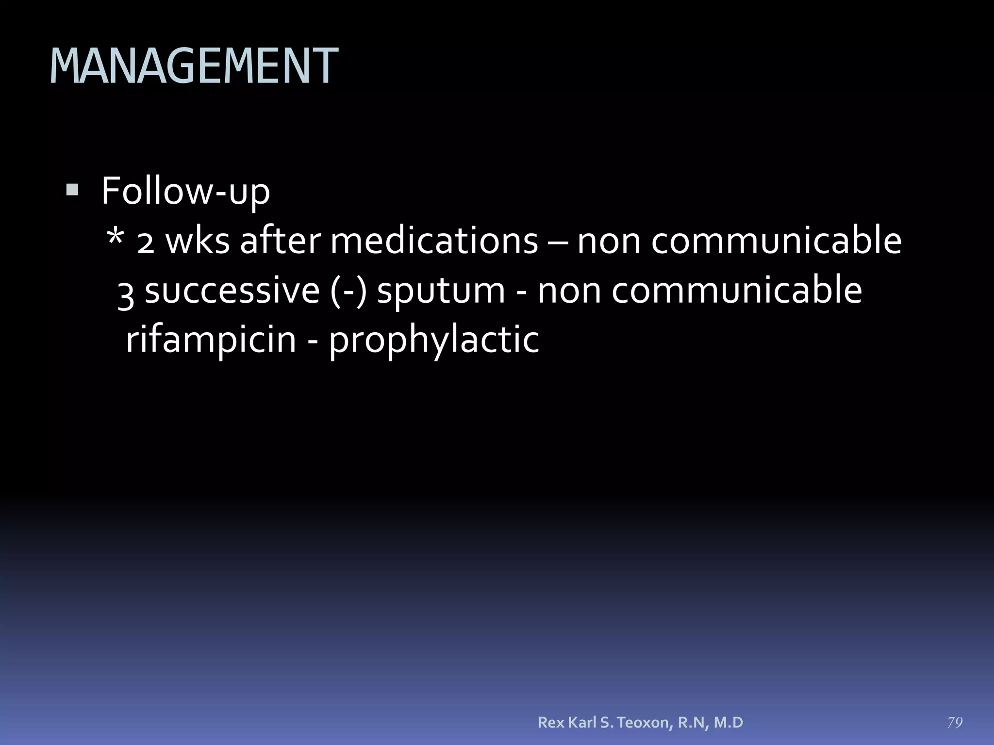
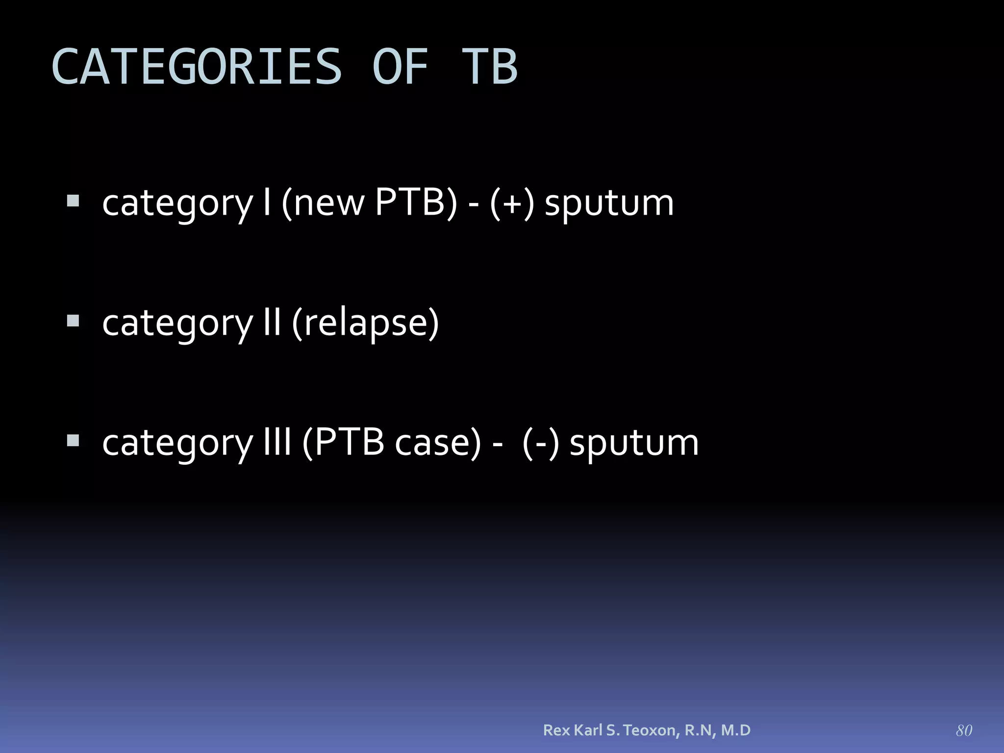
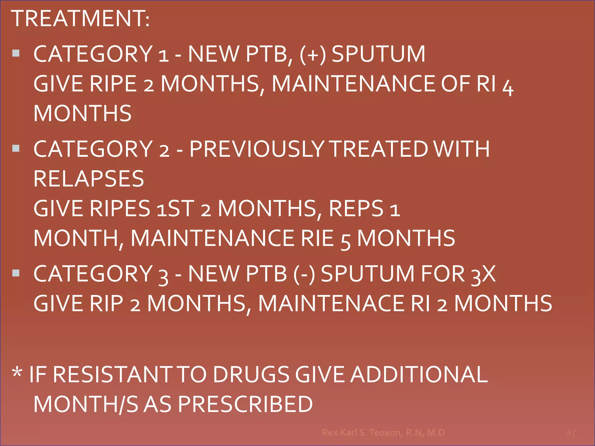
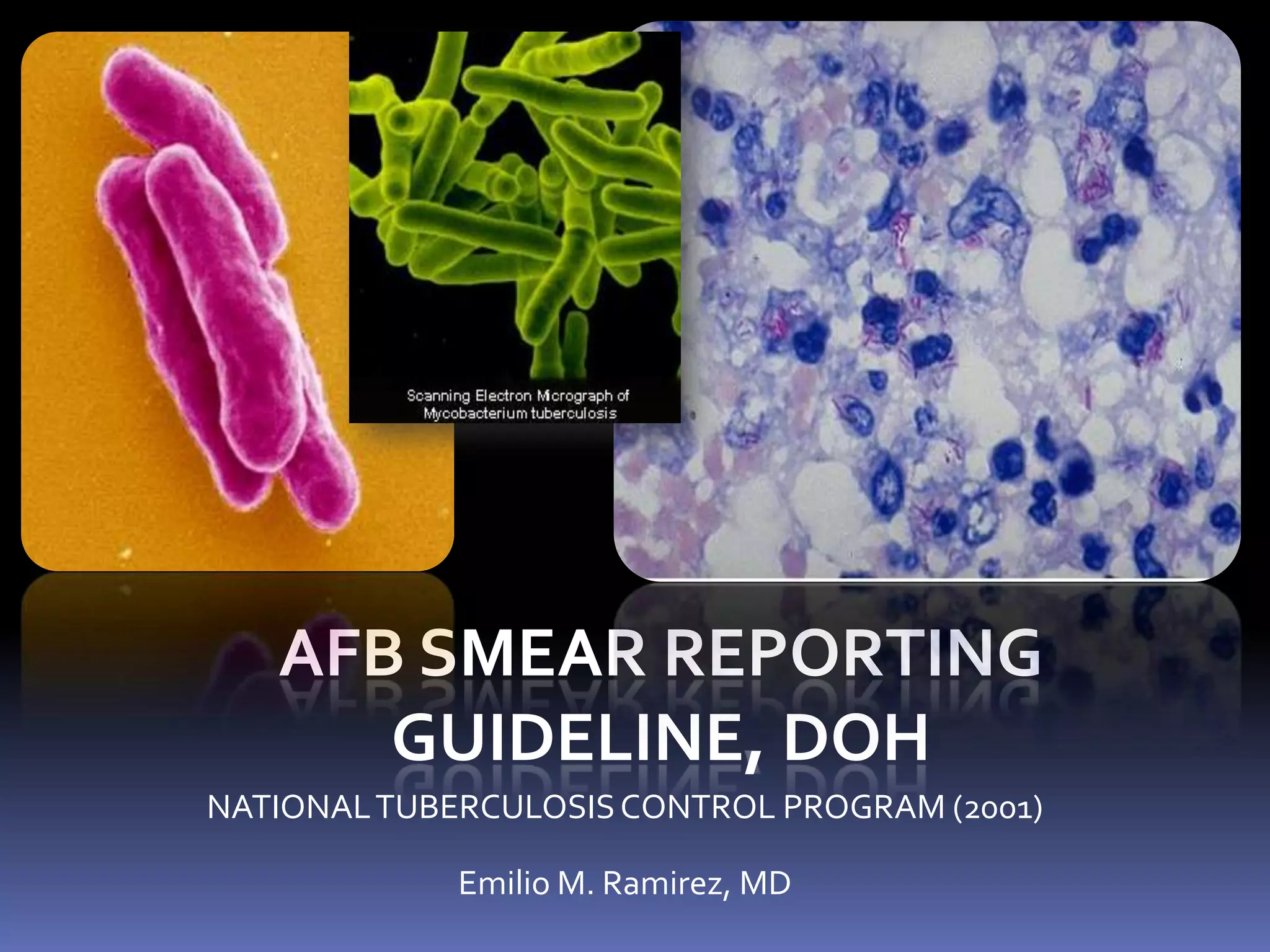
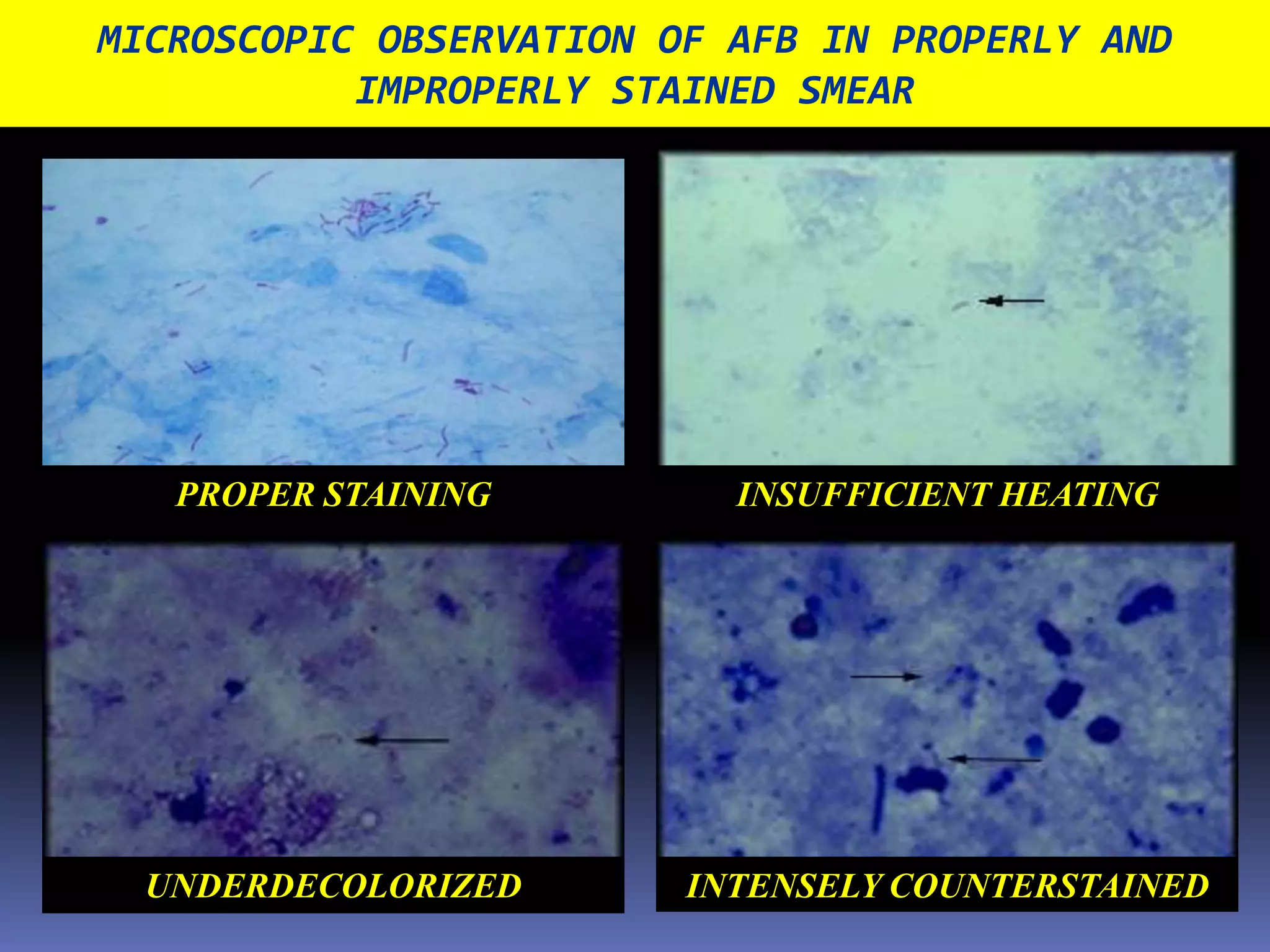
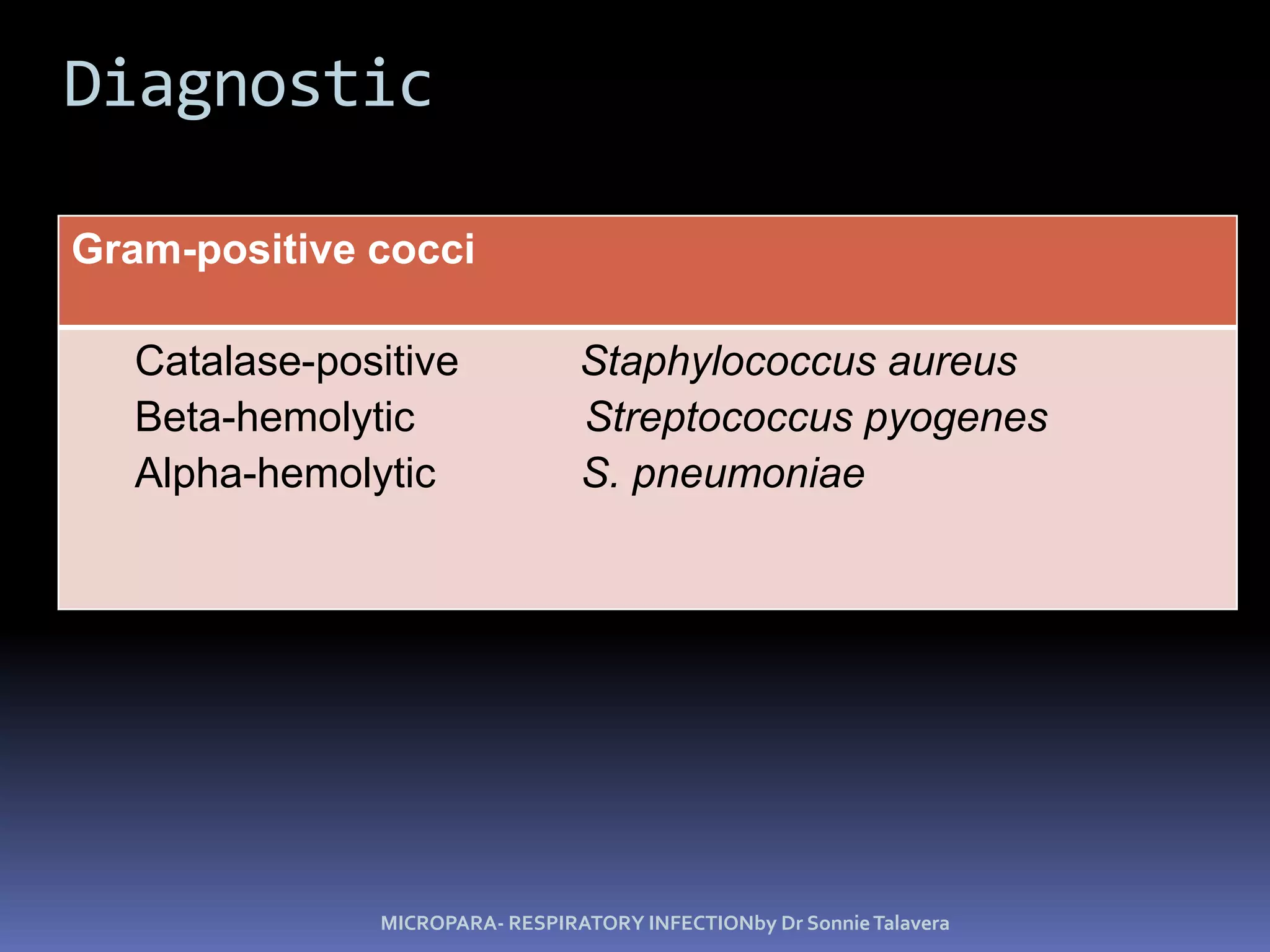
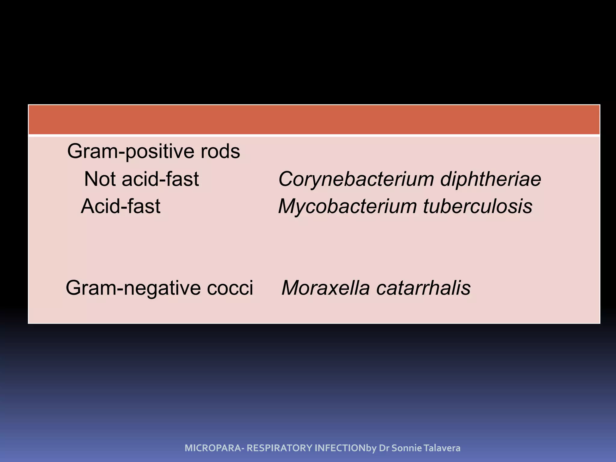
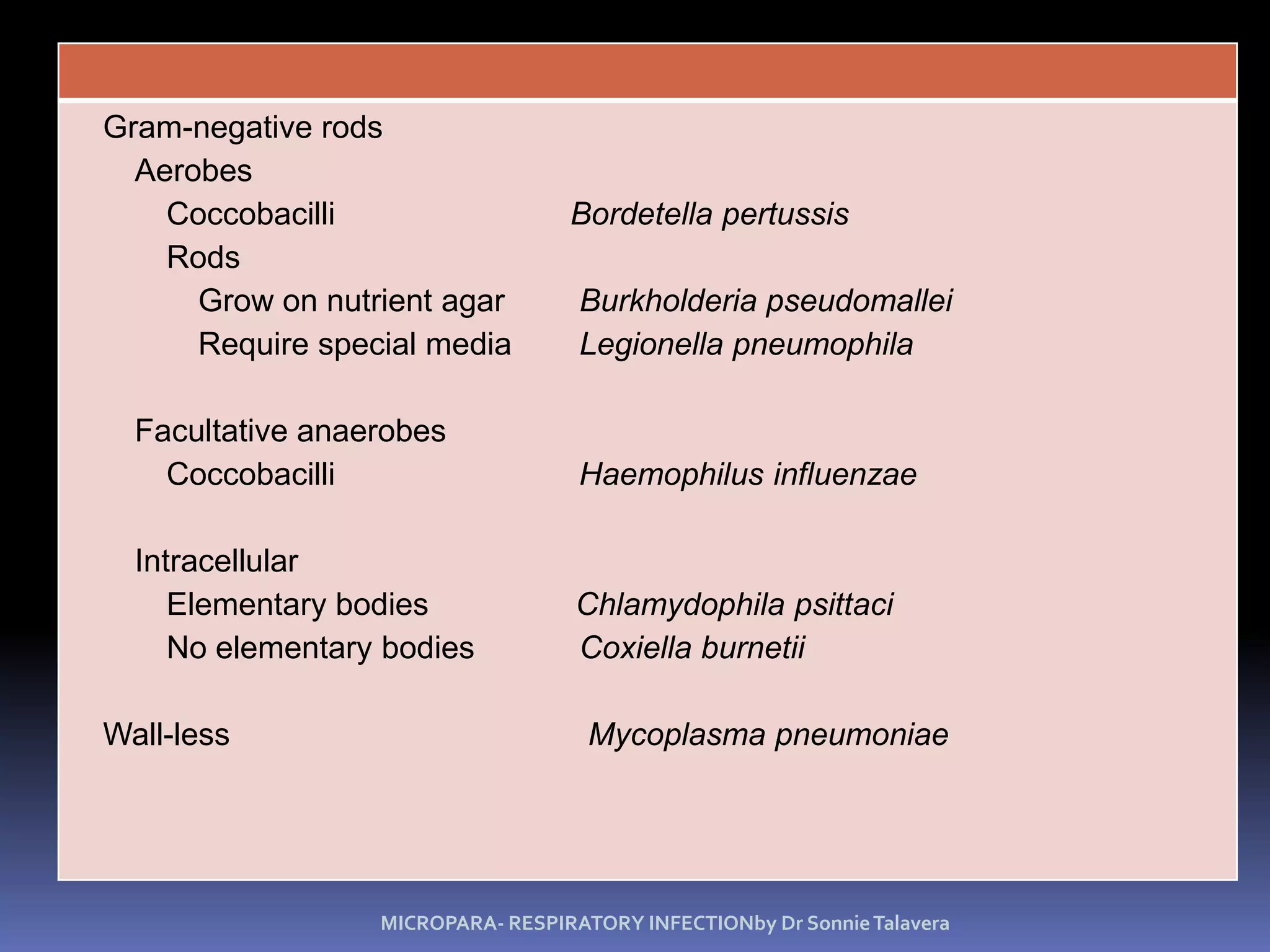
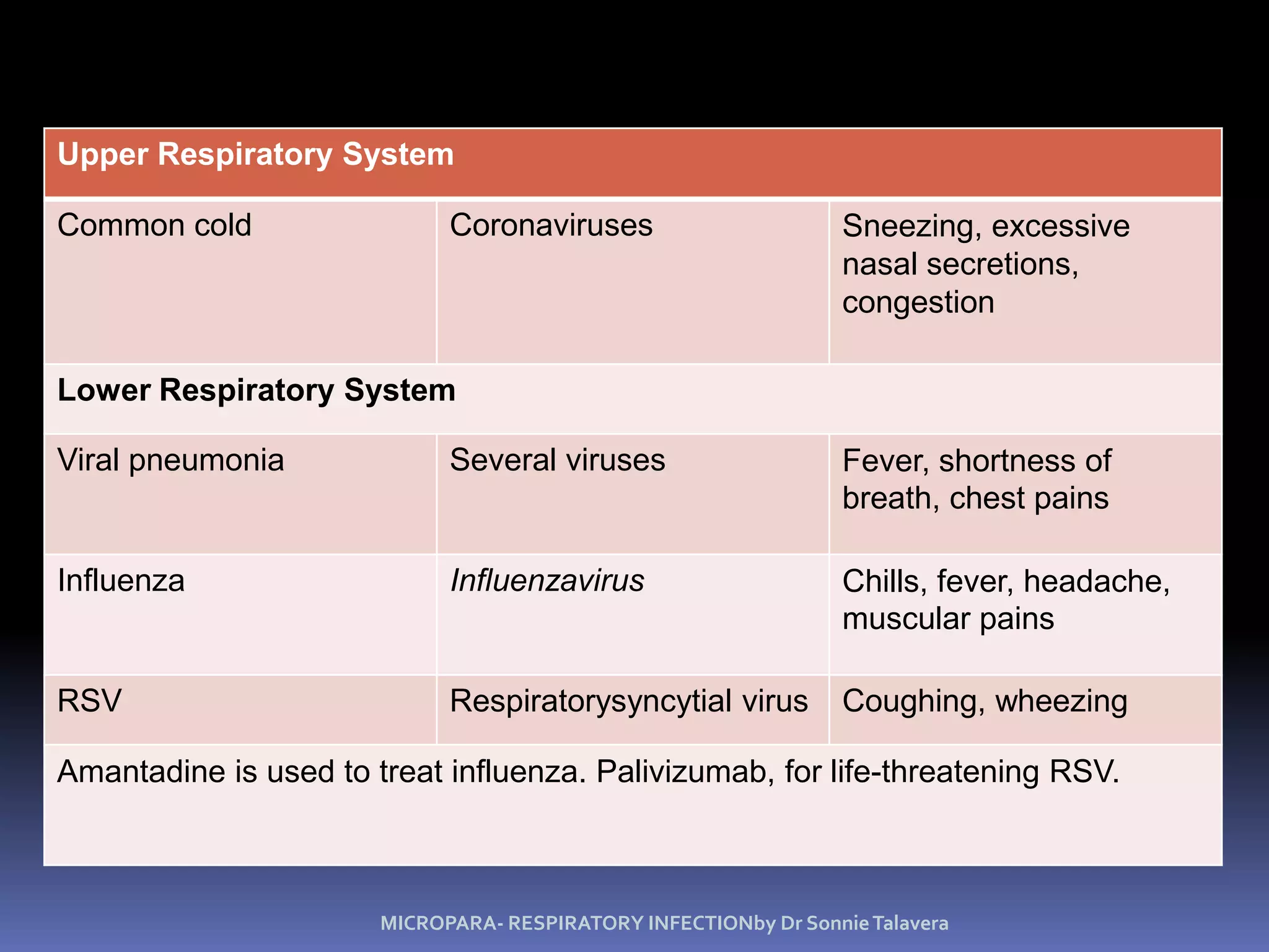
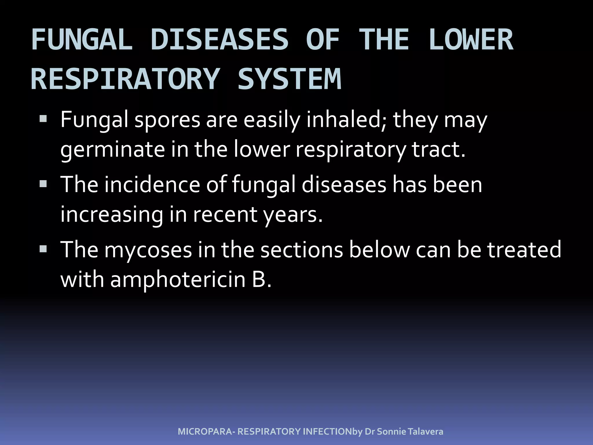
Call Girl Lucknow Mallika 7001305949 Independent Escort Service Lucknow
Â
Respimicro [recovered]
1. 24 Microbial Diseases of the Respiratory System MICROPARA- RESPIRATORY INFECTIONby Dr Sonnie Talavera
2. upper respiratory system The consists of the nose, pharynx, and associated structures, such as the middle ear and auditory tubes. Coarse hairs in the nose ciliated mucous membranes ( nose and throat ) Lymphoid tissue, tonsils, and adenoids MICROPARA- RESPIRATORY INFECTIONby Dr Sonnie Talavera
3. lower respiratory system consists of the larynx, trachea, bronchial tubes, and alveoli. ciliary escalator alveolar macrophages. Respiratory mucus contains IgA antibodies. MICROPARA- RESPIRATORY INFECTIONby Dr Sonnie Talavera
4. Normal Microbiota of the Respiratory System The normal microbiota of the nasal cavity and throat can include pathogenic microorganisms. The lower respiratory system is usually sterile because of the action of the ciliary escalator. MICROPARA- RESPIRATORY INFECTIONby Dr Sonnie Talavera
5. Upper Respiratory System Upper respiratory normal microbiota may include pathogens Figure 24.1 MICROPARA- RESPIRATORY INFECTIONby Dr Sonnie Talavera
6. Microbial Diseases of the Upper Respiratory System Laryngitis: S. pneumoniae, S. pyogenes, viruses Tonsillitis: S. pneumoniae, S. pyogenes, viruses Sinusitis: Bacteria Epiglottitis: H. influenzaeHib vaccine MICROPARA- RESPIRATORY INFECTIONby Dr Sonnie Talavera
7. Microbial Diseases of the Upper Respiratory System These infections may be caused by several bacteria and viruses, often in combination. Most respiratory tract infections are self-limiting. H. influenzae type b can cause epiglottitis. MICROPARA- RESPIRATORY INFECTIONby Dr Sonnie Talavera
8. Streptococcal Pharyngitis (Strep Throat) GAS- Streptococcus pyogenes Resistant to phagocytosis Streptokinaseslyse clots Streptolysins are cytotoxic Diagnosis by indirect agglutination/ EIA Figure 24.3 MICROPARA- RESPIRATORY INFECTIONby Dr Sonnie Talavera
9. Streptococcal Pharyngitis (Strep Throat)Â group A beta-hemolytic streptococci- Streptococcus pyogenes. Symptoms of this infection are inflammation of the mucous membrane and fever; tonsillitis and otitis media may also occur. MICROPARA- RESPIRATORY INFECTIONby Dr Sonnie Talavera
10. Streptococcal Pharyngitis (Strep Throat)Â Rapid diagnosis is made by enzyme immunoassays. Penicillin is used to treat streptococcal pharyngitis. Immunity to streptococcal infections is type-specific. Strep throat is usually transmitted by droplets. MICROPARA- RESPIRATORY INFECTIONby Dr Sonnie Talavera
11. Scarlet Fever Streptococcus pyogenes Pharyngitis Erythrogenic toxin produced by lysogenizedS. pyogenesby a phage. Figure 24.4 MICROPARA- RESPIRATORY INFECTIONby Dr Sonnie Talavera
12. Scarlet Fever  Strep throat, caused by an erythrogenic toxin-producing S. pyogenes, results in scarlet fever. starts general malaise and swelling of neck Symptoms include a red rash, high fever, and a SPOTTED STRABERRY like red, enlarged tongue. MICROPARA- RESPIRATORY INFECTIONby Dr Sonnie Talavera
14. Diphtheria Clinical Start as sore throat and fever followed by general malaise and swelling of neck Diphtheria (leather) tough grayish membrane of fibrin, dead tissue, and bacteria Diphtheria toxin produced by lysogenizedC. diphtheriae (highly virulent toxin) MICROPARA- RESPIRATORY INFECTIONby Dr Sonnie Talavera
16. Diphtheria  MICROPARA- RESPIRATORY INFECTIONby Dr Sonnie Talavera Vao day nghe bai nay di ban http://nhattruongquang.0catch.com
17. Diphtheria A membrane can block the passage of air. Exotoxin inhibits protein synthesis, and heart, kidney, or nerve damage may result (fatal) minimal dissemination of the exotoxin in the bloodstream. Antitoxin - neutralize the toxin Antibiotics- Penicillin and Erythromycin can stop growth of the bacteria. MICROPARA- RESPIRATORY INFECTIONby Dr Sonnie Talavera
18. Diphtheria Prevented by DTaP and Td vaccine (Diphtheria toxoid) Cutaneous diphtheria: Infected skin wound leads to slow healing ulcer MICROPARA- RESPIRATORY INFECTIONby Dr Sonnie Talavera
20. A 15 y/o male patient have chief complaint of difficulty of breathing with associated throat pain, difficulty of swallowing and fever. Physical exam reveals inflamed pharyngeal area covered by grayish thick mucus that is adherent. Patient was noted to have an incomplete set of vaccination. Grams staining reveals gram positive bacilli with endospore. MICROPARA- RESPIRATORY INFECTIONby Dr Sonnie Talavera
21.
22. Small, pleomorphic (club-shaped), gram-positive bacilli short chains (âVâ or âYâ configurations) or in clumps resembling âChinese lettersâ
26. otitis media with effusionA. Symptoms - fever, pain in the ear, dulled hearing.
27. Otitis Media S. pneumoniae (35%) H. influenzae (20-30%) M. catarrhalis (10-15%) S. pyogenes (8-10%) S. aureus (1-2%) RSV Affects 85% of children before age 3, and estimated 8 million cases/ year Figure 24.7 MICROPARA- RESPIRATORY INFECTIONby Dr Sonnie Talavera
28. Otitis Media Treated with broad-spectrum antibiotics Amoxicillin Incidence of S. pneumoniae reduced by vaccine Figure 24.7 MICROPARA- RESPIRATORY INFECTIONby Dr Sonnie Talavera
29. Otitis Media  Earache, or otitis media, can occur as a complication of nose and throat infections. Pus accumulation causes pressure on the eardrum causes inflammation and pain. Often in children because of shorter and more horizontal eustachian tube MICROPARA- RESPIRATORY INFECTIONby Dr Sonnie Talavera
30. B. DIAGNOSIS â 1. clinical presentation of fever and pain, especially following an URT infection such as a cold 2. otoscopic examination to see inflammation and/or fluid (pus); also loss of mobility with air pressure
34. The Common Cold Any one of approximately 200 different viruses can cause the common cold; rhinoviruses cause about 50% of all colds. Immunity is based on the ration of Ig A antibodies MICROPARA- RESPIRATORY INFECTIONby Dr Sonnie Talavera
35. The Common Cold Symptoms Sneezing nasal secretions congestion. Sinus infections, lower respiratory tract infections, laryngitis, and otitis media can occur as complications of a cold. MICROPARA- RESPIRATORY INFECTIONby Dr Sonnie Talavera
36. The Common Cold Colds are most often transmitted by indirect contact. Rhinoviruses grow best slightly below body temperature. The incidence of colds increases during cold weather Antibodies are produced against the specific viruses. MICROPARA- RESPIRATORY INFECTIONby Dr Sonnie Talavera
37. Microbial Diseases of the Lower Respiratory System Bacteria, viruses, and fungi cause Bronchitis Bronchiolitis Pneumonia MICROPARA- RESPIRATORY INFECTIONby Dr Sonnie Talavera
38. Lower Respiratory System The ciliary escalator keeps the lower respiratory system sterile. Figure 24.2 MICROPARA- RESPIRATORY INFECTIONby Dr Sonnie Talavera
43. Whooping cough Symptoms Severe coughing, spasms, inspiratory whoop Lymphocytosis Stages of disease Catarrhal -> Paroxysmal -> Convalescent Spread--highlycontagious Inhalation or direct contact with secretion Usually self-limiting Neurological sequelae Secondary respiratory infections Secondary aspiration pneumoniae leading cause of death
54. Situationer Leading causes of death world wide Up to a half of worldâs population infected, 95% in developing countries 8 million people get TB every year (WHO fact sheet 2007)
55. Philippines ranks 4th for # of TB cases worldwide, highest # per head in SEA 2/3 of Filipinos with TB (DOH, 2007)
56. Rex Karl S. Teoxon, R.N, M.D 46 PTB Mycobacterium tubercle, acid fast bacilli Airborne/droplets Pottâs disease â thoracolumbar Milliary TB â kidney, liver, lungs
61. TB symptoms Cough with two weeks or more Sputum expectoration Fever Significant weight loss Hemoptysis Chest and/or back pains
62. Rex Karl S. Teoxon, R.N, M.D 52 SIGNS AND SYMPTOMS Wt loss, night sweats, low fever, non productive to productive cough, anorexia, Pleural effusion and hypoxemia, cervical lymphadenopathy
63. Tuberculosis Mycobacterium tuberculosis: Acid-fast rod; transmitted from human to human. M. bovis: <1% U.S. cases, not transmitted from human to human. M. avium-intracellulare complex infects people with late stage HIV infection. Figure 24.9 MICROPARA- RESPIRATORY INFECTIONby Dr Sonnie Talavera
64. TuberculosisMycobacterium tuberculosis Acid fast-lipid, wax Slow growth (nutrient permeability) Resist to detergent and common antibiotics A leading cause of death by infectious disease 50% population infected, 3m death/yr Reemergence in 1980 (AIDS, homeless, immigrants) Diagnose PPD test Chest X-ray Sputum smear (for acid-fast bacilli) Sputum culture
72. Rex Karl S. Teoxon, R.N, M.D 57 DIAGNOSIS Chest x ray - cavitary lesion Sputum exam Sputum culture
73. Stages of disease Primary infection Asymptomatic to flu-like 3-5% develop TB Tubercle (granulomatous response) Reactivation (active TB) Years later, 10% experience LRT disease (pneumonia) Disseminated miliary TB Almost everywhere AIDS and antibiotic resistance
74. Stages of pathogenesis Encounter - respiratory droplet Entry - direct inhalation into LRT (ID=10) SPREAD - alveoli, but can spread throughout body seeding many tissues Multiplication Grows in phagosome of macrophage Strict aerobe Very slow in culture (24 hr doubling time) Evade defenses Inhibits phagolysosomal fusion
75. Tuberculosis Tuberculosis is caused by Mycobacterium tuberculosis. Large amounts of lipids in the cell wall account for the bacteriumâs acid-fast characteristic as well as its resistance to dryingnd disinfectants. MICROPARA- RESPIRATORY INFECTIONby Dr Sonnie Talavera
76. Tuberculosis M. tuberculosis may be ingested by alveolar macrophages; if not killed, the bacteria reproduce in the macrophages. MICROPARA- RESPIRATORY INFECTIONby Dr Sonnie Talavera
77. Tuberculosis Lesions formed by M. tuberculosis are called tubercles Dead macrophages and bacteria form the caseous lesion that might calcify and appear in an X ray as a Ghonâs complex. MICROPARA- RESPIRATORY INFECTIONby Dr Sonnie Talavera
78. Tuberculosis Liquefaction of the caseous lesion results in a tuberculous cavity in which M. tuberculosis can grow. MICROPARA- RESPIRATORY INFECTIONby Dr Sonnie Talavera
79. Tuberculosis New foci of infection can develop when a caseous lesion ruptures and releases bacteria into blood or lymph vessels; this is called miliary tuberculosis. MICROPARA- RESPIRATORY INFECTIONby Dr Sonnie Talavera
80.
81. M. tuberculosis Damage Host response to bacteria (cell-mediated immunity) Glycolipids (Freund adjuvant) Spread to new hosts Contagious by droplet, resistant to drying Vaccine - BCG Causes people to become PPD+, not very effective Infect AIDS Treatment Unusual set of antibiotics (isoniazid, ethambutol, rifampin) High mutation rate
82. Tuberculosis Miliary tuberculosis is characterized by weight loss, coughing, and loss of vigor. Chemotherapy usually involves 3 or 4 drugs taken for at least 6 months; multidrug-resistant M. tuberculosis is becoming prevalent. MICROPARA- RESPIRATORY INFECTIONby Dr Sonnie Talavera
83. Tuberculosis Positive tuberculin skin test an active case of TB prior infection vaccination immunity to the disease Induration and reddening at inoculation site within 48hours. Most accurate- Mantoux test MICROPARA- RESPIRATORY INFECTIONby Dr Sonnie Talavera
84. Tuberculosis Laboratory diagnosis is based on the presence of acid-fast bacilli and isolation of the bacteria, which requires incubation (3-6weeks) of up to 8 weeks (Lowenstein-Jensen Agar) MICROPARA- RESPIRATORY INFECTIONby Dr Sonnie Talavera
85. PPD Tuberculosis Skin Test Criteria PPD = Purified Protein Derivative from M. tuberculosis
89. Chest X-Ray of Patient with Active Pulmonary Tuberculosis
90. Tuberculosis Mycobacterium bovis causes bovine tuberculosis transmitted to humans by unpasteurized milk. affect the bones or lymphatic system. BCG vaccine -a live, avirulent culture of M. bovis M. avium-intracellulare complex infects patients in the late stages of HIV MICROPARA- RESPIRATORY INFECTIONby Dr Sonnie Talavera
91. Tuberculosis Treatment of tuberculosis: Prolonged treatment with multiple antibiotics. Vaccines: BCG, live, avirulent M. bovis; not widely used in United States. MICROPARA- RESPIRATORY INFECTIONby Dr Sonnie Talavera
93. Rex Karl S. Teoxon, R.N, M.D 78 MANAGEMENT short course â 6-9 months long course â 9-12 months DOTS- direct observe treatment short course Case finding Home meds (members of the family) Referrals Follow-up short course â 6-9 months long course â 9-12 months
94. Rex Karl S. Teoxon, R.N, M.D 79 MANAGEMENT Follow-up * 2 wks after medications â non communicable 3 successive (-) sputum - non communicable rifampicin - prophylactic
95. Rex Karl S. Teoxon, R.N, M.D 80 CATEGORIES OF TB category I (new PTB) - (+) sputum category II (relapse) category III (PTB case) - (-) sputum
96. Rex Karl S. Teoxon, R.N, M.D 81 TREATMENT: CATEGORY 1 - NEW PTB, (+) SPUTUMGIVE RIPE 2 MONTHS, MAINTENANCE OF RI 4 MONTHS CATEGORY 2 - PREVIOUSLY TREATED WITH RELAPSESGIVE RIPES 1ST 2 MONTHS, REPS 1 MONTH, MAINTENANCE RIE 5 MONTHS CATEGORY 3 - NEW PTB (-) SPUTUM FOR 3XGIVE RIP 2 MONTHS, MAINTENACE RI 2 MONTHS * IF RESISTANT TO DRUGS GIVE ADDITIONAL MONTH/S AS PRESCRIBED
97. Rex Karl S. Teoxon, R.N, M.D 82 MDT side effects r-orange urine i-neuritis and hepatitis p-hyperuricemia e-impairment of vision s-8th cranial nerve damage
98. AFB SMEAR REPORTING GUIDELINE, DOH NATIONAL TUBERCULOSIS CONTROL PROGRAM (2001) Emilio M. Ramirez, MD
100. Goal: Reduce TB mortality and prevalence through early case detection and treatment Target: identify at least 70% of new smear (+) cases, cure at least 85% TB patients discovered Strategy: DOTS (directly observed treatment short course chemotheapy)
108. DIRECT SMEAR PREPARATION LABELING THE SLIDES Write down the identification number of the sputum specimen on the end of a clean glass slide.
109. SMEARING SPREADING With a coconut midrib, fish out one (1) loopful of purulent, solid particles of the sputum. Spread the sputum evenly on the slide, approximately 2 x 3 cm in size.
111. SMEARING DRYING Allow the smear to dry completely at room temperature. Do not dry it under the sun or over the flame. Place used midribs in a bottle with alcohol and sand mixture or Lysol, or in a plastic containers and burn them later.
112. SMEARING FIXATION Fix the smear by passing it through the flame of an alcohol lamp 2 to 3 times, about 2-3 seconds each. Heat the back of smeared surface of the slide. Never scorch the smear.
113. STAINING FIXATION Fix the smear by passing it through the flame of an alcohol lamp 2 to 3 times, about 2-3 seconds each. Heat the back of smeared surface of the slide. Never scorch the smear.
114. STAINING INITIAL STAINING Arrange the slides on the staining bridge consecutively. Pour carbol fuchsin solution covering the whole surface of the slide.
115. STAINING HEATING OF THE SLIDE Heat the slide using an alcohol lamp or spirit cotton in a stick âtill steam comes off from the stain. Do not boil and do not allow the stain to dry. Leave it for five minutes.
116. STAINING WASHING OF THE SLIDE Tilt the slide to drain off excess stain. Wash the staining solution off with a gentle stream of running water.
117. STAINING DECOLOURIZATION Tilt the slide to drain off excess rinse water. Cover the whole slide with 3% hydrochloric acid-ethanol and leave it until solution runs clear.
118. Staining WASHING OF THE SLIDE Wash the slide with a gentle stream of running water. Tilt the slide to drain off excess rinse water.
119. Staining COUNTERSTAINING Pour 0.1% methylene blue to cover the whole surface of the smear and leave for 5-10 seconds. Tilt the slide to drain off excess methylene blue.
120. Staining WASHING AND DRYING Wash the slide with a gentle stream of running water. Tilt and place the slide on the slide rack to dry in the air. Donât place under the sun to dry.
125. INTERPRETATION OF LAB RESULTS POSITIVE - if all or at least two of the three specimens are positive NEGATIVE - if all (3) specimens are negative DOUBTFUL - if one of the three specimens is positive (sputum examination should be repeated)
126. Bacterial Pneumonias Typical pneumonia is caused by S. pneumoniae. Atypical pneumonias are caused by other microorganisms. Lobar pneumonia bronchopneumonia MICROPARA- RESPIRATORY INFECTIONby Dr Sonnie Talavera
133. Pneumomoccal Pneumonia Streptococcus pneumoniae: Gram-positive encapsulated diplococci Diagnosis is by culturing bacteria. Penicillin Fluoroquinolones is drug of choice. Figure 24.13 MICROPARA- RESPIRATORY INFECTIONby Dr Sonnie Talavera
134. Pneumococcal pneumonia(Streptococcus pneumoniae) Gram-positive diplococcus Encapsulated (>80 serotypes) Susceptible population Elderly Previously ill Phagocytic dysfunction (e.g., asplenic, sickle cell) Also cause meningitis, otitis media Sensitive to optichin; autolysis by bile
135. Pneumococcal Pneumonia The bacteria can be identified alpha-hemolysins, inhibition by optochin, bile solubility serological tests. MICROPARA- RESPIRATORY INFECTIONby Dr Sonnie Talavera
137. Pneumococcal Pneumonia  Symptoms Fever breathing difficulty chest pain rust-colored sputum. A vaccine consists of purified capsular material from 23 serotypes of S. pneumoniae. MICROPARA- RESPIRATORY INFECTIONby Dr Sonnie Talavera
138. Stages of pathogenesis Encounter - humans only, by respiratory droplet Entry - colonization of the oropharynx, aspiration into lung (pneumonia) Spread (extracellular) Pneumonia - blood culture can be positive Meningitis - penetration of mucous membrane Otitis media- eustachian tube to middle ear Multiplication Grows well in serous fluid in alveoli space Evade defenses Capsule--antiphagocytic sIgA protease
139. Haemophilus Influenzae Pneumonia Gram-negative coccobacillus Alcoholism, poor nutrition, cancer, or diabetes are predisposing factors. Second-generation cephalosporins MICROPARA- RESPIRATORY INFECTIONby Dr Sonnie Talavera
140. Mycoplasmal Pneumonia Mycoplasmapneumoniae causes mycoplasmal pneumonia; it is an endemic disease. Young adults and children Symptoms persist for 3 weeks and longer (low fever, cough and headaches) PRIMARY ATYPICAL/ WALKING PNEUMONIA MICROPARA- RESPIRATORY INFECTIONby Dr Sonnie Talavera
141. Mycoplasmal Pneumonia M. pneumoniae produces small âfried-eggâ colonies after two weeksâ incubation on enriched media containing horse serum and yeast extract. Diagnosis is by PCR or serological tests. MICROPARA- RESPIRATORY INFECTIONby Dr Sonnie Talavera
142. Atypical (walking) pneumoniaMycoplasma pneumonia Lacks peptidoglycan ď˘-lactam resistant Disease primarily in young adults Encounter - inhalation from human Entry - restricted to mucosal surface Terminal adhesin protein (P1) Multiplication - require sterols Damage Inflammation Damage and desquamation of ciliated epithelium Treatments Erythromycin, doxycycline, tetracyline
144. Legionellosis The disease is caused by the aerobic gram-negative rod Legionellapneumophila. High fever 40.5C, cough and general pneumonia symptoms The bacterium can grow in water, such as air-conditioning cooling towers, and then be disseminated in the air. MICROPARA- RESPIRATORY INFECTIONby Dr Sonnie Talavera
145. Legionnaire's disease/Pontiac FeverLegionella pneumophila Gram-negative rod Stains irregularly Silver stain Disease Pontiac Fever - flu-like in anyone (1968) Fever muscular ache and cough(self limiting) Legionnaire's disease - pneumonia Primarily in middle aged to older men who heavy smoker and drinker or chronically ill 1976 American Legion Convention in Philadelphia ( toll 182 cases/29 death)
147. Legionellosis This pneumonia does not appear to be transmitted from person to person. Bacterial culture, FA tests, and DNA probes are used for laboratory diagnosis. Prevention : Copper Ionizing procedure Treatment : Erythromycin, some macrolides like Azithromycin MICROPARA- RESPIRATORY INFECTIONby Dr Sonnie Talavera
148. Psittacosis (Ornithosis)Â Chlamydophilapsittaci â gram negative intracellular bacteria and is transmitted by contact with contaminated droppings and exudates of fowl. Elementary bodies allow the bacteria to survive outside a host. s/sx: fever, headache , chills, some with delirium and disorientation MICROPARA- RESPIRATORY INFECTIONby Dr Sonnie Talavera
149. Psittacosis (Ornithosis)Â Commercial bird handlers are most susceptible to this disease. The bacteria are isolated in embryonated eggs, mice, or cell culture; identification is based on FA staining. Tx: Tetracycline MICROPARA- RESPIRATORY INFECTIONby Dr Sonnie Talavera
150. Chlamydial Pneumonia Chlamydophilapneumoniae causes pneumonia; it is transmitted from person to person. Atherosclerosis-deposition of fats on arteries s/sx resemble mycoplasma pneumonia Tetracycline is used for treatment. MICROPARA- RESPIRATORY INFECTIONby Dr Sonnie Talavera
151. Q Fever Obligately parasitic, intracellular Coxiellaburnetii causes Q fever or X fever The disease is usually transmitted to humans through unpasteurized milk or inhalation of aerosols in dairy barns, cattle tick bites Laboratory diagnosis is made with the culture of bacteria in embryonated eggs or cell culture. MICROPARA- RESPIRATORY INFECTIONby Dr Sonnie Talavera
152. Q Fever Wide range of clinical symptoms 60% asymptomatic s/sx: High fever, muscle ache, headache and coughing Hepatitis and endocarditis (persist for months) Tx: Doxycycline , Chloroquine MICROPARA- RESPIRATORY INFECTIONby Dr Sonnie Talavera
153. Melioidosis Melioidosis, glanders disease (horses) caused by Burkholderiapseudomallei transmitted by inhalation, ingestion, or through puncture wounds. Symptoms include pneumonia, sepsis, and encephalitis. Tx: Ceftazidime MICROPARA- RESPIRATORY INFECTIONby Dr Sonnie Talavera
162. Viral Pneumonia  A number of viruses can cause pneumonia as a complication of infections such as influenza. The etiologies are not usually identified in a clinical laboratory because of the difficulty in isolating and identifying viruses. MICROPARA- RESPIRATORY INFECTIONby Dr Sonnie Talavera
163. Respiratory Syncytial Virus (RSV)Â Â RSV is the most common cause of pneumonia in infants 2-6months Life threatening- txRibavirin and Palizumab Coughing, wheezing last more than a week, fever by bacterial infection MICROPARA- RESPIRATORY INFECTIONby Dr Sonnie Talavera
164. Influenza Hemagglutinin (H) spikes used for attachment to host cells. Neuraminidase (N) spikes used to release virus from cell. Figure 24.16 MICROPARA- RESPIRATORY INFECTIONby Dr Sonnie Talavera
165. Influenza Antigenic shift Changes in H and N spikes Probably due to genetic recombination between different strains infecting the same cell Antigenic drift Mutations in genes encoding H or N spikes May involve only 1 amino acid. Allows virus to avoid mucosal IgA antibodies. MICROPARA- RESPIRATORY INFECTIONby Dr Sonnie Talavera
166. Influenza Serotypes A: Causes most epidemics, H3N2, H1N1, H2N2 B: Moderate, local outbreaks C: Mild disease MICROPARA- RESPIRATORY INFECTIONby Dr Sonnie Talavera
167. Influenza Deaths during epidemic - secondary bacterial infections. Multivalent vaccines for the elderly and other high-risk groups. Amantadine and rimantadine are effective prophylactic and curative drugs Zanamivir and oseltamivir MICROPARA- RESPIRATORY INFECTIONby Dr Sonnie Talavera
168. 152 SARS Coronavirus Severe acute respiratory syndrome IP: 2-7 days MOT: respiratory droplet/person to person contact
169. 153 RISK FACTORS history of recent travel to China, Hong Kong, or Taiwan or close contact w/ ill persons with a hx of recent travel to such areas, OR Is employed in an occupation at particular risk for SARS exposure, i.e. healthcare worker with direct patient contact or a worker in a laboratory that contains live SARS, OR Is part of a cluster of cases of atypical pneumonia without an alternative diagnosis
170. 154 SIGNS AND SYMPTOMS fever, chills, rigors, myalgia, headache, diarrhea, sore throat, rhinorrhea 2-7 days after onset of illness - shortness of breath and/or dry cough
176. Fungal Diseases of the Lower Respiratory System Fungal spores are easily inhaled; they may germinate in the lower respiratory tract. The incidence of fungal diseases has been increasing in recent years. The mycoses in the sections below can be treated with amphotericin B. MICROPARA- RESPIRATORY INFECTIONby Dr Sonnie Talavera
178. Â Histoplasmosis Resembles Tuberculosis Histoplasmacapsulatum causes a subclinical respiratory infection that only occasionally progresses to a severe, generalized disease. MICROPARA- RESPIRATORY INFECTIONby Dr Sonnie Talavera
179. Histoplasmosis Transmitted by airborne conidia from soil and thru bird droppings Diagnosis by culturing fungus Treatment: AmphotericinB or Itraconizole Figure 24.18 MICROPARA- RESPIRATORY INFECTIONby Dr Sonnie Talavera
181. Coccidioidomycosis  Valley Fever or San Joaquin Fever s/sx- fever, coughing and weigth loss Most cases are subclinical, but when there are predisposing factors such as fatigue and poor nutrition, a progressive disease resembling tuberculosis can result. MICROPARA- RESPIRATORY INFECTIONby Dr Sonnie Talavera
182. Coccidioidomycosis Transmitted by airborne arthrospores Diagnosis by serological tests or DNA probe Treatment: AmphotericinB Also Ketoconazole, Itraconazole Figure 24.20 MICROPARA- RESPIRATORY INFECTIONby Dr Sonnie Talavera
183. Pneumocystis Pneumonia Pneumocystis jiroveci (P. carinii) is found in healthy human lungs. Pneumonia occurs in newly infected infants and immunosuppressed individuals. Treatment: Timethoprim-sulfamethoxazole Figure 24.22a MICROPARA- RESPIRATORY INFECTIONby Dr Sonnie Talavera
184. Pneumocystis Pneumonia  P. jiroveci causes disease in immunosuppressed patients. Site - lining of alveoli DOC Trimetophrim -Sulfamethoxazole MICROPARA- RESPIRATORY INFECTIONby Dr Sonnie Talavera
186. Blastomycosis Blastomycesdermatitidis, dimorphic fungus Found in soil Can cause extensive tissue destruction, cutaneous lesions Treatment: Amphotericin B MICROPARA- RESPIRATORY INFECTIONby Dr Sonnie Talavera
187. Blastomycosis (North American Blastomycosis)Â The infection begins in the lungs and can spread to cause extensive abscesses. MICROPARA- RESPIRATORY INFECTIONby Dr Sonnie Talavera
188. Other Fungi Involved in Respiratory Disease  Opportunistic fungi can cause respiratory disease in immunosuppressed hosts, especially when large numbers of spores are inhaled. MICROPARA- RESPIRATORY INFECTIONby Dr Sonnie Talavera
189. Opportunistic Fungi Involved in Respiratory Disease Aspergillus Rhizopus Mucor Mucorindicus Figures 12.2b, 12.4 MICROPARA- RESPIRATORY INFECTIONby Dr Sonnie Talavera