Doppler ultrasound principles
•
3 likes•649 views

1) Doppler ultrasound measures differences in the time it takes for ultrasound signals to return from moving objects like blood cells, known as the Doppler frequency or phase shift. 2) The Doppler frequency increases if the flow velocity is higher, the ultrasound beam is more aligned with the direction of blood flow, or a higher transducer frequency is used. 3) Transcranial Doppler ultrasound can be used to diagnose conditions like stenosis of intracranial vessels, evaluate collateral circulation, cerebral hemodynamics, vasospasm, and microembolic signals. It can also monitor procedures like carotid endarterectomy.

Recommended
More Related Content
Viewers also liked
Similar to Doppler ultrasound principles
Similar to Doppler ultrasound principles (20)
More from Medicina Geriatrica Zanocchi
More from Medicina Geriatrica Zanocchi (20)
Recently uploaded
Recently uploaded (20)
Doppler ultrasound principles
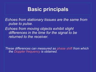
- 1. Basic principals Echoes from stationary tissues are the same from pulse to pulse. Echoes from moving objects exhibit slight differences in the time for the signal to be returned to the receiver. These differences can measured as phase shift from which the Doppler frequency is obtained.
- 2. (the angle q between the beam and the direction of flow becomes smaller). This is of the utmost importance in the use of Doppler ultrasound. Freq. qq The angle of insonation Flow velocity 3 2 1 Factors affecting doppler frequency
- 3. Basic Principals ‘Doppler frequency’ is obtained by measuring the time difference for the signal to be returned when reflected from moving scatterers . Doppler frequency increase if: 1. flow velocity increased . 2. beam is more aligned to the direction of flow. 3. higher transducer frequency is used.
- 4. (the angle q between the beam and the direction of flow becomes smaller). This is of the utmost importance in the use of Doppler ultrasound. beambeam (A)(A) is more aligned thanis more aligned than (B)(B) The beam/flow angle atThe beam/flow angle at (C)(C) is almost 90° and there is a very poor Doppler signalis almost 90° and there is a very poor Doppler signal The flow atThe flow at (D)(D) is away from the beam and there is a negative signal.is away from the beam and there is a negative signal.
- 10. Doppler indices
- 34. Blood flow velocities in the basal cerebral arteries according to age mean velocity [cm/s(95% CI)] artery velocity 20-39 y 40-59 y >60 y ACA peak 91 (87-95) 88 (83-93) 79 (75-84) mean 60 (57-62) 61 (57-64) 51 (48-54) ED 41 (39-43) 42 (40-45) 33 (31-35) MCA peak 113 (109-116) 106 (101-111) 92 (88-96) mean 74 (71-76) 72 (69-76) 58 (55-61) ED 51 (49-53) 47 (45-50) 35 (33-37) PCA (P1) peak 81 (78-84) 71 (68-74) 66 (63-69) mean 53 (51-55) 49 (48-51) 42 (40-45) ED 36 (35-38) 33 (31-35) 26 (24-28) CI indicated confidence interval; ACA, anterior cerebral artery, MCA, middle cerebral artery; PCA, posterior cerebral artery; ED, end-diastolic (all data from Martin et al. [1994]).
- 45. Classification of MCA degree of stenosis (Sliwka et al. 1997) peak flow velocity [cm/s] artery low- grade moderate severe MCA 140-180 181-220 > 220
- 51. TCD PREOPERATORIO / PREVISIONI SHUNT 4 CLASSI 1) DIMINUZIONE DELLA VELOCITA’ DI FLUSSO ALL’ MCA DEL 75% DOPO CHIUSURA DELLA CAROTIDE COMUNE + NON ATTIVAZIONE DEL CIRCOLO COLLATERALE + ANESTESIA GENERALE = PROBABILITA’ DI SHUNT DELL’ 88,9,9 % 2) DIMINUZIONE DELLA VELOCITA’ DI FLUSSO ALL’ MCA DEL 75% + ATTIVAZIONE DEL CIRCOLO COLLATERALE + ANESTESIA GENERALE = PROBABILITA’ DI SHUNT DELL’ 61,9 % 3) DIMINUZIONE DELLA VELOCITA’ DI FLUSSO ALL’ MCA DEL 75% + ATTIVAZIONE DEL CIRCOLO COLLATERALE + ANESTESIA LOCALE = PROBABILITA’ DI SHUNT DELL’ 34,4 % 4) DIMINUZIONE DELLA VELOCITA’ DI FLUSSO ALL’ MCA INFERIORE DEL 75% + ATTIVAZIONE DEL CIRCOLO COLLATERALE + ANESTESIA LOCALE = PROBABILITA’ DI SHUNT DELL’ 3,9 %
- 52. Pre-Anesthetic Baseline MCA Waveform Post-Clamping MCA Waveform Mean VMCA is 39 cm/sec at baseline and drops to 8 cm/sec after carotid clamping (21%). A shunt was inserted and the man VMCA increased to 22 cm/sec.
- 53. Baseline Waveform Post-Clamping Waveform Increased flow velocity after carotid clamping indicates that the posterior cerebral artery is being insonated. Note sample volume depth of 55 mm. The MCA signal was subsequently found at 50 mm.
- 56. A i r e m b o l i o c c u r r i n g Air emboli occurring immediately after release of the internal carotid artery clamp.
- 57. i g u r e 3 . M u l t i p l e p a r t i c u l a Multiple particulate embolic signatures in an MCA waveform.
- 61. Doppler Transcranico / Possibilità clinico diagnostiche Definite: •Stenosi emodinamiche dei vasi intracranici •Valutazione dei circoli collaterali •Valutazione dell’autoregolazione •Valutazione dell’emodinamica cerebrale in pz. con patologia vascolare extracranica •Diagnosi e follow-up del vasospasmo •Valutazione MAV e vasi coinvolti •Arresto del circolo cerebrale (diagnosi di morte cerebrale) •Monitoraggio intraoperatorio (TEA carotidea, BPAC) • Valutazione segnali microembolici
- 62. Doppler Transcranico / Possibilità clinico diagnostiche Non definite: •Studio dell’emicrania •Anemia a cellule falciformi •Infezioni meningee •Trombosi venosa cerebrale
Editor's Notes
- Emboli all’apertura dello shunt resosi necessario per l’azzeramento della velocità al clampaggio della carotide
- TAL 7