SlideShare a Scribd company logo
1 of 72
Biological Membrane & Transport
Table of contents
 Membrane character
 molecular constituent of
membrane
 supramolecular architecture of
membrane
 membrane motion
 membrane protein
 peripheral protein
 integral protein
 types of integral protein
 topology of integral protein
 cell-cell interaction & adhesion
 membrane fusion
 passive transport
 erythrocyte’s glucose transporter
 cotransport systems
 active transport
 types of active transporter
 Na+K+ATPase
 secondary active transport
 ion selective channels
Membrane (boundary)
 Flexible
 Self-sealing
 Selectively permeable
 Two-dimensional
Membrane components:
proteins, polar lipids and carbohydrates
Ratio of protein/lipids: depends on type & role of membrane
Supramolecular Architecture of Membranes
Common properties:
Impermeable
5 to 8 nm thick
Trilaminar
Asymmetric (structural & functional)
Fluid
Able to undergo fusion
Lipid aggregates: depending on the size of head & tail
Asymmetric Distribution of Phospholipids on Membrane
Membrane Motion
Conformational motion
Lateral diffusion
Flip-flop diffusion
Paracrystalline, transition temperature
Sterols moderate extremes of fluidity & solidity
High degree of regularity in one dimension &
Great mobility in the other
Transbilayer diffusion
flippases
Membrane proteins
Integral (intrinsic) proteins:
Firmly associated, removable only by agents that
interfere with hydrophobic interactions such as
detergents, organic solvents or denaturants.
Peripheral (extrinsic) proteins
Associated through electrostatic interactions &
hydrogen bonding with the hydrophilic domains of
integral proteins and with the polar head groups of
membrane lipids, readily removable by mild
treatments
Peripheral proteins anchored by
covalently attached lipids
Lipids with long-chain fatty acids, isoprenoids, or
glycosylated derivatives of phosphatidylinositol (GPI)
Proteins with GPI anchors are exclusively on the outer
face (extracellular), whereas other types of lipid-linked
proteins are found exclusively on the inner face
(cytosolic)
Integral proteins
Integral proteins are held in the membrane by hydrophobic
interactions with lipids, i.e., firmly fixed by interaction
between membrane lipids and hydrophobic domain of
proteins.
According to the spatial relationship of
protein domains to the lipid bilayer,
plasma membrane proteins fall into six
categories.
Types I & II have only one transmembrane
helix, the amino terminal domain is
outside the cell in type I proteins and
inside in type II.
Type III proteins have multiple
transmembrane helices in a single
polypeptide.
Type IV proteins have several transmembrane
domains from different polypeptide
chains to form a channel through the
membrane
Type V proteins are held to the bilayer
primarily by covalently linked lipid
Type VI proteins have both transmembrane
helices and lipid (GPI) anchors.
Topology of an integral protein
Hydropathy Plots
Glycophorin in the erythrocyte
For type III or IV proteins:
 -helical sequence of 20 to 25 amino acid residues
-sheet sequence of 7 to 9 amino acid residues
 -helical: average 3.6 amino acids for 0.54 nm
-sheet: average 2 amino acids for 0.65 to 0.7 nm
Porin FhuA, with 22 antiparallel  strands forming
channel for iron ion bound to the carrier ferrichrome
Cell-cell interaction &
adhesion
Integrins: heterodimeric, for binding collagen & fibronectin receptors &
signal transducers, and regulate platelet aggregation at the site
of a wound, tissue repair, activity of immune cells, and the
invasion of tissue by a tumor
Cadherins for homophilic interaction
Immunoglobulin-like protein for both homophilic & heterophilic interaction
Selectins for binding polysaccharides.
Membrane fusion
Specific fusion of two membranes requires:
a) They recognize each other
b) Their surfaces become closely apposed, which
requires the removal of water molecules normally
associated with the polar head groups of lipids
c) Their bilayer structures become locally disrupted
d) The two bilayers fuse to form a single continuous
bilayer
e) The fusion process is triggered at the appropriate
time or in response to a specific signal
Solute Transport across Membranes
Solute mediated by transmembrane channels, carriers or pumps
Passive transport & active transport
Passive Transport
Membrane potential (Vm) & electric gradient
Electrochemical gradient or electrochemical potential
Membrane: selectively permeable
The energy of activation for translocation of a polar solute across
the bilayer is so large that pure lipid bilayers are virtually
impermeable to polar and charged species over periods of time
relevant to cells.
Transmembrane passage of polar compounds and ions is made
possible by membrane proteins that lower the activation energy for
transport by providing an alternative path for specific solutes
through the lipid bilayer.
These proteins are called transporters or permeases.
Transporters span the lipid bilayers at least
once, and usually several times, forming a
transmembrane channel lined with hydrophilic
amino acid side chains. The channel provides
an alternative path for a specific substrate to
move across the lipid bilayer without its
having to dissolve in the bilayer, further
lowering the activation energy for
transmembrane diffusion.
Erythrocyte’s Glucose Transporter
Type III integral protein with MW of 45,000 and 12 hydrophobic
segments, each of which form a membrane-spanning helix
Hallmarks of passive transport
High rates of diffusion down a concentration gradient
Saturability
Specificity
Anion exchange protein
Obligatorily transport
one bicarbonate and
one chloride
Cotransport systems:
simultaneously carry two
solutes across a
membrane
Antiport systems: transporters that carry two substrates
moving in opposite directions
Symport systems: transporters that carry two substrates
moving simultaneously in the same direction.
Uniport systems: transporters that carry only one substrate
Active transport
Active transport results in solute movement against a
concentration or electrochemical gradient, thus it is
thermodynamically unfavorable or endergonic process,
and must be coupled with other energy releasing
processes.
Primary active transport (coupled with energy)
Secondary active transport (coupled with concentration flow)
(Major energy-consuming process)
Four types of transporters
Different in structure, mechanism, localization in specific tissues
and intracelluar compartments
1. P-type ATPase: ATP driven cation transporters, reversibly
phosphorylated by ATP during the transport cycle, with similar
amino acid sequence, can be inhibited by phosphate analog
vanadate. Generally have two types of integral protein
subunits. The  -subunit is essential, has Asp residue
phosphorylated during transport.
2. V-type ATPase: responsible for acidifying intracelluar
compartments in many organisms via proton-transporting, also
called proton pump. To acidify the vacuoles of fungi and higher
plants, as well as lysosomes, endosomes, the Golgi complex,
and secretory vesicles in animal cells. All have an integral
(transmembrane) domain as proton channel and a peripheral
domain containing the ATP-binding site and the ATPase activity.
3. F-type ATPase: central role in energy-conserving
reactions in bacteria, mitochondria and chloroplasts.
Catalyzes the uphill trans-membrane passage of
protons driven by ATP hydrolysis, as well as the
reverse reaction, in which downhill proton flow
drives ATP synthesis. (ATP synthases).
4. Multidrug transporter: responsible for removing
different drugs from tumor cell cytosol, preventing
their growth-inhibitory effect.
Na+K+ ATPase
P-type ATPase that cotransport Na+ and K+
Na+: lower in the cell than in the surrounding medium
K+: higher in the cell than in the surrounding medium
transmembrane potential of -50 to -70 mV
Na+K+ ATPase
Ion gradient for secondary active transport
Cells contain transport systems that couple the spontaneous,
downhill flow of Na+, H+ ions to the simultaneous uphill
pumping of another ion, sugar or amino acids.
For example, glucose:
Chemical potential
Electrical potential
Poison that collapses the
ion gradient across cellular
membrane
Ionophores, ion bearers
Ion selective channels
Move inorganic ions across membrane quickly.
Determine the plasma membrane’s permeability to specific
ions, and together with ion pumps such as Na/K ATPase,
regulate the cytosolic concentration of ions and the
membrane potential.
Characters: the rate of flux through channels can be orders
of magnitude greater than the turnover number for a
transporter, 107 to 108 ions per channel per second.
Not saturable
“Gated”, open or close in response to some cellular event
Ion channels
Ligand-gated channels: allosteric proteins change
conformation when bind to some extracellular or
intracellular small molecules
Acetylcholine receptor
Voltage-gated ion channels: response to a change in
transmembrane electrical potential
K+ channel
• LEHNINGER
• PRINCIPLES OF BIOCHEMISTRY
• Fifth Edition
David L. Nelson and Michael M. Cox
© 2008 W. H. Freeman and Company
CHAPTER 12
Biosignaling
Biosignaling
 Signal response pathways
 Types of signals
 Biosignaling characters
Signals from receptor to cell response
Autocrine: acting on the same cell that produces the signals
Paracrine: acting on a near neighbour
Endocrine: carried in the bloodstream from the producer cell to
a distant target cell.
The end result of a signaling pathway is the phosphorylation of
a few specific target-cell proteins, which changes their
activities and thus the activities of the cell
Characters of signal transduction
Ø Specificity: precise molecular complementarity
between the signal and receptor molecules,
mediated by weak forces occuring in the
enzyme-substrate, protein-ligand and antigen-
antibody interactions.
Ø Sensitivity
1) High affinity of receptors for signal molecules
2) Cooperativity in the ligand-receptor interaction
3) Amplification of the signal by enzyme cascades.
Ø Adaptation/Desensitization (saturation): When
receptor is continuously stimulated by
signal, the threshold would be leveled up.
ØIntegration: The ability of the system to receive
multiple signals and produce a unified
response appropriate to the needs of the cell
or organism.
membrane transport.ppt

More Related Content

Similar to membrane transport.ppt

Transport across cell membrane
Transport across cell membraneTransport across cell membrane
Transport across cell membraneJilsha Cecil
 
Ion Transport Through cell Membrane
Ion Transport Through cell MembraneIon Transport Through cell Membrane
Ion Transport Through cell MembraneMANISHSAHU106
 
Cell Biology Lecture 3
Cell Biology Lecture 3Cell Biology Lecture 3
Cell Biology Lecture 3Suk Namgoong
 
Transport mechanisms and their models.
Transport mechanisms and their models.Transport mechanisms and their models.
Transport mechanisms and their models.JyotiBishlay
 
Solute transport
Solute transport Solute transport
Solute transport satya patra
 
Unit 2-plasma membrane and membrane transport
Unit 2-plasma membrane and membrane transportUnit 2-plasma membrane and membrane transport
Unit 2-plasma membrane and membrane transportKomal Kp
 
Chapter 3 structure and functions
Chapter 3   structure and functionsChapter 3   structure and functions
Chapter 3 structure and functionsAlia Najiha
 
Cell membrane and transport
Cell membrane and transportCell membrane and transport
Cell membrane and transportAmit Kumar
 
Transport across cell membranes
Transport across cell membranesTransport across cell membranes
Transport across cell membranesOla Elgaddar
 
2227_Unit 1Transport accross membrane (7).pptx
2227_Unit 1Transport accross membrane (7).pptx2227_Unit 1Transport accross membrane (7).pptx
2227_Unit 1Transport accross membrane (7).pptxfarihahks
 
Membrane Dynamics, Part 2
Membrane Dynamics, Part 2Membrane Dynamics, Part 2
Membrane Dynamics, Part 2raj kumar
 
Membrane Dynamics, Part 2
Membrane Dynamics, Part 2Membrane Dynamics, Part 2
Membrane Dynamics, Part 2raj kumar
 
2. Absorption & Secretion Of Materials
2. Absorption & Secretion Of Materials2. Absorption & Secretion Of Materials
2. Absorption & Secretion Of Materialsrossbiology
 
Cell & cell membrane
Cell &  cell membraneCell &  cell membrane
Cell & cell membraneDrmohamed Badr
 
form4(BIOLOGY) chap3 pt1
form4(BIOLOGY) chap3 pt1form4(BIOLOGY) chap3 pt1
form4(BIOLOGY) chap3 pt1cikgushaik
 
Subtopic 3 1[1] FORM 5
Subtopic 3 1[1] FORM 5Subtopic 3 1[1] FORM 5
Subtopic 3 1[1] FORM 5cikgushaik
 
Structure, Chemical Properties, and Function of Proteins, Intracellular Traff...
Structure, Chemical Properties, and Function of Proteins, Intracellular Traff...Structure, Chemical Properties, and Function of Proteins, Intracellular Traff...
Structure, Chemical Properties, and Function of Proteins, Intracellular Traff...OMEED AKBAR
 
Membrane transport
Membrane transportMembrane transport
Membrane transportMir Ahmed
 

Similar to membrane transport.ppt (20)

Transport across cell membrane
Transport across cell membraneTransport across cell membrane
Transport across cell membrane
 
Cell transport
Cell transportCell transport
Cell transport
 
Ion Transport Through cell Membrane
Ion Transport Through cell MembraneIon Transport Through cell Membrane
Ion Transport Through cell Membrane
 
Cell Biology Lecture 3
Cell Biology Lecture 3Cell Biology Lecture 3
Cell Biology Lecture 3
 
Transport mechanisms and their models.
Transport mechanisms and their models.Transport mechanisms and their models.
Transport mechanisms and their models.
 
Solute transport
Solute transport Solute transport
Solute transport
 
Transport across Plasma Membrane
Transport across Plasma Membrane Transport across Plasma Membrane
Transport across Plasma Membrane
 
Unit 2-plasma membrane and membrane transport
Unit 2-plasma membrane and membrane transportUnit 2-plasma membrane and membrane transport
Unit 2-plasma membrane and membrane transport
 
Chapter 3 structure and functions
Chapter 3   structure and functionsChapter 3   structure and functions
Chapter 3 structure and functions
 
Cell membrane and transport
Cell membrane and transportCell membrane and transport
Cell membrane and transport
 
Transport across cell membranes
Transport across cell membranesTransport across cell membranes
Transport across cell membranes
 
2227_Unit 1Transport accross membrane (7).pptx
2227_Unit 1Transport accross membrane (7).pptx2227_Unit 1Transport accross membrane (7).pptx
2227_Unit 1Transport accross membrane (7).pptx
 
Membrane Dynamics, Part 2
Membrane Dynamics, Part 2Membrane Dynamics, Part 2
Membrane Dynamics, Part 2
 
Membrane Dynamics, Part 2
Membrane Dynamics, Part 2Membrane Dynamics, Part 2
Membrane Dynamics, Part 2
 
2. Absorption & Secretion Of Materials
2. Absorption & Secretion Of Materials2. Absorption & Secretion Of Materials
2. Absorption & Secretion Of Materials
 
Cell & cell membrane
Cell &  cell membraneCell &  cell membrane
Cell & cell membrane
 
form4(BIOLOGY) chap3 pt1
form4(BIOLOGY) chap3 pt1form4(BIOLOGY) chap3 pt1
form4(BIOLOGY) chap3 pt1
 
Subtopic 3 1[1] FORM 5
Subtopic 3 1[1] FORM 5Subtopic 3 1[1] FORM 5
Subtopic 3 1[1] FORM 5
 
Structure, Chemical Properties, and Function of Proteins, Intracellular Traff...
Structure, Chemical Properties, and Function of Proteins, Intracellular Traff...Structure, Chemical Properties, and Function of Proteins, Intracellular Traff...
Structure, Chemical Properties, and Function of Proteins, Intracellular Traff...
 
Membrane transport
Membrane transportMembrane transport
Membrane transport
 

Recently uploaded

Biogenic Sulfur Gases as Biosignatures on Temperate Sub-Neptune Waterworlds
Biogenic Sulfur Gases as Biosignatures on Temperate Sub-Neptune WaterworldsBiogenic Sulfur Gases as Biosignatures on Temperate Sub-Neptune Waterworlds
Biogenic Sulfur Gases as Biosignatures on Temperate Sub-Neptune WaterworldsSérgio Sacani
 
Terpineol and it's characterization pptx
Terpineol and it's characterization pptxTerpineol and it's characterization pptx
Terpineol and it's characterization pptxMuhammadRazzaq31
 
Dr. E. Muralinath_ Blood indices_clinical aspects
Dr. E. Muralinath_ Blood indices_clinical  aspectsDr. E. Muralinath_ Blood indices_clinical  aspects
Dr. E. Muralinath_ Blood indices_clinical aspectsmuralinath2
 
FAIRSpectra - Enabling the FAIRification of Analytical Science
FAIRSpectra - Enabling the FAIRification of Analytical ScienceFAIRSpectra - Enabling the FAIRification of Analytical Science
FAIRSpectra - Enabling the FAIRification of Analytical ScienceAlex Henderson
 
Genome Projects : Human, Rice,Wheat,E coli and Arabidopsis.
Genome Projects : Human, Rice,Wheat,E coli and Arabidopsis.Genome Projects : Human, Rice,Wheat,E coli and Arabidopsis.
Genome Projects : Human, Rice,Wheat,E coli and Arabidopsis.Cherry
 
TransientOffsetin14CAftertheCarringtonEventRecordedbyPolarTreeRings
TransientOffsetin14CAftertheCarringtonEventRecordedbyPolarTreeRingsTransientOffsetin14CAftertheCarringtonEventRecordedbyPolarTreeRings
TransientOffsetin14CAftertheCarringtonEventRecordedbyPolarTreeRingsSérgio Sacani
 
Thyroid Physiology_Dr.E. Muralinath_ Associate Professor
Thyroid Physiology_Dr.E. Muralinath_ Associate ProfessorThyroid Physiology_Dr.E. Muralinath_ Associate Professor
Thyroid Physiology_Dr.E. Muralinath_ Associate Professormuralinath2
 
GBSN - Biochemistry (Unit 2) Basic concept of organic chemistry
GBSN - Biochemistry (Unit 2) Basic concept of organic chemistry GBSN - Biochemistry (Unit 2) Basic concept of organic chemistry
GBSN - Biochemistry (Unit 2) Basic concept of organic chemistry Areesha Ahmad
 
Pteris : features, anatomy, morphology and lifecycle
Pteris : features, anatomy, morphology and lifecyclePteris : features, anatomy, morphology and lifecycle
Pteris : features, anatomy, morphology and lifecycleCherry
 
Role of AI in seed science Predictive modelling and Beyond.pptx
Role of AI in seed science  Predictive modelling and  Beyond.pptxRole of AI in seed science  Predictive modelling and  Beyond.pptx
Role of AI in seed science Predictive modelling and Beyond.pptxArvind Kumar
 
CURRENT SCENARIO OF POULTRY PRODUCTION IN INDIA
CURRENT SCENARIO OF POULTRY PRODUCTION IN INDIACURRENT SCENARIO OF POULTRY PRODUCTION IN INDIA
CURRENT SCENARIO OF POULTRY PRODUCTION IN INDIADr. TATHAGAT KHOBRAGADE
 
Call Girls Ahmedabad +917728919243 call me Independent Escort Service
Call Girls Ahmedabad +917728919243 call me Independent Escort ServiceCall Girls Ahmedabad +917728919243 call me Independent Escort Service
Call Girls Ahmedabad +917728919243 call me Independent Escort Serviceshivanisharma5244
 
Human & Veterinary Respiratory Physilogy_DR.E.Muralinath_Associate Professor....
Human & Veterinary Respiratory Physilogy_DR.E.Muralinath_Associate Professor....Human & Veterinary Respiratory Physilogy_DR.E.Muralinath_Associate Professor....
Human & Veterinary Respiratory Physilogy_DR.E.Muralinath_Associate Professor....muralinath2
 
COMPOSTING : types of compost, merits and demerits
COMPOSTING : types of compost, merits and demeritsCOMPOSTING : types of compost, merits and demerits
COMPOSTING : types of compost, merits and demeritsCherry
 
ONLINE VOTING SYSTEM SE Project for vote
ONLINE VOTING SYSTEM SE Project for voteONLINE VOTING SYSTEM SE Project for vote
ONLINE VOTING SYSTEM SE Project for voteRaunakRastogi4
 
Selaginella: features, morphology ,anatomy and reproduction.
Selaginella: features, morphology ,anatomy and reproduction.Selaginella: features, morphology ,anatomy and reproduction.
Selaginella: features, morphology ,anatomy and reproduction.Cherry
 
Bhiwandi Bhiwandi ❤CALL GIRL 7870993772 ❤CALL GIRLS ESCORT SERVICE In Bhiwan...
Bhiwandi Bhiwandi ❤CALL GIRL 7870993772 ❤CALL GIRLS  ESCORT SERVICE In Bhiwan...Bhiwandi Bhiwandi ❤CALL GIRL 7870993772 ❤CALL GIRLS  ESCORT SERVICE In Bhiwan...
Bhiwandi Bhiwandi ❤CALL GIRL 7870993772 ❤CALL GIRLS ESCORT SERVICE In Bhiwan...Monika Rani
 
Digital Dentistry.Digital Dentistryvv.pptx
Digital Dentistry.Digital Dentistryvv.pptxDigital Dentistry.Digital Dentistryvv.pptx
Digital Dentistry.Digital Dentistryvv.pptxMohamedFarag457087
 
CYTOGENETIC MAP................ ppt.pptx
CYTOGENETIC MAP................ ppt.pptxCYTOGENETIC MAP................ ppt.pptx
CYTOGENETIC MAP................ ppt.pptxCherry
 

Recently uploaded (20)

Biogenic Sulfur Gases as Biosignatures on Temperate Sub-Neptune Waterworlds
Biogenic Sulfur Gases as Biosignatures on Temperate Sub-Neptune WaterworldsBiogenic Sulfur Gases as Biosignatures on Temperate Sub-Neptune Waterworlds
Biogenic Sulfur Gases as Biosignatures on Temperate Sub-Neptune Waterworlds
 
PATNA CALL GIRLS 8617370543 LOW PRICE ESCORT SERVICE
PATNA CALL GIRLS 8617370543 LOW PRICE ESCORT SERVICEPATNA CALL GIRLS 8617370543 LOW PRICE ESCORT SERVICE
PATNA CALL GIRLS 8617370543 LOW PRICE ESCORT SERVICE
 
Terpineol and it's characterization pptx
Terpineol and it's characterization pptxTerpineol and it's characterization pptx
Terpineol and it's characterization pptx
 
Dr. E. Muralinath_ Blood indices_clinical aspects
Dr. E. Muralinath_ Blood indices_clinical  aspectsDr. E. Muralinath_ Blood indices_clinical  aspects
Dr. E. Muralinath_ Blood indices_clinical aspects
 
FAIRSpectra - Enabling the FAIRification of Analytical Science
FAIRSpectra - Enabling the FAIRification of Analytical ScienceFAIRSpectra - Enabling the FAIRification of Analytical Science
FAIRSpectra - Enabling the FAIRification of Analytical Science
 
Genome Projects : Human, Rice,Wheat,E coli and Arabidopsis.
Genome Projects : Human, Rice,Wheat,E coli and Arabidopsis.Genome Projects : Human, Rice,Wheat,E coli and Arabidopsis.
Genome Projects : Human, Rice,Wheat,E coli and Arabidopsis.
 
TransientOffsetin14CAftertheCarringtonEventRecordedbyPolarTreeRings
TransientOffsetin14CAftertheCarringtonEventRecordedbyPolarTreeRingsTransientOffsetin14CAftertheCarringtonEventRecordedbyPolarTreeRings
TransientOffsetin14CAftertheCarringtonEventRecordedbyPolarTreeRings
 
Thyroid Physiology_Dr.E. Muralinath_ Associate Professor
Thyroid Physiology_Dr.E. Muralinath_ Associate ProfessorThyroid Physiology_Dr.E. Muralinath_ Associate Professor
Thyroid Physiology_Dr.E. Muralinath_ Associate Professor
 
GBSN - Biochemistry (Unit 2) Basic concept of organic chemistry
GBSN - Biochemistry (Unit 2) Basic concept of organic chemistry GBSN - Biochemistry (Unit 2) Basic concept of organic chemistry
GBSN - Biochemistry (Unit 2) Basic concept of organic chemistry
 
Pteris : features, anatomy, morphology and lifecycle
Pteris : features, anatomy, morphology and lifecyclePteris : features, anatomy, morphology and lifecycle
Pteris : features, anatomy, morphology and lifecycle
 
Role of AI in seed science Predictive modelling and Beyond.pptx
Role of AI in seed science  Predictive modelling and  Beyond.pptxRole of AI in seed science  Predictive modelling and  Beyond.pptx
Role of AI in seed science Predictive modelling and Beyond.pptx
 
CURRENT SCENARIO OF POULTRY PRODUCTION IN INDIA
CURRENT SCENARIO OF POULTRY PRODUCTION IN INDIACURRENT SCENARIO OF POULTRY PRODUCTION IN INDIA
CURRENT SCENARIO OF POULTRY PRODUCTION IN INDIA
 
Call Girls Ahmedabad +917728919243 call me Independent Escort Service
Call Girls Ahmedabad +917728919243 call me Independent Escort ServiceCall Girls Ahmedabad +917728919243 call me Independent Escort Service
Call Girls Ahmedabad +917728919243 call me Independent Escort Service
 
Human & Veterinary Respiratory Physilogy_DR.E.Muralinath_Associate Professor....
Human & Veterinary Respiratory Physilogy_DR.E.Muralinath_Associate Professor....Human & Veterinary Respiratory Physilogy_DR.E.Muralinath_Associate Professor....
Human & Veterinary Respiratory Physilogy_DR.E.Muralinath_Associate Professor....
 
COMPOSTING : types of compost, merits and demerits
COMPOSTING : types of compost, merits and demeritsCOMPOSTING : types of compost, merits and demerits
COMPOSTING : types of compost, merits and demerits
 
ONLINE VOTING SYSTEM SE Project for vote
ONLINE VOTING SYSTEM SE Project for voteONLINE VOTING SYSTEM SE Project for vote
ONLINE VOTING SYSTEM SE Project for vote
 
Selaginella: features, morphology ,anatomy and reproduction.
Selaginella: features, morphology ,anatomy and reproduction.Selaginella: features, morphology ,anatomy and reproduction.
Selaginella: features, morphology ,anatomy and reproduction.
 
Bhiwandi Bhiwandi ❤CALL GIRL 7870993772 ❤CALL GIRLS ESCORT SERVICE In Bhiwan...
Bhiwandi Bhiwandi ❤CALL GIRL 7870993772 ❤CALL GIRLS  ESCORT SERVICE In Bhiwan...Bhiwandi Bhiwandi ❤CALL GIRL 7870993772 ❤CALL GIRLS  ESCORT SERVICE In Bhiwan...
Bhiwandi Bhiwandi ❤CALL GIRL 7870993772 ❤CALL GIRLS ESCORT SERVICE In Bhiwan...
 
Digital Dentistry.Digital Dentistryvv.pptx
Digital Dentistry.Digital Dentistryvv.pptxDigital Dentistry.Digital Dentistryvv.pptx
Digital Dentistry.Digital Dentistryvv.pptx
 
CYTOGENETIC MAP................ ppt.pptx
CYTOGENETIC MAP................ ppt.pptxCYTOGENETIC MAP................ ppt.pptx
CYTOGENETIC MAP................ ppt.pptx
 

membrane transport.ppt

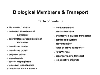
  • 1. Biological Membrane & Transport Table of contents  Membrane character  molecular constituent of membrane  supramolecular architecture of membrane  membrane motion  membrane protein  peripheral protein  integral protein  types of integral protein  topology of integral protein  cell-cell interaction & adhesion  membrane fusion  passive transport  erythrocyte’s glucose transporter  cotransport systems  active transport  types of active transporter  Na+K+ATPase  secondary active transport  ion selective channels
  • 2. Membrane (boundary)  Flexible  Self-sealing  Selectively permeable  Two-dimensional
  • 3.
  • 4. Membrane components: proteins, polar lipids and carbohydrates Ratio of protein/lipids: depends on type & role of membrane
  • 5.
  • 6. Supramolecular Architecture of Membranes Common properties: Impermeable 5 to 8 nm thick Trilaminar Asymmetric (structural & functional) Fluid Able to undergo fusion
  • 7. Lipid aggregates: depending on the size of head & tail
  • 8.
  • 9.
  • 10. Asymmetric Distribution of Phospholipids on Membrane
  • 11. Membrane Motion Conformational motion Lateral diffusion Flip-flop diffusion Paracrystalline, transition temperature Sterols moderate extremes of fluidity & solidity High degree of regularity in one dimension & Great mobility in the other Transbilayer diffusion flippases
  • 12.
  • 13.
  • 14.
  • 15.
  • 16.
  • 17.
  • 18. Membrane proteins Integral (intrinsic) proteins: Firmly associated, removable only by agents that interfere with hydrophobic interactions such as detergents, organic solvents or denaturants. Peripheral (extrinsic) proteins Associated through electrostatic interactions & hydrogen bonding with the hydrophilic domains of integral proteins and with the polar head groups of membrane lipids, readily removable by mild treatments
  • 19.
  • 20.
  • 21. Peripheral proteins anchored by covalently attached lipids Lipids with long-chain fatty acids, isoprenoids, or glycosylated derivatives of phosphatidylinositol (GPI) Proteins with GPI anchors are exclusively on the outer face (extracellular), whereas other types of lipid-linked proteins are found exclusively on the inner face (cytosolic)
  • 22.
  • 23. Integral proteins Integral proteins are held in the membrane by hydrophobic interactions with lipids, i.e., firmly fixed by interaction between membrane lipids and hydrophobic domain of proteins. According to the spatial relationship of protein domains to the lipid bilayer, plasma membrane proteins fall into six categories.
  • 24. Types I & II have only one transmembrane helix, the amino terminal domain is outside the cell in type I proteins and inside in type II. Type III proteins have multiple transmembrane helices in a single polypeptide. Type IV proteins have several transmembrane domains from different polypeptide chains to form a channel through the membrane Type V proteins are held to the bilayer primarily by covalently linked lipid Type VI proteins have both transmembrane helices and lipid (GPI) anchors.
  • 25. Topology of an integral protein
  • 27. Glycophorin in the erythrocyte
  • 28. For type III or IV proteins:  -helical sequence of 20 to 25 amino acid residues -sheet sequence of 7 to 9 amino acid residues  -helical: average 3.6 amino acids for 0.54 nm -sheet: average 2 amino acids for 0.65 to 0.7 nm
  • 29. Porin FhuA, with 22 antiparallel  strands forming channel for iron ion bound to the carrier ferrichrome
  • 30. Cell-cell interaction & adhesion Integrins: heterodimeric, for binding collagen & fibronectin receptors & signal transducers, and regulate platelet aggregation at the site of a wound, tissue repair, activity of immune cells, and the invasion of tissue by a tumor Cadherins for homophilic interaction Immunoglobulin-like protein for both homophilic & heterophilic interaction Selectins for binding polysaccharides.
  • 31. Membrane fusion Specific fusion of two membranes requires: a) They recognize each other b) Their surfaces become closely apposed, which requires the removal of water molecules normally associated with the polar head groups of lipids c) Their bilayer structures become locally disrupted d) The two bilayers fuse to form a single continuous bilayer e) The fusion process is triggered at the appropriate time or in response to a specific signal
  • 32.
  • 33. Solute Transport across Membranes Solute mediated by transmembrane channels, carriers or pumps Passive transport & active transport
  • 34. Passive Transport Membrane potential (Vm) & electric gradient Electrochemical gradient or electrochemical potential
  • 35.
  • 36.
  • 37. Membrane: selectively permeable The energy of activation for translocation of a polar solute across the bilayer is so large that pure lipid bilayers are virtually impermeable to polar and charged species over periods of time relevant to cells. Transmembrane passage of polar compounds and ions is made possible by membrane proteins that lower the activation energy for transport by providing an alternative path for specific solutes through the lipid bilayer. These proteins are called transporters or permeases.
  • 38. Transporters span the lipid bilayers at least once, and usually several times, forming a transmembrane channel lined with hydrophilic amino acid side chains. The channel provides an alternative path for a specific substrate to move across the lipid bilayer without its having to dissolve in the bilayer, further lowering the activation energy for transmembrane diffusion.
  • 39.
  • 40.
  • 41.
  • 42. Erythrocyte’s Glucose Transporter Type III integral protein with MW of 45,000 and 12 hydrophobic segments, each of which form a membrane-spanning helix
  • 43.
  • 44.
  • 45.
  • 46.
  • 47.
  • 48. Hallmarks of passive transport High rates of diffusion down a concentration gradient Saturability Specificity
  • 49. Anion exchange protein Obligatorily transport one bicarbonate and one chloride Cotransport systems: simultaneously carry two solutes across a membrane
  • 50.
  • 51. Antiport systems: transporters that carry two substrates moving in opposite directions Symport systems: transporters that carry two substrates moving simultaneously in the same direction. Uniport systems: transporters that carry only one substrate
  • 52. Active transport Active transport results in solute movement against a concentration or electrochemical gradient, thus it is thermodynamically unfavorable or endergonic process, and must be coupled with other energy releasing processes. Primary active transport (coupled with energy) Secondary active transport (coupled with concentration flow) (Major energy-consuming process)
  • 53. Four types of transporters Different in structure, mechanism, localization in specific tissues and intracelluar compartments
  • 54. 1. P-type ATPase: ATP driven cation transporters, reversibly phosphorylated by ATP during the transport cycle, with similar amino acid sequence, can be inhibited by phosphate analog vanadate. Generally have two types of integral protein subunits. The  -subunit is essential, has Asp residue phosphorylated during transport. 2. V-type ATPase: responsible for acidifying intracelluar compartments in many organisms via proton-transporting, also called proton pump. To acidify the vacuoles of fungi and higher plants, as well as lysosomes, endosomes, the Golgi complex, and secretory vesicles in animal cells. All have an integral (transmembrane) domain as proton channel and a peripheral domain containing the ATP-binding site and the ATPase activity.
  • 55. 3. F-type ATPase: central role in energy-conserving reactions in bacteria, mitochondria and chloroplasts. Catalyzes the uphill trans-membrane passage of protons driven by ATP hydrolysis, as well as the reverse reaction, in which downhill proton flow drives ATP synthesis. (ATP synthases). 4. Multidrug transporter: responsible for removing different drugs from tumor cell cytosol, preventing their growth-inhibitory effect.
  • 56. Na+K+ ATPase P-type ATPase that cotransport Na+ and K+ Na+: lower in the cell than in the surrounding medium K+: higher in the cell than in the surrounding medium transmembrane potential of -50 to -70 mV
  • 58.
  • 59. Ion gradient for secondary active transport Cells contain transport systems that couple the spontaneous, downhill flow of Na+, H+ ions to the simultaneous uphill pumping of another ion, sugar or amino acids. For example, glucose: Chemical potential Electrical potential Poison that collapses the ion gradient across cellular membrane Ionophores, ion bearers
  • 60. Ion selective channels Move inorganic ions across membrane quickly. Determine the plasma membrane’s permeability to specific ions, and together with ion pumps such as Na/K ATPase, regulate the cytosolic concentration of ions and the membrane potential. Characters: the rate of flux through channels can be orders of magnitude greater than the turnover number for a transporter, 107 to 108 ions per channel per second. Not saturable “Gated”, open or close in response to some cellular event
  • 61. Ion channels Ligand-gated channels: allosteric proteins change conformation when bind to some extracellular or intracellular small molecules Acetylcholine receptor Voltage-gated ion channels: response to a change in transmembrane electrical potential K+ channel
  • 62. • LEHNINGER • PRINCIPLES OF BIOCHEMISTRY • Fifth Edition David L. Nelson and Michael M. Cox © 2008 W. H. Freeman and Company CHAPTER 12 Biosignaling
  • 63. Biosignaling  Signal response pathways  Types of signals  Biosignaling characters
  • 64. Signals from receptor to cell response Autocrine: acting on the same cell that produces the signals Paracrine: acting on a near neighbour Endocrine: carried in the bloodstream from the producer cell to a distant target cell.
  • 65.
  • 66. The end result of a signaling pathway is the phosphorylation of a few specific target-cell proteins, which changes their activities and thus the activities of the cell
  • 67. Characters of signal transduction Ø Specificity: precise molecular complementarity between the signal and receptor molecules, mediated by weak forces occuring in the enzyme-substrate, protein-ligand and antigen- antibody interactions. Ø Sensitivity 1) High affinity of receptors for signal molecules 2) Cooperativity in the ligand-receptor interaction 3) Amplification of the signal by enzyme cascades.
  • 68. Ø Adaptation/Desensitization (saturation): When receptor is continuously stimulated by signal, the threshold would be leveled up. ØIntegration: The ability of the system to receive multiple signals and produce a unified response appropriate to the needs of the cell or organism.

Editor's Notes

  1. FIGURE 11-3 Fluid mosaic model for membrane structure. The fatty acyl chains in the interior of the membrane form a fluid, hydrophobic region. Integral proteins float in this sea of lipid, held by hydrophobic interactions with their nonpolar amino acid side chains. Both proteins and lipids are free to move laterally in the plane of the bilayer, but movement of either from one leaflet of the bilayer to the other is restricted. The carbohydrate moieties attached to some proteins and lipids of the plasma membrane are exposed on the extracellular surface of the membrane.
  2. FIGURE 11-2 Lipid composition of the plasma membrane and organelle membranes of a rat hepatocyte. The functional specialization of each membrane type is reflected in its unique lipid composition. Cholesterol is prominent in plasma membranes but barely detectable in mitochondrial membranes. Cardiolipin is a major component of the inner mitochondrial membrane but not of the plasma membrane. Phosphatidylserine, phosphatidylinositol, and phosphatidylglycerol are relatively minor components (yellow) of most membranes but serve critical functions; phosphatidylinositol and its derivatives, for example, are important in signal transductions triggered by hormones. Sphingolipids, phosphatidylcholine, and phosphatidylethanolamine are present in most membranes, but in varying proportions. Glycolipids, which are major components of the chloroplast membranes of plants, are virtually absent from animal cells.
  3. FIGURE 11-4a Amphipathic lipid aggregates that form in water. (a) In micelles, the hydrophobic chains of the fatty acids are sequestered at the core of the sphere. There is virtually no water in the hydrophobic interior.
  4. FIGURE 11-4b Amphipathic lipid aggregates that form in water. (b) In an open bilayer, all acyl side chains except those at the edges of the sheet are protected from interaction with water.
  5. FIGURE 11-4c Amphipathic lipid aggregates that form in water. (c) When a two-dimensional bilayer folds on itself, it forms a closed bilayer, a three-dimensional hollow vesicle (liposome) enclosing an aqueous cavity.
  6. FIGURE 11-5 Asymmetric distribution of phospholipids between the inner and outer monolayers of the erythrocyte plasma membrane. The distribution of a specific phospholipid is determined by treating the intact cell with phospholipase C, which cannot reach lipids in the inner monolayer (leaflet) but removes the head groups of lipids in the outer monolayer. The proportion of each head group released provides an estimate of the fraction of each lipid in the outer monolayer.
  7. FIGURE 11-15 Two extreme states of bilayer lipids. (a) In the paracrystalline state, or gel phase, polar head groups are uniformly arrayed at the surface, and the acyl chains are nearly motionless and packed with regular geometry. (b) In the liquid-disordered state, or fluid state, acyl chains undergo much thermal motion and have no regular organization. Intermediate between these extremes is the liquid-ordered state, in which individual phospholipid molecules can diffuse laterally but the acyl groups remain extended and more or less ordered.
  8. FIGURE 11-16 Motion of single phospholipids in a bilayer. (a) Uncatalyzed movement from one leaflet to the other is very slow, but (b) lateral diffusion within the leaflet is very rapid, requiring no catalysis. (c) Three types of phospholipid translocaters in the plasma membrane. Flippases translocate primarily aminophospholipids (phosphatidylethanolamine (PE), phosphatidylserine (PS)) from the outer (exoplasmic) leaflet to the inner (cytosolic) leaflet; they require ATP and are members of the P-type ATPase family. Floppases move phospholipids from the cytosolic to the outer leaflet, require ATP, and are members of the ABC transporter family. Scramblases equilibrate phospholipids across both leaflets; they do not require ATP but are activated by Ca2+.
  9. FIGURE 11-16a Motion of single phospholipids in a bilayer. (a) Uncatalyzed movement from one leaflet to the other is very slow, but (b) lateral diffusion within the leaflet is very rapid, requiring no catalysis.
  10. FIGURE 11-16b Motion of single phospholipids in a bilayer. (a) Uncatalyzed movement from one leaflet to the other is very slow, but (b) lateral diffusion within the leaflet is very rapid, requiring no catalysis.
  11. FIGURE 11-16c Motion of single phospholipids in a bilayer. (c) Three types of phospholipid translocaters in the plasma membrane. Flippases translocate primarily aminophospholipids (phosphatidylethanolamine (PE), phosphatidylserine (PS)) from the outer (exoplasmic) leaflet to the inner (cytosolic) leaflet; they require ATP and are members of the P-type ATPase family. Floppases move phospholipids from the cytosolic to the outer leaflet, require ATP, and are members of the ABC transporter family. Scramblases equilibrate phospholipids across both leaflets; they do not require ATP but are activated by Ca2+.
  12. FIGURE 11-6 Peripheral, integral, and amphitropic proteins. Membrane proteins can be operationally distinguished by the conditions required to release them from the membrane. Most peripheral proteins are released by changes in pH or ionic strength, removal of Ca2+ by a chelating agent, or addition of urea or carbonate. Integral proteins are extractable with detergents, which disrupt the hydrophobic interactions with the lipid bilayer and form micelle-like clusters around individual protein molecules. Integral proteins covalently attached to a membrane lipid, such as a glycosyl phosphatidylinositol (GPI; see Figure 11-14), can be released by treatment with phospholipase C. Amphitropic proteins are sometimes associated with membranes and sometimes not, depending on some type of regulatory process such as reversible palmitoylation.
  13. FIGURE 11-7 Transbilayer disposition of glycophorin in an erythrocyte. One hydrophilic domain, containing all the sugar residues, is on the outer surface, and another hydrophilic domain protrudes from the inner face of the membrane. Each red hexagon represents a tetrasaccharide (containing two Neu5Ac (sialic acid), Gal, and GalNAc) Olinked to a Ser or Thr residue; the blue hexagon represents an oligosaccharide N-linked to an Asn residue. The relative size of the oligosaccharide units is larger than shown here. A segment of 19 hydrophobic residues (residues 75 to 93) forms an α helix that traverses the membrane bilayer (see Figure 11-11a). The segment from residues 64 to 74 has some hydrophobic residues and probably penetrates the outer face of the lipid bilayer, as shown.
  14. FIGURE 11-8 Integral membrane proteins. For known proteins of the plasma membrane, the spatial relationships of protein domains to the lipid bilayer fall into six categories. Types I and II have a single transmembrane helix; the amino-terminal domain is outside the cell in type I proteins and inside in type II. Type III proteins have multiple transmembrane helices in a single polypeptide. In type IV proteins, transmembrane domains of several different polypeptides assemble to form a channel through the membrane. Type V proteins are held to the bilayer primarily by covalently linked lipids (see Figure 11-14), and type VI proteins have both transmembrane helices and lipid (GPI) anchors. In this figure, and in figures throughout the book, we represent transmembrane protein segments in their most likely conformations: as α helices of six to seven turns. Sometimes these helices are shown simply as cylinders. As relatively few membrane protein structures have been deduced by x-ray crystallography, our representation of the extramembrane domains is arbitrary and not necessarily to scale.
  15. FIGURE 11-11 Hydropathy plots. Hydropathy index (see Table 3-1) is plotted against residue number for two integral membrane proteins. The hydropathy index for each amino acid residue in a sequence of defined length, or "window," is used to calculate the average hydropathy for that window. The horizontal axis shows the residue number in the middle of the window. (a) Glycophorin from human erythrocytes has a single hydrophobic sequence between residues 75 and 93 (yellow); compare this with Figure 11-7. (b) Bacteriorhodopsin, known from independent physical studies to have seven transmembrane helices (see Figure 11-9), has seven hydrophobic regions. Note, however, that the hydropathy plot is ambiguous in the region of segments 6 and 7. X-ray crystallography has confirmed that this region has two transmembrane segments.
  16. FIGURE 11-22 Membrane fusion. The fusion of two membranes is central to a variety of cellular processes involving organelles and the plasma membrane.
  17. FIGURE 11-26a Movement of solutes across a permeable membrane. (a) Net movement of an electrically neutral solute is toward the side of lower solute concentration until equilibrium is achieved. The solute concentrations on the left and right sides of the membrane are designated C1 and C2. The rate of transmembrane movement (indicated by the arrows) is proportional to the concentration gradient, C2/C1.
  18. FIGURE 11-26b Movement of solutes across a permeable membrane. (b) Net movement of an electrically charged solute is dictated by a combination of the electrical potential (Vm) and the chemical concentration difference (C2/C1) across the membrane; net ion movement continues until this electrochemical potential reaches zero.
  19. FIGURE 11-27 Energy changes accompanying passage of a hydrophilic solute through the lipid bilayer of a biological membrane. (a) In simple diffusion, removal of the hydration shell is highly endergonic, and the energy of activation (ΔG‡) for diffusion through the bilayer is very high. (b) A transporter protein reduces the ΔG‡ for transmembrane diffusion of the solute. It does this by forming noncovalent interactions with the dehydrated solute to replace the hydrogen bonding with water and by providing a hydrophilic transmembrane pathway.
  20. FIGURE 11-28 Classification of transporters
  21. FIGURE 11-29a Proposed structure of GLUT1. (a) Transmembrane helices are represented here as oblique (angled) rows of three or four amino acid residues, each row depicting one turn of the α helix. Nine of the 12 helices contain three or more polar or charged residues (blue or red), often separated by several hydrophobic residues (yellow). This representation of topology is not intended to represent three-dimensional structure.
  22. FIGURE 11-29b Proposed structure of GLUT1. (b) A helical wheel diagram shows the distribution of polar and nonpolar residues on the surface of a helical segment. The helix is diagrammed as though observed along its axis from the amino terminus. Adjacent residues in the linear sequence are connected, and each residue is placed around the wheel in the position it occupies in the helix; recall that 3.6 residues are required to make one complete turn of the α helix. In this example, the polar residues (blue) are on one side of the helix and the hydrophobic residues (yellow) on the other. This is, by definition, an amphipathic helix.
  23. FIGURE 11-29c Proposed structure of GLUT1. (c) Side-by-side association of four amphipathic helices, each with its polar face oriented toward the central cavity, can produce a transmembrane channel lined with polar (and charged) residues. This channel provides many opportunities for hydrogen bonding with glucose as it moves through.
  24. FIGURE 11-30a Kinetics of glucose transport into erythrocytes. (a) The initial rate of glucose entry into an erythrocyte, V0, depends on the initial concentration of glucose on the outside, [S]out.
  25. FIGURE 11-30b Kinetics of glucose transport into erythrocytes. (b) Double-reciprocal plot of the data in (a). The kinetics of facilitated diffusion is analogous to the kinetics of an enzyme-catalyzed reaction. Compare these plots with Figure 6-11, and with Figure 1 in Box 6-1. Note that Kt is analogous to Km, the Michaelis constant.
  26. FIGURE 11-31 Model of glucose transport into erythrocytes by GLUT1. The transporter exists in two conformations: T1, with the glucose-binding site exposed on the outer surface of the plasma membrane, and T2, with the binding site exposed on the inner surface. Glucose transport occurs in four steps. 1 Glucose in blood plasma binds to a stereospecific site on T1; this lowers the activation energy for 2 a conformational change from glucoseout • T1 to glucosein • T2, effecting the transmembrane passage of the glucose. 3 Glucose is released from T2 into the cytoplasm, and 4 the transporter returns to the T1 conformation, ready to transport another glucose molecule.
  27. FIGURE 11-33 Three general classes of transport systems. Transporters differ in the number of solutes (substrates) transported and the direction in which each solute moves. Examples of all three types of transporters are discussed in the text. Note that this classification tells us nothing about whether these are energy-requiring (active transport) or energy-independent (passive transport) processes.
  28. FIGURE 11-37 Postulated mechanism of the Na+K+ ATPase.
  29. FIGURE 12-1a Four features of signal-transducing systems.
  30. FIGURE 12-1b Four features of signal-transducing systems.
  31. FIGURE 12-1c Four features of signal-transducing systems.
  32. FIGURE 12-1d Four features of signal-transducing systems.