Plasma Membrane Structure and Transport in 40 Characters
•Download as DOC, PDF•
4 likes•106 views

The plasma membrane envelops the cell and maintains its structure and integrity. It is composed of a lipid bilayer with embedded and associated proteins. The lipid bilayer is 7.5 nm thick and consists of phospholipids, glycolipids, and cholesterol arranged in a fluid mosaic. Integral proteins span the membrane or are anchored to one leaflet. Peripheral proteins are attached to the cytoplasmic side. The membrane regulates the movement of molecules via transport proteins and allows the cell to interact with its environment.

Recommended
More Related Content
What's hot
What's hot (20)
Similar to Plasma Membrane Structure and Transport in 40 Characters
Similar to Plasma Membrane Structure and Transport in 40 Characters (20)
More from Nawfal Aldujaily
More from Nawfal Aldujaily (20)
Recently uploaded
Recently uploaded (20)
Plasma Membrane Structure and Transport in 40 Characters
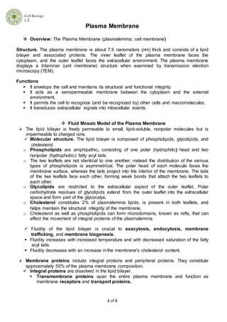
- 1. Cell Biology L 2. 1 of 5 Plasma Membrane Overview: The Plasma Membrane (plasmalemma; cell membrane) Structure. The plasma membrane is about 7.5 nanometers (nm) thick and consists of a lipid bilayer and associated proteins. The inner leaflet of the plasma membrane faces the cytoplasm, and the outer leaflet faces the extracellular environment. The plasma membrane displays a trilaminar (unit membrane) structure when examined by transmission electron microscopy (TEM). Functions It envelops the cell and maintains its structural and functional integrity. It acts as a semipermeable membrane between the cytoplasm and the external environment. It permits the cell to recognize (and be recognized by) other cells and macromolecules. It transduces extracellular signals into intracellular events. Fluid Mosaic Model of the Plasma Membrane The lipid bilayer is freely permeable to small, lipid-soluble, nonpolar molecules but is impermeable to charged ions. Molecular structure. The lipid bilayer is composed of phospholipids, glycolipids, and cholesterol. o Phospholipids are amphipathic, consisting of one polar (hydrophilic) head and two nonpolar (hydrophobic) fatty acyl tails. o The two leaflets are not identical to one another; instead the distribution of the various types of phospholipids is asymmetrical. The polar head of each molecule faces the membrane surface, whereas the tails project into the interior of the membrane. The tails of the two leaflets face each other, forming weak bonds that attach the two leaflets to each other. o Glycolipids are restricted to the extracellular aspect of the outer leaflet. Polar carbohydrate residues of glycolipids extend from the outer leaflet into the extracellular space and form part of the glycocalyx. o Cholesterol constitutes 2% of plasmalemma lipids, is present in both leaflets, and helps maintain the structural integrity of the membrane. o Cholesterol as well as phospholipids can form microdomains, known as rafts, that can affect the movement of integral proteins of the plasmalemma. Fluidity of the lipid bilayer is crucial to exocytosis, endocytosis, membrane trafficking, and membrane biogenesis. Fluidity increases with increased temperature and with decreased saturation of the fatty acyl tails. Fluidity decreases with an increase in the membrane's cholesterol content. Membrane proteins include integral proteins and peripheral proteins. They constitute approximately 50% of the plasma membrane composition. Integral proteins are dissolved in the lipid bilayer. Transmembrane proteins span the entire plasma membrane and function as membrane receptors and transport proteins.
- 2. Cell Biology L 2. 2 of 5 Most transmembrane proteins are glycoproteins. Transmembrane proteins are amphipathic and contain hydrophilic and hydrophobic amino acids, some of which interact with the hydrocarbon tails of the membrane phospholipids. Most transmembrane proteins are folded so that they pass back and forth across the plasmalemma; therefore, they are also known as multipass proteins. Integral proteins may also be anchored to the inner (or occasionally outer) leaflet via fatty acyl or prenyl groups. In freeze-fracture preparations, integral proteins remain preferentially attached to the P-face, the external surface of the inner leaflet, rather than the E-face. Peripheral proteins do not extend into the lipid bilayer. These proteins are located on the cytoplasmic aspect of the inner leaflet. The outer leaflets of some cells possess covalently linked glycolipids to which peripheral proteins are anchored; these peripheral proteins thus project into the extracellular space. Peripheral proteins bond to the phospholipid polar groups or integral proteins of the membrane via noncovalent interactions. They usually function as part of the cytoskeleton or as part of an intracellular second messenger system. They include a group of anionic, calcium-dependent, lipid-binding proteins known as annexins, which act to modify the relationships of other peripheral proteins with the lipid bilayer. Functional characteristics of membrane proteins The lipid-to-protein ratio (by weight) in plasma membranes ranges from 1:1 in most cells to 4:1 in myelin. Some membrane proteins diffuse laterally in the lipid bilayer; others are immobile and are held in place by cytoskeletal components.
- 3. Cell Biology L 2. 3 of 5 Glycocalyx (cell coat) is the sugar coat located on the outer surface of the outer leaflet of the plasmalemma. When examined by TEM, it varies in appearance (fuzziness) and thickness (up to 50 nm). Composition. The glycocalyx consists of polar oligosaccharide side chains linked covalently to most proteins and some lipids (glycolipids) of the plasmalemma. It also contains proteoglycans (glycosaminoglycans bound to integral proteins). Function o The glycocalyx aids in attachment of some cells (e.g., fibroblasts but not epithelial cells) to extracellular matrix components. o It binds antigens and enzymes to the cell surface. o It facilitates cell-cell recognition and interaction. o It protects cells from injury by preventing contact with inappropriate substances. o It assists T cells and antigen-presenting cells in aligning with each other in the proper fashion and aids in preventing inappropriate enzymatic cleavage of receptors and ligands. Plasma Membrane Transport Processes. These processes include transport of a single molecule (uniport) or cotransport of two different molecules in the same (symport) or opposite (antiport) direction. Passive transport includes simple and facilitated diffusion .Neither of these processes requires energy because molecules move across the plasma membrane down a concentration or electrochemical gradient. o Simple diffusion transports small nonpolar molecules (e.g., 0 2 and N2) and small, uncharged, polar molecules (e.g., H20, CO2, and glycerol). It exhibits little specificity, and the diffusion rate is proportional to the concentration gradient of the diffusing molecule.
- 4. Cell Biology L 2. 4 of 5 o Facilitated diffusion occurs via ion channel and/or carrier proteins, structures that exhibit specificity for the transported molecules. It is faster than simple diffusion; ions and large polar molecules are thus capable of traversing membranes that would otherwise be impermeable to them. Ion channel proteins are multipass transmembrane proteins that form small aqueous pores across membranes through which specific small water-soluble molecules and ions pass down an electrochemical gradient (passive transport). Carrier proteins are multipass transmembrane proteins that undergo reversible conformational changes to transport specific molecules across the membrane; these proteins function in both passive transport and active transport. Active transport is an energy-requiring process that transports a molecule against an electrochemical gradient via carrier proteins. o Na+-K+ pump Mechanism. The Na+-K+ pump involves the antiport transport of Na+ and K+ ions mediated by the carrier protein, Na+-K+ adenosine triphosphatase (ATPase). Three Na+ ions are pumped out of the cell and two K+ ions are pumped into the cell. The hydrolysis of a single ATP molecule by the Na+-K+ ATPase is required to transport five ions. Function:The primary function is to maintain constant cell volume by decreasing the intracellular ion concentration (and thus the osmotic pressure) and increasing the extracellular ion concentration, thus decreasing the flow of water into the cell. The Na+-K+ pump also plays a minor role in the maintenance of a potential difference across the plasma membrane. These differences in ion concentration and electrical charge are important in the functioning of nerve and muscle cells in animals. o Glucose transport involves the symport movement of glucose across an epithelium (transepithelial transport). Transport is frequently powered by an electrochemical Na+ gradient, which drives carrier proteins located at specific regions of the cell surface.
- 5. Cell Biology L 2. 5 of 5 Facilitated diffusion of ions can occur via ion channel proteins or ionophores. Selective ion channel proteins permit only certain ions to traverse them. o K+ leak channels are the most common ion channels. These channels are ungated and leak K+, the ions most responsible for establishing a potential difference across the plasmalemma. o Gated ion channels open only transiently in response to various stimuli. They include the following types: Voltage-gated channels open when the potential difference across the membrane changes (e.g., voltage-gated Na+ channels, which function in the generation of action potentials. Mechanically gated channels open in response to a mechanical stimulus (e.g., the tactile response of the hair cells in the inner ear). Ligand-gated channels open in response to the binding of a signaling molecule or ion. These channels include neurotransmitter-gated channels, nucleotide-gated channels, and G-protein gated KA- channels of cardiac muscle cells. Ionophores are molecules that form a complex with ions and insert into the lipid bilayer to transport those ions across the membrane.