Selaginella: features, morphology ,anatomy and reproduction.
Glial cells final group presentation
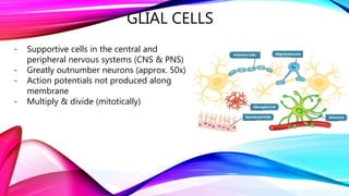
1. GLIAL CELLS
- Supportive cells in the central and
peripheral nervous systems (CNS & PNS)
- Greatly outnumber neurons (approx. 50x)
- Action potentials not produced along
membrane
- Multiply & divide (mitotically)
2. - Formed from oligodendrocyte progenitor cells
(OPCs)
- Found in CNS
- Myelinating oligodendrocytes in white matter
- Non-Myelinating oligodendrocytes in grey
matter
3. ASTROCYTES
Astrocytes are ‘star-shaped’ cells present
throughout the CNS
- Involved in the homeostasis of the interstitial
fluid (IF)
- Carry out the Glutamate-Glutamine cycle
- Part of the Blood-brain barrier (BBB)
- Provide neurons with energy
4. MICROGLIA
- Immune cell for the CNS
- Vital for brain maintenance
- Phagocytosis of pathogens and foreign materials
- Communicate with other cells
- Eliminates unnecessary synapses
- Promotes inflammation of infected areas
5. - Made up of: depending on its location determines its singular
layer of ciliated squamous, columnar or cuboidal cells.
- Amount: uknown definite amount, but what is known is the
approx. cell length - ‘13um’ (micrometre)
- Found: only in central nervous system (brain and spinal cord),
lining cavities in the brain.
- Function: to create, surround and secrete cerebrospinal fluid in
the ventricles/cavities. A constant flow - ‘Around 500 ml is
produced each day, with around 150-250 ml being present in the
body at any one time.’
Ependymal cells
6. SUMMARY
- Glial cells are a key component of the
CNS
- Further research needs to be done to
develop understanding of glial cell
function and importance in the CNS
Editor's Notes
CNS – Oligodendrocytes, Astrocytes, Microglia and Ependymal cells (focusing on these in this presentation)
PNS – Satellite cells and Schwann cells
- Each have a role in aiding neuronal functioning
- Structure: formed from oligodendrocyte progenitor cells (OPCs)
- Function: Myelinating cell of CNS in white matter, metabolic support in grey matter
- Structure - They have a cell body and processes (with end-feet)
- Two different types – fibrous astrocytes (white matter) and protoplasmic astrocytes (grey matter)
Homeostasis of IF – ions/pH/water
Glutamate-Glutamine cycle
BBB stops specific particles from entering the IF/brain tissue. End-feet attach to BBB. Means astrocytes are connected to blood vessels and neurons
Converts glycogen stores to lactate – released and taken up by neurons – used for energy
Huntington’s disease, Alzheimer’s disease, Parkinson’s diseases, ALS
Reactive astrogliosis
Type of immune cells, macrophages, found in the brain that acts as a removal for dead and damaged neurons prevent infections in the nervous system
Found in the brain and the spine
Key to brain maintenance. First and key immune cell
Sensitive to any pathological changes due to a potassium channels that detect any extracellular potassium found in the body
Microglia acknowledge present pathogens and engulf the cells and act as an antigen presenting cell which stimulates the production and activation of T cells
Acts as a housekeeper of the brain and removes foreign material, damaged cells, apoptotic cells, neurofibrillary tangles, DNA fragments, or plaques
Promote inflammation in infected areas by complicated cell signalling which activates more microglias and other cells such as astrocytes and t cells.
Originally thought they were produced in the bone marrow by hematopoietic stem cells, but recent studies suggest they are produced in the yolk sac during embryonic development and make up the brain mesenchyme. Are able to reproduce
Use phagocytic and cytotoxic mechanisms to break down foreign bodies
After too much phagocytosis, microglia become ‘full’ of cell debris and form gitter cells which form a granular corpuscle