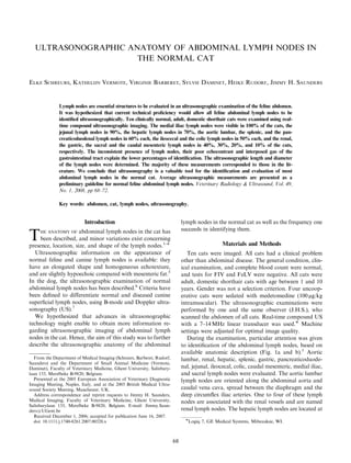
Anatomila dos linfonodos em gatos Schreurs2008.pdf
1. ULTRASONOGRAPHIC ANATOMY OF ABDOMINAL LYMPH NODES IN
THE NORMAL CAT
ELKE SCHREURS, KATHELIJN VERMOTE, VIRGINIE BARBERET, SYLVIE DAMINET, HEIKE RUDORF, JIMMY H. SAUNDERS
Lymph nodes are essential structures to be evaluated in an ultrasonographic examination of the feline abdomen.
It was hypothesized that current technical proficiency would allow all feline abdominal lymph nodes to be
identified ultrasonographically. Ten clinically normal, adult, domestic shorthair cats were examined using real-
time compound ultrasonographic imaging. The medial iliac lymph nodes were visible in 100% of the cats, the
jejunal lymph nodes in 90%, the hepatic lymph nodes in 70%, the aortic lumbar, the splenic, and the pan-
creaticoduodenal lymph nodes in 60% each, the ileocecal and the colic lymph nodes in 50% each, and the renal,
the gastric, the sacral and the caudal mesenteric lymph nodes in 40%, 30%, 20%, and 10% of the cats,
respectively. The inconsistent presence of lymph nodes, their poor echocontrast and interposed gas of the
gastrointestinal tract explain the lower percentages of identification. The ultrasonographic length and diameter
of the lymph nodes were determined. The majority of these measurements corresponded to those in the lit-
erature. We conclude that ultrasonography is a valuable tool for the identification and evaluation of most
abdominal lymph nodes in the normal cat. Average ultrasonographic measurements are presented as a
preliminary guideline for normal feline abdominal lymph nodes. Veterinary Radiology & Ultrasound, Vol. 49,
No. 1, 2008, pp 68–72.
Key words: abdomen, cat, lymph nodes, ultrasonography.
Introduction
THE ANATOMY OF abdominal lymph nodes in the cat has
been described, and minor variations exist concerning
presence, location, size, and shape of the lymph nodes.1–4
Ultrasonographic information on the appearance of
normal feline and canine lymph nodes is available: they
have an elongated shape and homogeneous echotexture,
and are slightly hypoechoic compared with mesenteric fat.5
In the dog, the ultrasonographic examination of normal
abdominal lymph nodes has been described.6
Criteria have
been defined to differentiate normal and diseased canine
superficial lymph nodes, using B-mode and Doppler ultra-
sonography (US).7
We hypothesized that advances in ultrasonographic
technology might enable to obtain more information re-
garding ultrasonographic imaging of abdominal lymph
nodes in the cat. Hence, the aim of this study was to further
describe the ultrasonographic anatomy of the abdominal
lymph nodes in the normal cat as well as the frequency one
succeeds in identifying them.
Materials and Methods
Ten cats were imaged. All cats had a clinical problem
other than abdominal disease. The general condition, clin-
ical examination, and complete blood count were normal,
and tests for FIV and FeLV were negative. All cats were
adult, domestic shorthair cats with age between 1 and 10
years. Gender was not a selection criterion. Four uncoop-
erative cats were sedated with medetomedine (100mg/kg
intramuscular). The ultrasonographic examinations were
performed by one and the same observer (J.H.S.), who
scanned the abdomen of all cats. Real-time compound US
with a 7–14 MHz linear transducer was used. Machine
settings were adjusted for optimal image quality.
During the examination, particular attention was given
to identification of the abdominal lymph nodes, based on
available anatomic description (Fig. 1a and b).1
Aortic
lumbar, renal, hepatic, splenic, gastric, pancreaticoduode-
nal, jejunal, ileocecal, colic, caudal mesenteric, medial iliac,
and sacral lymph nodes were evaluated. The aortic lumbar
lymph nodes are oriented along the abdominal aorta and
caudal vena cava, spread between the diaphragm and the
deep circumflex iliac arteries. One to four of these lymph
nodes are associated with the renal vessels and are named
renal lymph nodes. The hepatic lymph nodes are located at
Presented at the 2005 European Association of Veterinary Diagnostic
Imaging Meeting, Naples, Italy, and at the 2005 British Medical Ultra-
sound Society Meeting, Manchester, UK.
Address correspondence and reprint requests to Jimmy H. Saunders,
Medical Imaging, Faculty of Veterinary Medicine, Ghent University,
Salisburylaan 133, Merelbeke B-9820, Belgium. E-mail: Jimmy.Saun-
ders@UGent.be
Received December 1, 2006; accepted for publication June 16, 2007.
doi: 10.1111/j.1740-8261.2007.00320.x
From the Department of Medical Imaging (Schreurs, Barberet, Rudorf,
Saunders) and the Department of Small Animal Medicine (Vermote,
Daminet), Faculty of Veterinary Medicine, Ghent University, Salisbury-
laan 133, Merelbeke B-9820, Belgium.
Logiq 7, GE Medical Systems, Milwaukee, WI.
68
2. the junction of the splenic and gastroduodenal veins with
the portal vein and in the hilus of the liver. The splenic
lymph nodes can be found adjacent to the splenic vessels in
the hilus of the spleen. The gastric lymph nodes are em-
bedded in the lesser omentum along the lesser curvature of
the stomach, adjacent to the cardia or occasionally adja-
cent to the pylorus. The pancreaticoduodenal lymph nodes
are oriented at the caudal aspect of the pylorus, where the
cranial pancreaticoduodenal and the right gastroepiploic
veins meet. One or two pancreaticoduodenal lymph nodes
may be present adjacent to the right pancreatic lobe. Mul-
tiple jejunal lymph nodes are located adjacent to the cranial
mesenteric artery and the origin of the jejunal arteries at
the root of the mesentery. In 50% of the cats, some lymph
nodes can be found along the jejunal vessels in the more
distal part of the mesentery, near the jejunum and the
ileum. The ileocecal lymph nodes are embedded in the
ileocecal fold at both sides of the concavity of the cecum.
The mesocolon contains the colic lymph nodes near the
ascending and transverse colon, and the caudal mesenteric
lymph nodes near the descending colon. The medial iliac
lymph nodes are found adjacent to the abdominal aorta
and the caudal vena cava. They are caudal to the deep
circumflex iliac artery and vein, and cranial to the external
iliac artery and common iliac vein. The sacral lymph nodes
are located caudal to the origin of the internal iliac arteries
and at the origin of the median sacral artery. They may
follow the course of these vessels.
A normal lymph node was recognized ultrasonograph-
ically as a hypoechoic, homogeneous, elongated structure
in its expected anatomic location. A record was made of
the number of cats in which a particular lymph node could
be identified. For each lymph node, the maximal length
and the maximal diameter were measured once in each cat.
The maximal diameter was defined to be perpendicular to
the maximal length. Depending on where the parameters
were largest, length and diameter were identified on the
same image or on two separate images. Subsequently, the
mean maximal length and the mean maximal diameter of
each lymph node were calculated by averaging the mea-
surements of the 10 cats. If more than one lymph node was
identified in one location, the largest one was measured.
If only one lymph node was visible, it was considered
representative of the group of lymph nodes. As soon as one
lymph node was identified during the ultrasonographic
examination, no further attempt was made to find other
lymph nodes of the same group.
Results
The results are summarized in Table 1 and compared
with the range of normal anatomic values.1–4
The medial iliac and the jejunal lymph nodes were the
most frequently identified lymph nodes, visible, respective-
ly, in 100% and 90% of the cats. The caudal mesenteric
and the sacral lymph nodes had the lowest detection fre-
quencies, which were 10% and 20%, respectively. Other
detection frequencies ranged between 30% and 80%.
The length of all lymph nodes was in accordance with
the anatomy literature,1–4
except for one ileocecal lymph
node. This lymph node had a length of 23.2mm, which
exceeds the reported 15mm.
Fig. 1. (A) Feline parietal abdominal lymph nodes with surrounding anatomy. Right is to the left of the image, cranial is to the top of the image. LN, lymph
node(s). (B) Feline visceral abdominal lymph nodes with surrounding anatomy. Adapted from Barone1
with permission. Right is to the left of the image, cranial
is to the top of the image. LN, lymph node(s).
69
ULTRASOUND OF FELINE ABDOMINAL LYMPH NODES
Vol. 49, No. 1
3. The ranges for normal lymph node diameters are avail-
able in anatomy texts for aortic lumbar, splenic, jejunal,
ileocecal, caudal mesenteric, and medial iliac lymph
nodes1–4
and matched the measured diameter in 23/36
individual lymph nodes (diameter of one ileocecal lymph
node was lost during the procedure). In 11/36 lymph nodes,
the diameter was below the anatomic range, and 2/36
lymph nodes had a diameter above the anatomic range.
While the reported approximate diameters of the hepatic
and the pancreaticoduodenal lymph nodes are 10 and
5 mm, respectively, the corresponding ranges in our study
were 2.5–3.6 mm, for six hepatic lymph nodes (diameter of
one hepatic lymph node was lost during procedure), and
3.6–6.2 mm, for six pancreaticoduodenal lymph nodes.
Discussion
Real-time compound imaging, a technique based on
multiple angles of insonation per scan plane, was used
in this study. Compared with conventional B-mode US,
it produces superior border definition of rounded struc-
tures, less image speckle and improved soft tissue
contrast.8
These advantages are beneficial in imaging
lymph nodes, known for having poor contrast with sur-
rounding tissue.
Few reports are available on the ultrasonographic char-
acteristics of normal abdominal lymph nodes in the cat.
The appearance of feline gastric lymph nodes has been
described in connection with the normal pancreas.9
In that
study, only one gastric lymph node was found craniome-
dial to the pylorus. Its largest size was 10 6 mm, while
our largest measurement was 6.4 1.9 mm. In the same
study, the gastric lymph node was identified in six of 20
normal cats, which is similar to our study.
Regarding the ileocecocolic region in normal cats, all of
31 cats had at least two colic lymph nodes identified in this
area.10
This is higher than the 50% frequency reported in
our study. Nevertheless, the diameter of both lymph nodes
ranged between 1.9 and 4.9 mm, which was similar to our
range of maximal diameter, 1.9–5.2mm.
In the dog, normal abdominal lymph nodes are difficult
to identify due to their small size and as their echogenicity
is similar to those of the surrounding tissues.6
Only the
medial iliac and the jejunal lymph nodes are regularly
identified. In another study, the medial iliac lymph nodes
were identified in 45% (left) and 82% (right) of 11 normal
dogs.11
In our study, at least one medial iliac lymph node
was found in each cat and a jejunal lymph node was visible
in nine of 10 cats.
Not all abdominal lymph nodes were consistently found
in cats in the present study. According to anatomic de-
scriptions, most abdominal lymph nodes are present in
individual cats, although their number may be variable.
Individual cats may occasionally be missing the aortic
lumbar, renal, splenic, gastric, ileocecal, or sacral lymph
nodes.1–3
However, in this study, they were identified, re-
spectively, in 60%, 40%, 60%, 30%, 50%, and 20% of the
cats. It is not known if the lymph nodes that could not be
seen in our 10 cats were actually absent or just not detected
during the ultrasonographic examination, as no gold stan-
dard for deciding on the presence or absence of the nodes is
available. There was no tendency for the lymph nodes with
the lowest detection frequencies to be identified within the
same individual cat, which would suggest a better visibility
of the abdominal lymph nodes in these specific cats.
Visceral and vascular landmarks can be used to locate
abdominal lymph nodes ultrasonographically. In the liter-
ature, an ultrasonographic map of abdominal vessels is
available for the dog, but not for the cat.12
In our study, we
used both visceral and vascular landmarks. Color or power
Doppler sonography was used whenever necessary (Figs.
2–6).
Table 1. Ultrasonographic Frequency of Detection and Measurements of Feline Abdominal Lymph Nodes
Frequency (%) US Length (mm) Anatomic Length (mm) US Diameter (mm) Anatomic Diameter (mm)
Aortic lumbar 60 9.9 (2.1–16.7) 0.5–18 3.2 (0.3–7.4) 3–4
Renal 40 6.1 (4.7–7.7) 0.5–14.5 3.5 (2.9–4.1) Not available
Hepatic 70 7.6 (5.9–9.5) 1.5–30.5 2.9 (2.5–3.6) 10
Splenic 60 8.4 (5.0–11.2) 2–22 3.2 (1.9–4.8) 2–22
Gastric 30 5.1 (4.6–6.4) 1–20 1.9 (1.9–1.9) Not available
Pancreaticoduodenal 60 8.4 (6.6–13.0) 3–15.5 4.6 (3.6–6.2) 5
Jejunal 90 20.1 (11.4–39.0) 5–80 5.0 (2.8–7.2) Max 10
Ileocecal 50 11.8 (6.7–23.2) 3–15 4.1 (2.7–4.8) 5–9
Colic 50 9.0 (4.6–12.1) 1–30 3.1 (1.9–5.2) Not available
Caudal mesenteric 10 6.0 (6.0–6.0) 5–15 2.1 (2.1–2.1) 5–15
Medial iliac 100 13.5 (5.0–23.3) 1–28 4.5 (1.3–14.0) 2–7
Sacral 20 9.6 (9.2–10.0) 1–28 2.2 (1.7–2.7) Not available
Results of the ultrasonographic examination of the abdomen of 10 cats. The ultrasonographically measured maximal length and diameter are
represented as mean and range. The ranges of anatomic measurements out of literature are available for comparison.
Ultrasonographic.
70 SCHREURS ET AL. 2008
4. In humans, interposed gas makes perigastric and
perisplenic lymph nodes difficult to access ultrasonogra-
phically.13
A similar problem was noted in our study, with
gas obscuring lymph nodes near the stomach, cecum, and
colon (gastric, pancreaticoduodenal, ileocecal, colic, and
caudal mesenteric lymph nodes). Small intestinal loops
were not detrimental to lymph node identification, as they
mostly contained mucus and could be displaced during
scanning.
Apart from one ileocecal lymph node, all lymph nodes
had their lengths falling within the published range.1–4
Not
all diameters of the individual lymph nodes were in har-
mony with the literature, although the mean diameters of
lumbar aortic, splenic, jejunal, and medial iliac lymph
nodes were. The maximal diameters of the individual il-
eocecal and the caudal mesenteric lymph nodes were con-
sistently lower than those of the anatomic references. Also,
the maximal diameter of the individual hepatic lymph
nodes did not approach the reference value of 10mm. No
anatomic reference values are available for diameters of
renal, gastric, colic, and sacral lymph nodes.
To our knowledge, the accuracy of ultrasonographic
measurement of lymph nodes has not been validated. We
propose our measurements as a preliminary guideline of
the expected sizes of abdominal lymph nodes in normal
Fig. 2. The aortic lumbar lymph nodes area located along the abdominal
aorta (AA). Length and diameter are indicated by the callipers. Cranial is to
the left of the image.
Fig. 3. The hepatic lymph nodes are located in the hilus of the liver (L)
and near the junction of the splenic and gastroduodenal veins with the portal
vein. The latter are not in the field of view in this image. Length is indicated
by the callipers. Cranial is to the left of the image. S, stomach.
Fig. 4. The jejunal lymph nodes are located adjacent to the cranial mes-
enteric artery (CMA) and the origin of the jejunal arteries at the root of the
mesentery. Length and diameter are indicated by the callipers. Cranial is to
the left of the image. SI, small intestines.
Fig. 5. The ileocecal lymph nodes are located on both sides of the cecum
(C). Length and diameter are indicated by the callipers. Cranial is to the left
of the image. I, ileum.
71
ULTRASOUND OF FELINE ABDOMINAL LYMPH NODES
Vol. 49, No. 1
5. cats. This is especially important due to the similar body
conformation among adult cats of most breeds.
Pathologic abdominal lymph nodes are easier to image
ultrasonographically because they are usually enlarged,
have decreased echogenicity and are more rounded.5,6,14
In
advanced disease, they may become irregularly shaped,
heterogeneous, and poorly marginated.15
A good optimal
knowledge of the lymph node drainage pattern is impor-
tant to increase specificity of detected abdominal abnor-
malities.1–4
Attempts to characterize abnormal lymph
nodes as benign or malignant have lead to numerous
ultrasonographic parameters in humans and animals.7,16
It
is concluded that in humans the Doppler characteristics
described for superficial lymph nodes are difficult to apply
to abdominal lymph nodes because of the deeper location
of the latter ones.17
To conclude, most of the abdominal lymph nodes in the
normal cat can be assessed ultrasonographically. We sug-
gest using the proposed ultrasonographic measurements as
a guideline for the assessment of abdominal lymph nodes
in normal cats.
ACKNOWLEDGEMENT
The authors thank Karen Bonte, DVM, for her enthusiastic support
at the start of this study.
REFERENCES
1. Barone R. Système lymphatique du chat. In: Barone R (ed): Ana-
tomie Comparée des Mammifères Domestiques. Tome 5 Angiologie. Paris:
Editions Vigot, 1996;833–844.
2. Saar LI, Getty R. Carnivore lymphatic system. Part II: feline. In:
Getty R (ed): Sisson and Grossman’s the anatomy of the domestic animals,
5th ed. Philadelphia: W.B. Saunders Company, 1975;1661–1669.
3. Tompkins MB. Lymphoid system. In: Hudson LC, Hamilton WP
(eds): Atlas of feline anatomy for veterinarians. Philadelphia: W.B. Saunders
Company, 1993;113–126.
4. Vollmerhaus B. Lymphknoten und Lymphsämmelgänge der
Katze. In: Nickel R, Schemmer A, Seiferle E (eds): Lehrbuch der
Anatomie der Haustiere, Band III, 2nd ed. Berlin: Verlag Paul Parey,
1984;366–376.
5. Lamb CR. Recent developments in diagnostic imaging of the gas-
trointestinal tract of the dog and cat. Vet Clin North Am Small Anim Pract
1999;29:307–342.
6. Pugh CR. Ultrasonographic examination of abdominal lymph nodes
in the dog. Vet Radiol Ultrasound 1994;35:110–115.
7. Nyman HT, Kristensen AT, Skovgaard IM, McEvoy FJ. Charac-
terization of normal and abnormal canine superficial lymph nodes using
gray-scale B-mode, color flow mapping, power, and spectral Doppler
ultrasonography: a multivariate study. Vet Radiol Ultrasound 2005;46:
404–410.
8. Entrekin RR, Porter BA, Sillesen HH, Wong AD, Cooperberg PL,
Fix CH. Real-time spatial compound imaging: application to breast,
vascular, and musculoskeletal ultrasound. Semin Ultrasound CT MRI
2001;22:50–64.
9. Etue SM, Penninck DG, Labato MA, Pearson S, Tidwell A. Ultra-
sonography of the normal feline pancreas and associated anatomic land-
marks: a prospective study of 20 cats. Vet Radiol Ultrasound 2001;42:
330–336.
10. Besso JG, Rault D, Begon D. Feline cecum and ileocecocolic junc-
tion: normal ultrasonographic features and clinical applications [abstract].
Vet Radiol Ultrasound 2004;45:599.
11. Llabrés-Dı́az FJ. Ultrasonography of the medial iliac lymph nodes in
the dog. Vet Radiol Ultrasound 2004;45:156–165.
12. Spaulding KA. A review of sonographic identification of abdominal
blood vessels and juxtavascular organs. Vet Radiol Ultrasound 1997;38:
4–23.
13. Delorme S, van Kaick G. Imaging of abdominal nodal spread in
malignant disease. Eur Radiol 1996;6:262–274.
14. Lüerssen D, Janthur M. Lymph nodes. In: Nautrup CP, Tobias R,
Cartee RE (eds): An atlas and diagnostic textbook of diagnostic ultrasono-
graphy of the dog and cat, 2nd ed. London: Manson Publishing, 2001;
244–247.
15. Mattoon JS, Nyland TG. Abdominal fluid, lymph nodes, masses,
peritoneal cavity, and great vessel thrombosis. In: Nyland TG, Mattoon JS
(eds): Small animal diagnostic ultrasound, 2nd ed. Philadelphia: W. B.
Saunders Company, 2002;82–91.
16. Nyman HT, Kristensen AT, Flagstad A, McEvoy FJ. A review of
the sonographic assessment of tumor metastases in liver and superficial
lymph nodes. Vet Radiol Ultrasound 2004;45:438–448.
17. De Gaetano AM, Vecchioli A, Minordi LM, et al. Role of diagnostic
imaging in abdominal lymphadenopathy. Rays 2000;25:463–484.
Fig. 6. The sacral lymph nodes are located caudal to the origin of the
internal iliac arteries (IIA). Length and diameter are indicated by the calli-
pers. Cranial is to the left of the image.
72 SCHREURS ET AL. 2008