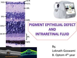
Pigment epithelial defect and intraretinal fluid
•Download as PPTX, PDF•
5 likes•2,592 views

A simple and informative presentation on PED & IRF with pathophysiology, clinical examination, diagnostic imaging and one case study each for both PED & IRF

Recommended
More Related Content
What's hot
What's hot (20)
Similar to Pigment epithelial defect and intraretinal fluid
Similar to Pigment epithelial defect and intraretinal fluid (20)
More from Loknath Goswami
Recently uploaded
Recently uploaded (20)
Pigment epithelial defect and intraretinal fluid
- 1. By, Loknath Goswami B. Optom 4th year
- 2. Introduction • Retinal pigment epithelial detachments (PEDs) are characterized by separation between the RPE and the inner most aspect of Bruch's membrane.
- 3. Introduction
- 4. Introduction
- 5. Introduction • The space created by this separation is occupied by blood, serous exudate, drusenoid material, fibrovascular tissue or a combination.
- 7. Types • Drusenoid PEDs are seen mostly in dry AMD • Serous PEDs are typically associated with the wet form of AMD • Vascularized PEDs associated with Type 1 (sub-RPE) neovascularization
- 9. Ocular Diseases • PEDs are present in several chorioretinal diseases including: – Vogt-Koyanagi-Harada (VKH) Syndrome – Central Serous Chorioretinopathy (CSC) – Polypoidal Choroidal Vasculopathy (PCV) – Exudative/Non-Exudative Age-Related Macular Degeneration (AMD).
- 10. Systemic Diseases • PEDs have also been associated with certain systemic conditions including: – Sarcoidosis – Neurosyphilis – Cryoglobulinemia – Large cell non–Hodgkin lymphoma
- 11. Pathophysiology • The retinal pigment epithelium (RPE) monolayer, extending from the optic disk margin uninterrupted through to the ciliary body epithelium, is bounded apically by the apical surface of the retina and on its basal surface by the collagenous layer of Bruch’s membrane.
- 12. Pathophysiology • Proper anatomical apposition between the retina, the RPE, and Bruch’s membrane is crucial for nutritional support of the photoreceptors, retinol metabolism, phagocytosis of the photoreceptors outer segments, and formation of the outer blood-retinal barrier.
- 13. Pathophysiology • Under normal conditions, there exists a net bulk flow of fluid towards the choroid from the vitreous, with its generation dependent upon hydrostatic and osmotic forces within the two bodies. Both the RPE and the retina produce resistance to this fluid flow. • The RPE has greater resistance due to its limited hydraulic conductivity, subsequently, a vector force is generated pushing it against Bruch’s membrane.
- 14. Pathophysiology • The attachment of the RPE basement membrane to Bruch’s membrane is possibly supplemented by regions of hemidesmosomes containing fine filaments of laminin, proteoglycans and collagen types IV and V. • Age-related deposition of lipids, such as cholesterol esters, triglycerides, and fatty acids, in Bruch membrane may change its permeability altering retinochoroidal flow. • Fluid may accumulate in the sub- RPE space, unable to pass through Bruch membrane, resulting in RPE elevation.
- 15. Diagnosing
- 16. Diagnosis History Physical examination Imaging tests Diagnosing Diagnosing PEDs relies on careful history and physical exam with further information provided by various imaging modalities. Treatment
- 17. History • Patients will typically present with painless blurred vision or partial vision loss. • Others have described a dark shadowing effect or sensation that a curtain has been pulled in front of their vision.
- 18. Physical Exam • Often PEDs will transilluminate if they are filled predominantly with serous fluid when observed at the slit lamp. Pigment figures can also indicate chronicity of disease. • Examination reveals a reticulated pattern of increased pigmentation extending radially over the PED, likely due to migration of RPE cells into the outer retinal space however it is unclear whether these carry a prognostic significance.
- 19. Drusenoid PED • Drusenoid PEDs appear as well-circumscribed yellow or yellow–white elevations of the RPE that are usually found within the macula. They may have scalloped borders and a slightly irregular surface. It is not uncommon to observe a speckled or stellate pattern of brown or gray pigmentation on their surface.
- 20. Drusenoid PED Yellow or yellow–white elevations Scalloped borders Stellate pattern of brown or gray pigmentation on their surface
- 21. Serous PED • Serous PED appears as a distinct circular or oval-like detachment of the RPE. Clear or yellowish–orange in color, this dome-shaped elevation of the RPE has a sharply demarcated border.
- 22. Serous PED Yellowish–orange in colour A sharply demarcated border
- 23. Vascular PED • Fibrovascular PED’s indicate the growth of a choroidal neovascular membrane (CNVM) in the sub-RPE space; this is also known as occult choroidal neovascularisation. • Gass reported that a flattened or notched border of the PED is a frequent and important sign of hidden associated CNV
- 24. Vascular PED
- 27. Drusenoid PED • Drusenoid PEDs demonstrate faint hyperfluorescence in the early phase that increases throughout the transit stage of the study without late leakage. • The correlation of FA findings with SD-OCT and occasionally ICGA may help differentiate drusenoid from vascularized PEDs.
- 28. Drusenoid PED
- 29. Serous PED • Serous PEDs demonstrate intense early hyperfluorescence and brisk, progressive pooling within the PED in a homogeneous and well-demarcated manner • Late staining of serous PEDs is typical and may make it difficult to differentiate these PEDs from those that are vascularized based on FA alone
- 30. Serous PED
- 31. Vascular PED • From the analysis of fundus photographs of the macula and FA, the Macular Photocoagulation Study identified two main patterns of CNV: Classic Occult
- 32. Vascular PED • Classic CNV is characterized by a well-defined area of early typically lacy hyperfluorescence with progressive leakage in the late stages of the study.
- 34. Vascular PED • The term “occult” refers to a type of CNV that is difficult to visualize, analyse and localize on FA
- 35. Vascular PED
- 37. Drusenoid PED • Using a confocal scanning laser ophthalmoscope (SLO) system and ICGA, the content of the drusenoid PED will block the fluorescence emitted from the underlying choroidal vasculature and, therefore, the PED will appear as a homogeneous hypofluorescent lesion during the early phase and remain hypofluorescent throughout the transit.
- 38. Drusenoid PED
- 39. Serous PED • With an infrared fundus camera, the ICGA reveals only variable, minimal blockage of normal choroidal vessels by the serous PEDs in the late phase. Using a confocal SLO system, the ICGA reveals hypofluorescence in both the early and the late phases of the ICGA study with complete blockage of the normal choroidal vasculature.
- 40. Serous PED
- 41. Vascular PED • ICGA is very useful for diagnosing and classifying CNV associated to serous PED, due to its capacity to distinguish between the serous and the vascular component
- 42. Vascular PED The cases involve OCNV, where ICGA can show hot spots on the black image of the PED associated to RAP (yellow arrows) or polyps (red arrows).
- 44. Drusenoid PED • Drusenoid PEDs usually show a smooth contour of the detached hyperreflective RPE band that may demonstrate an undulating appearance. • The material beneath the RPE band typically exhibits a dense homogeneous appearance with moderate or high hyperreflectivity. • Drusenoid PEDs are typically not associated with overlying subretinal or intraretinal fluid.
- 45. Drusenoid PED
- 46. Drusenoid PED
- 47. Serous PED • On OCT, serous PEDs appear as well- demarcated, abrupt elevations of the RPE with a homogenously hyporeflective sub-RPE space. • Enhanced depth imaging (EDI) OCT is useful to determine whether serous PED is caused by AMD (normal subfoveal choroidal thickness) or by CSC (increased subfoveal choroidal thickness).
- 48. Serous PED
- 49. Serous PED
- 50. Vascular PED • Optical coherence tomography allows better visualization of the exact relationship between neovascular membranes and PEDs. • Enhanced depth imaging OCT enables better visualization of the contents of PEDs. Untreated PEDs demonstrate evidence of fibrovascular proliferation, often coursing along the back surface of the detached RPE.
- 51. Vascular PED
- 52. Vascular PED
- 53. Management • Depending on the etiology of the PED, different treatment modalities have been explored to prevent vision loss.
- 54. Treatment • Several strategies, have being used to treat vascularized PEDs, including laser photocoagulation, photodynamic therapy (PDT), intravitreal steroids and anti-VEGF therapy. • The results from the trial indicated that PDT could significantly reduce the risk of moderate and severe vision loss among patients with subfoveal occult CNV.
- 55. Treatment • Another treatment modality, described recently by Costa et al as a pilot trial, is photothrombosis at the neovascular ingrowth site using ICG injection followed by laser application. Occlusion of the feeder vessel with cessation of leakage, restoration of macular architecture and visual improvement were induced in two patients with CNV associated with PEDs.
- 56. Treatment • Currently no treatment for serous PED is proven effective, nor are recommendations for treatment guidelines established.
- 57. Prognosis • The location of the PED is important in determining prognosis. • Patients with extrafoveal PEDs tend to preserve good visual acuity, whereas patients with subfoveal PEDs can have worse visual outcomes. • The course of PEDs also varies in CSC versus AMD.
- 58. Prognosis • Mudvari et al demonstrated with a mean follow-up of 49 months that 65% of PEDs in CSC completely resolved and the other 35% PEDs remained persistent. • Retinal pigment epithelium atrophy was evident in 86% of patients over the area of the resolved PED.
- 59. Prognosis • The natural course of Type 1 or occult CNV can vary considerably. Type 1 CNV patients can appear relatively asymptomatic and may never experience vision loss despite continued growth of the neovascular lesion. • On the other hand, large vascularized or hemorrhagic PEDs are typically associated with significant vision loss. Additionally, Type 1 CNV can erode through the RPE, becoming Type 2 CNV and follow a more aggressive course with more progressive and severe vision loss.
- 60. Case study • An 81-year-old female patient presented for a routine eye examination with no ocular complaints. • There were no general health concerns and no family general or ocular health history. • Refraction showed no change and visual acuity (VA) in both eyes was stable at 6/7.5 • Binocular indirect examination of the right fundus showed extensive exudates between the macula and disc • OCT examination revealed the presence of a large pigment epithelial detachment (PED)
- 61. Case study
- 62. Case study • In this case, the PED displays OCT characteristics consistent with a fibrovascular PED • The RPE is broadly and irregularly elevated with the PED appearing to be filled with solid layers of medium reflective material, separated by hyporeflective clefts.
- 63. Case study • While vision remained stable in this patient – likely due to the absence of sub-retinal and intraretinal fluid and the maintained integrity of the photoreceptor complex – as wet AMD was suspected the patient was referred via a fast track macular service. • Fluorescein angiography examination, which was performed within 10 days of the original referral, confirmed OCT findings of a right occult CNVM.
- 64. Case study • However, with VA of 6/7.5, the patient fell outside NICE guidance for treatment with the anti-VEGF therapy, Lucentis (ranibuzumab), which states that VA must be between 6/12 and 6/96. • The patient was advised to self-monitor with an Amsler grid and would be reassessed on a monthly basis with OCT.
- 65. Case study • As this patient has wet AMD in her right eye and dry AMD in the left eye, it would be advisable to offer nutritional supplementation. • In a patient with these characteristics, supplementation with the AREDS 2 formula (10mg lutein, 25mg zinc, 2mg copper, 500mg vitamin C and 400IU vitamin E) could reduce risk of progression to advanced AMD by 18–25%, depending on dietary intake of nutrients.
- 66. Case study • Nutritional supplementation with antioxidants is believed to reduce the risk of AMD progression by reducing the level of oxidative stress at the retina, one of the most popular hypotheses regarding AMD development and progression.
- 68. Introduction • It normally appears above outer plexiform layer • Intraretinal fluid either can occur diffusely and cause increased retinal thickness and reduced retinal reflectivity or be localized or non- reflective cysts (cystic edema) • It is mainly seen in cystoid macular edema
- 69. Cystoid macular edema • Cystoid Macular Edema (CME) is retinal thickening of the macula due to a disruption of the normal blood-retinal barrier; this causes leakage from the perifoveal retinal capillaries and accumulation of fluid within the intracellular spaces of the retina, primarily in the outer plexiform layer. • Visual loss occurs from retinal thickening and fluid collection that distorts the architecture of the photoreceptors.
- 70. Pathophysiology • A delicate exchange of homeostatic mechanisms is in place with the vitreous, retina, retinal pigment epithelium (RPE), and choroid receiving their circulation through the retinal and choroidal vasculature.
- 71. Pathophysiology • There is an intrinsic balance amongst the osmotic force, hydrostatic force, capillary permeability, and tissue compliance that occur within the vasculature. Specifically, the capillary filtration rate should equal the rate of fluid removal from extracellular retinal tissue, such as glial and RPE cells.
- 72. Pathophysiology • Once these forces are disrupted an imbalance occurs, accumulation of fluid is seen in cystoid spaces within the inner layers of the retina, most commonly the outer plexiform layer (OPL). • Accumulation of the fluid commonly occurs in the Henle’s fiber layer causing the classic petaloid pattern.
- 73. Pathophysiology • A common factor that can cause CME is vitreomacular traction (VMT)
- 74. Signs • Using slit lamp or direct/indirect ophthalmoscopy, clinically significant foveal edema and retinal thickening more than 300 μm can be seen as a loss of foveal reflex; this is better visualized using green light to outline the cystic spaces. • Subclinical foveal edema is described as edema less than 300 μm and is better seen through retinal imaging. • Vitritis and optic nerve head swelling can also be seen in clinical examination.
- 75. Symptoms • Decrease in visual acuity that is associated with retinal edema • Loss of contrast sensitivity and color vision • Metamorphopsia that can be demonstrated on Amsler grid, micropsia, and central scotoma
- 76. Risk Factors • Diabetes • Epinephrine • Pars Planitis/Uveitis • Retinitis Pigmentosa (RP) • Vein Occlusion • Nicotinic acid and Niacin • Surgery
- 78. Color Fundus Photography (CFP) • It depicts intraretinal cysts within the foveal region of the macula in Henle's layer in a honey comb pattern
- 79. Fluorescein Angiography (FA) • It studies the circulation of the retina and choroid. In the early phase of FA, capillary dilation in the perifoveal region is appreciated
- 80. Fluorescein Angiography (FA) • In the late phase (may take 5-15 minutes) of FA, leakage into the cystoid spaces is distributed radially in Henle’s layer forming the classic petaloid leakage pattern or expansile dot appearance
- 81. Optical coherence tomography (OCT) • OCT can depict the mechanical forces induced by vitreomacular interface abnormalities, such as VMT or epiretinal membrane (ERM), via a hyperreflective band on the inner surface of the retina
- 83. General treatment • Therapeutic approaches, whether medical or surgical, in treating CME are dependent on the underlying etiology. • Most cases are self-limiting within 3-4 months. • If CME persists then medical or surgical therapy is warranted.
- 84. Medical therapy • NSAIDS • Corticosteroids • Carbonic anhydrase inhibitors (CAIs) • Anti-VEGF agents • Pharmacologic vitreolysis agents
- 85. Surgery • Pars plana vitrectomy can help to relieve macula edema due to tractional or nontractional components • Side effects of vitrectomy include cataract, retinal detachment, vitreous hemorrhage, and a rise in intraocular pressure.
- 86. Case study • MRD No : 152337 • Gender : Male • Age : 70 Years • Main complaints : OD – Floaters since 2Year • No h/o ocular or head injury • Past ocular history : Nil
- 87. Case study • Past medical history: – Diabetes mellitus - not using Rx. – Hypertension systemic - take tablets • Family history: Nil • Allergy: Not aware of • Current medication: Nil
- 88. Case study • Vision (aided) : – OD = Dist. 6/12 Near N6 – OS = Dist. 6/7.5 Near N6
- 89. Oct report
- 90. Case study • Diagnosis: – Occlusion branch retinal vein (BRVO) OD – OU – Cataract immature • Medications : – OD- Extragat eyedrops-4times/day x 1 week Remarks :- start one day before inj. • Advice – Avastin (OD)
- 91. Review after 26 days • Vision remained constant • OD – Macular edema reduced
- 92. OCT report after Avastin inj.
- 93. Comparison Pre op Post op
- 94. Review after 26 days • Medications – Gatilox Hx eye drops – 3times/day x 1 week Remarks: Start one day before inj. • Advice :- OD (Avastin)
- 95. References • http://www.amdbook.org/content/serous-ped-0 • http://eyewiki.aao.org/Pigment_Epithelial_Detachment • http://eyewiki.aao.org/Cystoid_Macular_Edema • http://www.amdbook.org/book/export/html/484 • Mudvari, S. S., Goff, M. J., Fu, A. D., McDONALD, H. R., Johnson, R. N., Ai, E., & Jumper, J. M. (2007). The natural history of pigment epithelial detachment associated with central serous chorioretinopathy. Retina, 27(9), 1168-1173. • Costa, R. A., Rocha, K. M., Calucci, D., Cardillo, J. A., & Farah, M. E. (2003). Neovascular ingrowth site photothrombosis in choroidal neovascularization associated with retinal pigment epithelial detachment. Graefe's archive for clinical and experimental ophthalmology, 241(3), 245-250
Editor's Notes
- The classification of PEDs in AMD can be divided based on their contents. Categories include drusenoid, serous, vascularized, or mixed components.