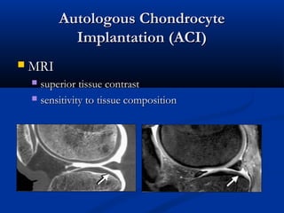
SlideShare a Scribd company logo
1 of 36
Autologous ChondrocyteAutologous Chondrocyte
Implantation (ACI)Implantation (ACI)
 MRIMRI
 superior tissue contrastsuperior tissue contrast
 sensitivity to tissue compositionsensitivity to tissue composition
MRIMRI
 tremendous potential in the study oftremendous potential in the study of cartilage repaircartilage repair
 help to estimate thehelp to estimate the size, nature, and locationsize, nature, and location of lesionsof lesions
preoperatively, in orderpreoperatively, in order to optimize surgical planningto optimize surgical planning
 help to evaluate thehelp to evaluate the quality and success of tissue repairquality and success of tissue repair
processesprocesses after surgicalafter surgical treatmenttreatment
 allow one toallow one to monitor degenerative changesmonitor degenerative changes in the jointin the joint
after cartilage repair, potentially in comparison to patientsafter cartilage repair, potentially in comparison to patients
who have not been treated forwho have not been treated for cartilage lesionscartilage lesions
MRIMRI
 MRIMRI
 less invasive methodless invasive method
 directly depictsdirectly depicts
 Cartilage interiorCartilage interior
 subchondral bone and bone marrowsubchondral bone and bone marrow
 ArthroscopyArthroscopy
 cartilage surface (minor surface abnormalities)cartilage surface (minor surface abnormalities)
 tissue biopsy for histologic assessmenttissue biopsy for histologic assessment of theof the
implantimplant
MRIMRI
 MRIMRI
 MR arthrographyMR arthrography
 outline cartilage defectsoutline cartilage defects
 improveimprove the conspicuity of lesionsthe conspicuity of lesions
 Direct: Intra-articular GdDirect: Intra-articular Gd
 Indirect: IV GdIndirect: IV Gd
MRIMRI
 Morphological assessment:Morphological assessment:
 SurfaceSurface
 ThicknessThickness
 VolumeVolume
 Subchondral boneSubchondral bone
 Biochemical statusBiochemical status
 Biomechanical statusBiomechanical status
Preoperative estimation of lesion size, nature,Preoperative estimation of lesion size, nature,
and location: morphologyand location: morphology
 MR technique and sequencesMR technique and sequences
 fat-suppressed three-dimensional gradient echo (3D-fat-suppressed three-dimensional gradient echo (3D-
GRE): T1GRE): T1
 exact depiction of the thickness and surface of cartilageexact depiction of the thickness and surface of cartilage
 Intermediate-weightedIntermediate-weighted fast spin echo (FSE)fast spin echo (FSE)
techniques with ortechniques with or without fat-suppression: T2without fat-suppression: T2
 normal and abnormal internal structure of hyalinenormal and abnormal internal structure of hyaline
cartilagecartilage
Preoperative estimation of lesion size, nature,Preoperative estimation of lesion size, nature,
and location:and location: morphologymorphology
IM TSE IM TSE FS
Preoperative estimation of lesion size, nature,Preoperative estimation of lesion size, nature,
and location:and location: morphologymorphology
3D GRE FS3D GRE FS
DESSDESS TRUEFISPTRUEFISP
Preoperative estimation of lesion size, nature,Preoperative estimation of lesion size, nature,
and location:and location: morphologymorphology
 MR technique and sequencesMR technique and sequences
 voxel size under 300 μm is required to reveal frayingvoxel size under 300 μm is required to reveal fraying
of the articular surface of cartilageof the articular surface of cartilage
 high-resolution threedimensional (3D) isotropic cartilage-high-resolution threedimensional (3D) isotropic cartilage-
sensitive sequencessensitive sequences
 High-field MRI scanners and new coil technologies:High-field MRI scanners and new coil technologies:
mult-element design with parallel imagingmult-element design with parallel imaging
 scan times can be kept well below 10 minscan times can be kept well below 10 min
 signal-to-noise ratiosignal-to-noise ratio
Preoperative estimation of lesion size, nature,Preoperative estimation of lesion size, nature,
and location:and location: morphologymorphology
 Scoring methodsScoring methods
 MRI classification system (Yulish et al)MRI classification system (Yulish et al)
 Grade 1: abnormal intrachondral signal with a normalGrade 1: abnormal intrachondral signal with a normal
chondral surfacechondral surface
 Grade 2: mild surface irregularity and/or focal loss of lessGrade 2: mild surface irregularity and/or focal loss of less
than 50% of the cartilage thicknessthan 50% of the cartilage thickness
 Grade 3: severe surface irregularity with focal loss of 50%Grade 3: severe surface irregularity with focal loss of 50%
to 100% of the cartilage thicknessto 100% of the cartilage thickness
 Grade 4: complete loss of articular cartilage, withGrade 4: complete loss of articular cartilage, with
exposure of subchondral boneexposure of subchondral bone
Preoperative estimation of lesion size, nature,Preoperative estimation of lesion size, nature,
and location:and location: morphologymorphology
 AccuracyAccuracy
 sensitivity of 93% and a specificity of 99% insensitivity of 93% and a specificity of 99% in
detecting chondral lesions with MRI when axial anddetecting chondral lesions with MRI when axial and
coronal images were combined,and values of 94 andcoronal images were combined,and values of 94 and
99% when images in all three planes99% when images in all three planes were usedwere used
 accuracy was highest for severe cartilage lesions andaccuracy was highest for severe cartilage lesions and
lowest for smaller lesionslowest for smaller lesions
Bredella MA, Tirman PF, Peterfy CG et al (1999) Accuracy of T2-weighted fast spin-echo MR imaging with fat saturation inBredella MA, Tirman PF, Peterfy CG et al (1999) Accuracy of T2-weighted fast spin-echo MR imaging with fat saturation in
detecting cartilage defects in the knee: comparison with arthroscopy in 130 patients. AJR Am J Roentgenol 172:1073–1080detecting cartilage defects in the knee: comparison with arthroscopy in 130 patients. AJR Am J Roentgenol 172:1073–1080
Evaluation of the quality of tissue-repairEvaluation of the quality of tissue-repair
processesprocesses after surgical treatment: morphologyafter surgical treatment: morphology
 Important to know:Important to know:
 Type of cartilage repairType of cartilage repair
 Size and location within the jointSize and location within the joint
 Concomitant procedures (eg osteotomy or ligament repair)Concomitant procedures (eg osteotomy or ligament repair)
 The MR sequences commonly used for evaluatingThe MR sequences commonly used for evaluating
the morphology of cartilage repair are identical withthe morphology of cartilage repair are identical with
those used for evaluating cartilage lesionsthose used for evaluating cartilage lesions
 Specific grading systemsSpecific grading systems
Evaluation of the quality of tissue-repair processesEvaluation of the quality of tissue-repair processes
after surgical treatment: morphologyafter surgical treatment: morphology
 MOCART:MOCART:
 filling of the defectfilling of the defect
 integration of the border zone to the adjacent cartilageintegration of the border zone to the adjacent cartilage
 surface of the repair tissuesurface of the repair tissue
 structurestructure of the repair tissueof the repair tissue
 signal intensity of the repair tissuesignal intensity of the repair tissue
 intactness of the subchondral laminaintactness of the subchondral lamina
 intactness of the subchondral boneintactness of the subchondral bone
 adhesionsadhesions
 effusioneffusion
Evaluation of the quality of tissue-repair processesEvaluation of the quality of tissue-repair processes
after surgical treatment: morphologyafter surgical treatment: morphology
 3D MOCART3D MOCART
 Defect fillDefect fill
 Cartilage interfaceCartilage interface
 Bone interfaceBone interface
 SurfaceSurface
 StructureStructure
 Signal intensitySignal intensity
 Subchondral laminaSubchondral lamina
 Chondral osteophytesChondral osteophytes
 Bone marrow edemaBone marrow edema
 Subchondral boneSubchondral bone
 EffusionEffusion
Evaluation of the quality of tissue-repair processesEvaluation of the quality of tissue-repair processes
after surgical treatment: morphologyafter surgical treatment: morphology
 MOCARTMOCART
 Almost perfect agreement between readersAlmost perfect agreement between readers
 Comparing the MRI scores with clinical outcome (knee-Comparing the MRI scores with clinical outcome (knee-
related quality of life) 2 years after ACI: a statisticallyrelated quality of life) 2 years after ACI: a statistically
significant correlation was found forsignificant correlation was found for
 filling of the defectfilling of the defect
 structure of the repair tissuestructure of the repair tissue
 changes in the subchondral bonechanges in the subchondral bone
 signal intensities of the repair issuesignal intensities of the repair issue
Evaluation of the quality of tissue-repair processesEvaluation of the quality of tissue-repair processes
after surgical treatment: morphologyafter surgical treatment: morphology
 filling of the defectfilling of the defect
 volume of repair tissue generally decreases slightlyvolume of repair tissue generally decreases slightly
after the immediate postoperative periodafter the immediate postoperative period
 Stabilization repair tissue: approximately 3 monthsStabilization repair tissue: approximately 3 months
after AICafter AIC
Evaluation of the quality of tissue-repair processesEvaluation of the quality of tissue-repair processes
after surgical treatment: morphologyafter surgical treatment: morphology
 filling:filling:
 CompleteComplete
 Hypertrophy: thickness greater than that of theHypertrophy: thickness greater than that of the
native cartilagenative cartilage
 IncompleteIncomplete
 >50%>50%
 <50%<50%
 subchondral bone exposedsubchondral bone exposed
Evaluation of the quality of tissue-repair processesEvaluation of the quality of tissue-repair processes
after surgical treatment: morphologyafter surgical treatment: morphology
Complete Incomplete HypertrophyComplete Incomplete Hypertrophy
Evaluation of the quality of tissue-repair processesEvaluation of the quality of tissue-repair processes
after surgical treatment: morphologyafter surgical treatment: morphology
 Integration to border zoneIntegration to border zone
 Integration between repair tissue andIntegration between repair tissue and
subchondral bonesubchondral bone
Evaluation of the quality of tissue-repair processesEvaluation of the quality of tissue-repair processes
after surgical treatment: morphologyafter surgical treatment: morphology
 Signal Intensity of Repair TissueSignal Intensity of Repair Tissue
 3D spoiled GRE imaging3D spoiled GRE imaging
 low SI of healthy repair tissue immediately afterlow SI of healthy repair tissue immediately after
autologous chondrocyte implantationautologous chondrocyte implantation
 SI increases with time and, 6–9 months later, resemblesSI increases with time and, 6–9 months later, resembles
that of native cartilagethat of native cartilage
 9–12 months after ACI, the signal intensity of normal9–12 months after ACI, the signal intensity of normal
repair tissue reaches a plateaurepair tissue reaches a plateau
Evaluation of the quality of tissue-repair processesEvaluation of the quality of tissue-repair processes
after surgical treatment: morphologyafter surgical treatment: morphology
 Signal Intensity of Repair TissueSignal Intensity of Repair Tissue
Evaluation of the quality of tissue-repair processesEvaluation of the quality of tissue-repair processes
after surgical treatment: morphologyafter surgical treatment: morphology
 Surface of the repair tissueSurface of the repair tissue
Evaluation of the quality of tissue-repair processesEvaluation of the quality of tissue-repair processes
after surgical treatment: morphologyafter surgical treatment: morphology
 Change in subchondral laminaChange in subchondral lamina
 Change in subchondral boneChange in subchondral bone
 Edema-like signal intensity isEdema-like signal intensity is
common in the earlycommon in the early
postoperative periodpostoperative period
 Persistence or progression of mayPersistence or progression of may
indicate a failure of graftindicate a failure of graft
incorporationincorporation
Evaluation of the quality of tissue-repair processesEvaluation of the quality of tissue-repair processes
after surgical treatmentafter surgical treatment:: biochemical structurebiochemical structure
 quantitative MRI techniques give the option ofquantitative MRI techniques give the option of
studying the composition of the cartilage matrixstudying the composition of the cartilage matrix
ultrastructure and can therefore be consideredultrastructure and can therefore be considered
molecular-imaging techniquesmolecular-imaging techniques
 particular interest for the study of cartilageparticular interest for the study of cartilage
repair: potential to evaluaterepair: potential to evaluate
 cartilage maturationcartilage maturation
 cartilage adaptation after surgery in vivocartilage adaptation after surgery in vivo
Evaluation of the quality of tissue-repair processesEvaluation of the quality of tissue-repair processes
after surgical treatment: biochemical structureafter surgical treatment: biochemical structure
 most promising techniques:most promising techniques:
 the longitudinal relaxation time T1 in the presence ofthe longitudinal relaxation time T1 in the presence of
gadolinium:T1Gd =gadolinium:T1Gd = dGEMRICdGEMRIC indexindex
 transverse relaxation time T2:transverse relaxation time T2: T2 mappingT2 mapping
 DWIDWI: diffusion-weighted: diffusion-weighted imagingimaging
 validation research on native cartilage tissue,validation research on native cartilage tissue,
limited validation inlimited validation in cartilage repair tissuecartilage repair tissue
Evaluation of the quality of tissue-repair processesEvaluation of the quality of tissue-repair processes
after surgical treatment: biochemical structureafter surgical treatment: biochemical structure
 dGEMRICdGEMRIC (delayed Gd enhanced MRI of cartilage):(delayed Gd enhanced MRI of cartilage):
detect proteoglycan depletion in articular cartilagedetect proteoglycan depletion in articular cartilage
 IV Gd-DTPA2-IV Gd-DTPA2-
 Diffuses in the cartilage layerDiffuses in the cartilage layer
 Equilibrates in inverse relation to the FCSEquilibrates in inverse relation to the FCS
(fixed charge density)(fixed charge density)
 Directly relates to the GAGDirectly relates to the GAG
(glucosaminoglycans) concentration(glucosaminoglycans) concentration
Evaluation of the quality of tissue-repair processesEvaluation of the quality of tissue-repair processes
after surgical treatment: biochemical structureafter surgical treatment: biochemical structure
 dGEMRICdGEMRIC
 T1 mapping:T1 mapping:
 T1 values high in normal cartilageT1 values high in normal cartilage
 T1 values low in GAG-depleted degenerative cartilageT1 values low in GAG-depleted degenerative cartilage
Evaluation of the quality of tissue-repair processesEvaluation of the quality of tissue-repair processes
after surgical treatment: biochemical structureafter surgical treatment: biochemical structure
 dGEMRICdGEMRIC
 Double dose Gd IVDouble dose Gd IV
 Moderate exercise (10-20 minutes joint movementModerate exercise (10-20 minutes joint movement
eg walking up and down stairs)eg walking up and down stairs)
 T1 imaging 90 minutes after injectionT1 imaging 90 minutes after injection
Evaluation of the quality of tissue-repair processesEvaluation of the quality of tissue-repair processes
after surgical treatment: biochemical structureafter surgical treatment: biochemical structure
 dGEMRICdGEMRIC
 repair tissue: heterogeneous T1 values compared torepair tissue: heterogeneous T1 values compared to
normal cartilage prior to the administration of Gdnormal cartilage prior to the administration of Gd
 postcontrast T1 mapping does not correlate directlypostcontrast T1 mapping does not correlate directly
with GAG content, but the difference between pre-with GAG content, but the difference between pre-
and postcontrast imaging doesand postcontrast imaging does
Evaluation of the quality of tissue-repair processesEvaluation of the quality of tissue-repair processes
after surgical treatment: biochemical structureafter surgical treatment: biochemical structure
 dGEMRICdGEMRIC
 In cartilage repair both pre- and postcontrastIn cartilage repair both pre- and postcontrast
measurements are currently considered necessary formeasurements are currently considered necessary for
a maximum sensitivity of the techniquea maximum sensitivity of the technique
 overall examination time of 2 h diminishes theoverall examination time of 2 h diminishes the
attractiveness for clinical useattractiveness for clinical use
 dGEMRIC can be considered to be the currentdGEMRIC can be considered to be the current
gold standard in cartilage ultrastructure MRIgold standard in cartilage ultrastructure MRI
Evaluation of the quality of tissue-repair processesEvaluation of the quality of tissue-repair processes
after surgical treatment: biochemical structureafter surgical treatment: biochemical structure
 T2-mappingT2-mapping
 Quantitative T2 mapping correlates toQuantitative T2 mapping correlates to
 Collagen orientationCollagen orientation
 Collagen concentrationCollagen concentration
 Free waterFree water
 Native hyaline cartilage: depth wise variationNative hyaline cartilage: depth wise variation
 Radial zone: collagen highly ordered – shorter T2 valuesRadial zone: collagen highly ordered – shorter T2 values
 Transitional zone: less organization of the collagen –Transitional zone: less organization of the collagen –
longer T2 valueslonger T2 values
Evaluation of the quality of tissue-repair processesEvaluation of the quality of tissue-repair processes
after surgical treatment: biochemical structureafter surgical treatment: biochemical structure
 T2-mappingT2-mapping
 assess the repair tissue organization and identify sites
of early-stage degeneration (early disruption of the
collagen matrix) in cartilage
 visualize tissue remodeling over time with
eventual success signaled by the emergence of a
collagen network that has a shape and overall and
zonal organization similar to those seen in normal
cartilage.
T2-mappingT2-mapping
Evaluation of the quality of tissue-repair processesEvaluation of the quality of tissue-repair processes
after surgical treatment: biochemical structureafter surgical treatment: biochemical structure
 DWIDWI: diffusion weighted imaging: diffusion weighted imaging
 molecular motion thatmolecular motion that is influenced byis influenced by
intra- and extracellular barriers: Brownianintra- and extracellular barriers: Brownian
motion of water molecules in tissuemotion of water molecules in tissue
 it is possible to estimate biochemicalit is possible to estimate biochemical
structure and architecture of the tissue bystructure and architecture of the tissue by
measuring molecular movementmeasuring molecular movement
Evaluation of the quality of tissue-repair processesEvaluation of the quality of tissue-repair processes
after surgical treatment: biochemical structureafter surgical treatment: biochemical structure
 DWI: diffusion weighted imagingDWI: diffusion weighted imaging
 healthy cartilage: diffusion of water molecules restricted byhealthy cartilage: diffusion of water molecules restricted by
cartilage componentscartilage components
 disruption of the cartilage matrix results in enhanced waterdisruption of the cartilage matrix results in enhanced water
mobilitymobility
Evaluation of the quality of tissue-repair processesEvaluation of the quality of tissue-repair processes
after surgical treatment: biochemical structureafter surgical treatment: biochemical structure
 DWI: diffusion weighted imagingDWI: diffusion weighted imaging
 In comparison with dGEMRICIn comparison with dGEMRIC
 no contrast medium is neededno contrast medium is needed
 the anatomical coverage is largerthe anatomical coverage is larger
 The spatial resolution higherThe spatial resolution higher
 the scan times are shorterthe scan times are shorter
 Diffusion:Diffusion:
 promising tool for compositional evaluation of cartilagepromising tool for compositional evaluation of cartilage
transplants in the futuretransplants in the future
 may be added to dGEMRIC and T2 mapping in a clinicalmay be added to dGEMRIC and T2 mapping in a clinical
setting for evaluation of cartilage repair outcomessetting for evaluation of cartilage repair outcomes

More Related Content

What's hot

Kyphoplasty
KyphoplastyKyphoplasty
Kyphoplasty
yury
 
Cartilage Injury in Sports I Dr.RAJAT JANGIR JAIPUR
Cartilage Injury in Sports  I Dr.RAJAT JANGIR JAIPURCartilage Injury in Sports  I Dr.RAJAT JANGIR JAIPUR
Cartilage Injury in Sports I Dr.RAJAT JANGIR JAIPUR
Dr.RAJAT JANGIR Orthopaedic surgeon Jaipur
 
Dorsal Anatomic Plantar Plate Repair
Dorsal Anatomic Plantar Plate RepairDorsal Anatomic Plantar Plate Repair
Dorsal Anatomic Plantar Plate Repair
Wenjay Sung
 
Rotator cuff patches literature review 2012 - fraser taylor
Rotator cuff patches   literature review 2012 - fraser taylorRotator cuff patches   literature review 2012 - fraser taylor
Rotator cuff patches literature review 2012 - fraser taylor
Lennard Funk
 

What's hot (20)

Limb salvage surgery
Limb salvage surgery Limb salvage surgery
Limb salvage surgery
 
Non Union
Non UnionNon Union
Non Union
 
Kyphoplasty
KyphoplastyKyphoplasty
Kyphoplasty
 
Delayed Union and non union fractures
Delayed Union and non union fracturesDelayed Union and non union fractures
Delayed Union and non union fractures
 
Ilizarov Methods versus Masquelet’s Technique in Management of Segmental Skel...
Ilizarov Methods versus Masquelet’s Technique in Management of Segmental Skel...Ilizarov Methods versus Masquelet’s Technique in Management of Segmental Skel...
Ilizarov Methods versus Masquelet’s Technique in Management of Segmental Skel...
 
Salvage of bone defects
Salvage of bone defectsSalvage of bone defects
Salvage of bone defects
 
Meniscus Transplant and Replacement
Meniscus Transplant and ReplacementMeniscus Transplant and Replacement
Meniscus Transplant and Replacement
 
Post-Traumatic Tibial Defects Treated by the Ilizarov Method
Post-Traumatic Tibial Defects Treated by the Ilizarov MethodPost-Traumatic Tibial Defects Treated by the Ilizarov Method
Post-Traumatic Tibial Defects Treated by the Ilizarov Method
 
Management of Cartilage injuries
Management of Cartilage injuriesManagement of Cartilage injuries
Management of Cartilage injuries
 
Cable techniques for bone transport in massive bone defects #dr_azanki
Cable techniques for bone transport in massive bone defects  #dr_azankiCable techniques for bone transport in massive bone defects  #dr_azanki
Cable techniques for bone transport in massive bone defects #dr_azanki
 
Ligamentotaxis in the Intraarticular and Juxta Articular Fracture of Wrist
Ligamentotaxis in the Intraarticular and Juxta Articular Fracture of WristLigamentotaxis in the Intraarticular and Juxta Articular Fracture of Wrist
Ligamentotaxis in the Intraarticular and Juxta Articular Fracture of Wrist
 
BioPoly - ISAKOS Cartilage Symposium - Shanghai June 2017
BioPoly - ISAKOS Cartilage Symposium - Shanghai June 2017BioPoly - ISAKOS Cartilage Symposium - Shanghai June 2017
BioPoly - ISAKOS Cartilage Symposium - Shanghai June 2017
 
Articular cartilage injuries
Articular cartilage injuriesArticular cartilage injuries
Articular cartilage injuries
 
Cartilage Injury in Sports I Dr.RAJAT JANGIR JAIPUR
Cartilage Injury in Sports  I Dr.RAJAT JANGIR JAIPURCartilage Injury in Sports  I Dr.RAJAT JANGIR JAIPUR
Cartilage Injury in Sports I Dr.RAJAT JANGIR JAIPUR
 
CT assesment of Ilizarov treatment
CT assesment of Ilizarov treatmentCT assesment of Ilizarov treatment
CT assesment of Ilizarov treatment
 
Dorsal Anatomic Plantar Plate Repair
Dorsal Anatomic Plantar Plate RepairDorsal Anatomic Plantar Plate Repair
Dorsal Anatomic Plantar Plate Repair
 
Rotator cuff patches literature review 2012 - fraser taylor
Rotator cuff patches   literature review 2012 - fraser taylorRotator cuff patches   literature review 2012 - fraser taylor
Rotator cuff patches literature review 2012 - fraser taylor
 
Pedicle screw fixation in osteoporotic fractures
Pedicle screw fixation in osteoporotic fracturesPedicle screw fixation in osteoporotic fractures
Pedicle screw fixation in osteoporotic fractures
 
Bobic Vladimir - OATS - ICRS Gothenburg 290617
Bobic Vladimir - OATS - ICRS Gothenburg  290617Bobic Vladimir - OATS - ICRS Gothenburg  290617
Bobic Vladimir - OATS - ICRS Gothenburg 290617
 
Kyphoplasty mahgoub presentation
Kyphoplasty mahgoub presentationKyphoplasty mahgoub presentation
Kyphoplasty mahgoub presentation
 

Viewers also liked

Pt slab design philosophy with slides and pictures showing benefit
Pt slab design philosophy  with slides and pictures showing benefitPt slab design philosophy  with slides and pictures showing benefit
Pt slab design philosophy with slides and pictures showing benefit
Perwez Ahmad
 
Xvii design of two way slab
Xvii design of two way slabXvii design of two way slab
Xvii design of two way slab
Chhay Teng
 
Guide to the design and construction of reinforced concrete flat slabs (1)
Guide to the design and construction of reinforced concrete flat slabs (1)Guide to the design and construction of reinforced concrete flat slabs (1)
Guide to the design and construction of reinforced concrete flat slabs (1)
abbdou001
 
ENGINEERING MATERIALS AND METALLURGY Part - I
ENGINEERING MATERIALS AND METALLURGY Part - IENGINEERING MATERIALS AND METALLURGY Part - I
ENGINEERING MATERIALS AND METALLURGY Part - I
Prof. S.Rajendiran
 
Plastic Deformation And Alloys
Plastic Deformation And AlloysPlastic Deformation And Alloys
Plastic Deformation And Alloys
Jutka Czirok
 
Flat plate slab design (10.01.03.052)
Flat plate slab design (10.01.03.052)Flat plate slab design (10.01.03.052)
Flat plate slab design (10.01.03.052)
Yeasir Gerrard
 

Viewers also liked (20)

Parametric study of flat slab building with and without shear wall to seismic...
Parametric study of flat slab building with and without shear wall to seismic...Parametric study of flat slab building with and without shear wall to seismic...
Parametric study of flat slab building with and without shear wall to seismic...
 
Lec 3 design problem of flat plate slab
Lec 3 design problem of flat plate slabLec 3 design problem of flat plate slab
Lec 3 design problem of flat plate slab
 
Sachpazis: Flat slab design to bs8110 part 1-1997
Sachpazis: Flat slab design to bs8110 part 1-1997Sachpazis: Flat slab design to bs8110 part 1-1997
Sachpazis: Flat slab design to bs8110 part 1-1997
 
Pt slab design philosophy with slides and pictures showing benefit
Pt slab design philosophy  with slides and pictures showing benefitPt slab design philosophy  with slides and pictures showing benefit
Pt slab design philosophy with slides and pictures showing benefit
 
Slab Design-(BNBC & ACI)
Slab Design-(BNBC & ACI)Slab Design-(BNBC & ACI)
Slab Design-(BNBC & ACI)
 
Solid state physics lec 1
Solid state physics lec 1Solid state physics lec 1
Solid state physics lec 1
 
Design of flat plate slab and its Punching Shear Reinf.
Design of flat plate slab and its Punching Shear Reinf.Design of flat plate slab and its Punching Shear Reinf.
Design of flat plate slab and its Punching Shear Reinf.
 
design flat slab
design flat slabdesign flat slab
design flat slab
 
Flat_Slab_Punching shear Design ACI 318-08 - تسليح القص بالثقب فلات سلاب – ال...
Flat_Slab_Punching shear Design ACI 318-08 - تسليح القص بالثقب فلات سلاب – ال...Flat_Slab_Punching shear Design ACI 318-08 - تسليح القص بالثقب فلات سلاب – ال...
Flat_Slab_Punching shear Design ACI 318-08 - تسليح القص بالثقب فلات سلاب – ال...
 
Xvii design of two way slab
Xvii design of two way slabXvii design of two way slab
Xvii design of two way slab
 
Guide to the design and construction of reinforced concrete flat slabs (1)
Guide to the design and construction of reinforced concrete flat slabs (1)Guide to the design and construction of reinforced concrete flat slabs (1)
Guide to the design and construction of reinforced concrete flat slabs (1)
 
Ch02
Ch02Ch02
Ch02
 
ENGINEERING MATERIALS AND METALLURGY Part - I
ENGINEERING MATERIALS AND METALLURGY Part - IENGINEERING MATERIALS AND METALLURGY Part - I
ENGINEERING MATERIALS AND METALLURGY Part - I
 
9 design-of-flat-slabs
9 design-of-flat-slabs9 design-of-flat-slabs
9 design-of-flat-slabs
 
Plastic Deformation And Alloys
Plastic Deformation And AlloysPlastic Deformation And Alloys
Plastic Deformation And Alloys
 
Flat slab
Flat slabFlat slab
Flat slab
 
Unit i-crystal structure
Unit i-crystal structureUnit i-crystal structure
Unit i-crystal structure
 
Design and detailing of flat slabs
Design and detailing of flat slabs Design and detailing of flat slabs
Design and detailing of flat slabs
 
Flat slab ppt
Flat slab pptFlat slab ppt
Flat slab ppt
 
Flat plate slab design (10.01.03.052)
Flat plate slab design (10.01.03.052)Flat plate slab design (10.01.03.052)
Flat plate slab design (10.01.03.052)
 

Similar to Aci

kent_komine_assip_poster
kent_komine_assip_posterkent_komine_assip_poster
kent_komine_assip_poster
Kent Komine
 
Presentation1.pptx. imaging of the cartilage.
Presentation1.pptx. imaging of the cartilage.Presentation1.pptx. imaging of the cartilage.
Presentation1.pptx. imaging of the cartilage.
Abdellah Nazeer
 
Mri in corellation to surgery
Mri in corellation to surgeryMri in corellation to surgery
Mri in corellation to surgery
Shoulder Library
 

Similar to Aci (20)

Cartilage imaging simplified
Cartilage imaging simplifiedCartilage imaging simplified
Cartilage imaging simplified
 
Assessment Of Glenoid Bone Loss In Recurrent Shoulder Dislocation
Assessment Of Glenoid Bone LossIn Recurrent Shoulder Dislocation Assessment Of Glenoid Bone LossIn Recurrent Shoulder Dislocation
Assessment Of Glenoid Bone Loss In Recurrent Shoulder Dislocation
 
kent_komine_assip_poster
kent_komine_assip_posterkent_komine_assip_poster
kent_komine_assip_poster
 
Bobic - 2023 Update on Knee OA - Chester Uni 020323.pdf
Bobic - 2023 Update on Knee OA - Chester Uni 020323.pdfBobic - 2023 Update on Knee OA - Chester Uni 020323.pdf
Bobic - 2023 Update on Knee OA - Chester Uni 020323.pdf
 
Presentation1.pptx. imaging of the cartilage.
Presentation1.pptx. imaging of the cartilage.Presentation1.pptx. imaging of the cartilage.
Presentation1.pptx. imaging of the cartilage.
 
Residual ridge reduction in removable prosthodontics
Residual ridge reduction in removable prosthodonticsResidual ridge reduction in removable prosthodontics
Residual ridge reduction in removable prosthodontics
 
A Prospective Comparative Study Correlating Arthroscopic Findings And Magneti...
A Prospective Comparative Study Correlating Arthroscopic Findings And Magneti...A Prospective Comparative Study Correlating Arthroscopic Findings And Magneti...
A Prospective Comparative Study Correlating Arthroscopic Findings And Magneti...
 
MRI kolano cz.1
MRI kolano cz.1MRI kolano cz.1
MRI kolano cz.1
 
3D-MRI Evaluation of the Anterolateral Ligament: An Evaluation of ACL Deficie...
3D-MRI Evaluation of the Anterolateral Ligament: An Evaluation of ACL Deficie...3D-MRI Evaluation of the Anterolateral Ligament: An Evaluation of ACL Deficie...
3D-MRI Evaluation of the Anterolateral Ligament: An Evaluation of ACL Deficie...
 
Update on Knee OA - Vladimir Bobic - Chester University 280324.pdf
Update on Knee OA - Vladimir Bobic - Chester University 280324.pdfUpdate on Knee OA - Vladimir Bobic - Chester University 280324.pdf
Update on Knee OA - Vladimir Bobic - Chester University 280324.pdf
 
Update on Knee OA - Vladimir Bobic - Chester University 280324.pdf
Update on Knee OA - Vladimir Bobic - Chester University 280324.pdfUpdate on Knee OA - Vladimir Bobic - Chester University 280324.pdf
Update on Knee OA - Vladimir Bobic - Chester University 280324.pdf
 
Mri in corellation to surgery
Mri in corellation to surgeryMri in corellation to surgery
Mri in corellation to surgery
 
Short Term Analysis of Clinical, Functional Radiological Outcome of Total Kne...
Short Term Analysis of Clinical, Functional Radiological Outcome of Total Kne...Short Term Analysis of Clinical, Functional Radiological Outcome of Total Kne...
Short Term Analysis of Clinical, Functional Radiological Outcome of Total Kne...
 
Management of Fractures
Management of FracturesManagement of Fractures
Management of Fractures
 
Orofacial implant
Orofacial implantOrofacial implant
Orofacial implant
 
Arthroscopic Transosseous(No Implant) Rotator Cuff Repair-Dr. Raghuveer Reddy .K
Arthroscopic Transosseous(No Implant) Rotator Cuff Repair-Dr. Raghuveer Reddy .KArthroscopic Transosseous(No Implant) Rotator Cuff Repair-Dr. Raghuveer Reddy .K
Arthroscopic Transosseous(No Implant) Rotator Cuff Repair-Dr. Raghuveer Reddy .K
 
Current management of ACL injury 2017
Current management of ACL injury 2017Current management of ACL injury 2017
Current management of ACL injury 2017
 
Ablation of osteoid osteoma - البروفيسور فريح ابوحسان – استشاري جراحة العظام ...
Ablation of osteoid osteoma - البروفيسور فريح ابوحسان – استشاري جراحة العظام ...Ablation of osteoid osteoma - البروفيسور فريح ابوحسان – استشاري جراحة العظام ...
Ablation of osteoid osteoma - البروفيسور فريح ابوحسان – استشاري جراحة العظام ...
 
Tecnica MACI versus le tecniche one step
Tecnica MACI versus le tecniche one stepTecnica MACI versus le tecniche one step
Tecnica MACI versus le tecniche one step
 
Evaluation and management of cervical spine injury
Evaluation and management of cervical spine injuryEvaluation and management of cervical spine injury
Evaluation and management of cervical spine injury
 

Recently uploaded

Recently uploaded (20)

Strategies for Unlocking Knowledge Management in Microsoft 365 in the Copilot...
Strategies for Unlocking Knowledge Management in Microsoft 365 in the Copilot...Strategies for Unlocking Knowledge Management in Microsoft 365 in the Copilot...
Strategies for Unlocking Knowledge Management in Microsoft 365 in the Copilot...
 
Strategize a Smooth Tenant-to-tenant Migration and Copilot Takeoff
Strategize a Smooth Tenant-to-tenant Migration and Copilot TakeoffStrategize a Smooth Tenant-to-tenant Migration and Copilot Takeoff
Strategize a Smooth Tenant-to-tenant Migration and Copilot Takeoff
 
Understanding Discord NSFW Servers A Guide for Responsible Users.pdf
Understanding Discord NSFW Servers A Guide for Responsible Users.pdfUnderstanding Discord NSFW Servers A Guide for Responsible Users.pdf
Understanding Discord NSFW Servers A Guide for Responsible Users.pdf
 
Axa Assurance Maroc - Insurer Innovation Award 2024
Axa Assurance Maroc - Insurer Innovation Award 2024Axa Assurance Maroc - Insurer Innovation Award 2024
Axa Assurance Maroc - Insurer Innovation Award 2024
 
Strategies for Landing an Oracle DBA Job as a Fresher
Strategies for Landing an Oracle DBA Job as a FresherStrategies for Landing an Oracle DBA Job as a Fresher
Strategies for Landing an Oracle DBA Job as a Fresher
 
Tata AIG General Insurance Company - Insurer Innovation Award 2024
Tata AIG General Insurance Company - Insurer Innovation Award 2024Tata AIG General Insurance Company - Insurer Innovation Award 2024
Tata AIG General Insurance Company - Insurer Innovation Award 2024
 
Data Cloud, More than a CDP by Matt Robison
Data Cloud, More than a CDP by Matt RobisonData Cloud, More than a CDP by Matt Robison
Data Cloud, More than a CDP by Matt Robison
 
Finology Group – Insurtech Innovation Award 2024
Finology Group – Insurtech Innovation Award 2024Finology Group – Insurtech Innovation Award 2024
Finology Group – Insurtech Innovation Award 2024
 
TrustArc Webinar - Stay Ahead of US State Data Privacy Law Developments
TrustArc Webinar - Stay Ahead of US State Data Privacy Law DevelopmentsTrustArc Webinar - Stay Ahead of US State Data Privacy Law Developments
TrustArc Webinar - Stay Ahead of US State Data Privacy Law Developments
 
04-2024-HHUG-Sales-and-Marketing-Alignment.pptx
04-2024-HHUG-Sales-and-Marketing-Alignment.pptx04-2024-HHUG-Sales-and-Marketing-Alignment.pptx
04-2024-HHUG-Sales-and-Marketing-Alignment.pptx
 
Real Time Object Detection Using Open CV
Real Time Object Detection Using Open CVReal Time Object Detection Using Open CV
Real Time Object Detection Using Open CV
 
AWS Community Day CPH - Three problems of Terraform
AWS Community Day CPH - Three problems of TerraformAWS Community Day CPH - Three problems of Terraform
AWS Community Day CPH - Three problems of Terraform
 
Bajaj Allianz Life Insurance Company - Insurer Innovation Award 2024
Bajaj Allianz Life Insurance Company - Insurer Innovation Award 2024Bajaj Allianz Life Insurance Company - Insurer Innovation Award 2024
Bajaj Allianz Life Insurance Company - Insurer Innovation Award 2024
 
The 7 Things I Know About Cyber Security After 25 Years | April 2024
The 7 Things I Know About Cyber Security After 25 Years | April 2024The 7 Things I Know About Cyber Security After 25 Years | April 2024
The 7 Things I Know About Cyber Security After 25 Years | April 2024
 
Workshop - Best of Both Worlds_ Combine KG and Vector search for enhanced R...
Workshop - Best of Both Worlds_ Combine  KG and Vector search for  enhanced R...Workshop - Best of Both Worlds_ Combine  KG and Vector search for  enhanced R...
Workshop - Best of Both Worlds_ Combine KG and Vector search for enhanced R...
 
TrustArc Webinar - Unlock the Power of AI-Driven Data Discovery
TrustArc Webinar - Unlock the Power of AI-Driven Data DiscoveryTrustArc Webinar - Unlock the Power of AI-Driven Data Discovery
TrustArc Webinar - Unlock the Power of AI-Driven Data Discovery
 
Boost Fertility New Invention Ups Success Rates.pdf
Boost Fertility New Invention Ups Success Rates.pdfBoost Fertility New Invention Ups Success Rates.pdf
Boost Fertility New Invention Ups Success Rates.pdf
 
Scaling API-first – The story of a global engineering organization
Scaling API-first – The story of a global engineering organizationScaling API-first – The story of a global engineering organization
Scaling API-first – The story of a global engineering organization
 
Handwritten Text Recognition for manuscripts and early printed texts
Handwritten Text Recognition for manuscripts and early printed textsHandwritten Text Recognition for manuscripts and early printed texts
Handwritten Text Recognition for manuscripts and early printed texts
 
A Year of the Servo Reboot: Where Are We Now?
A Year of the Servo Reboot: Where Are We Now?A Year of the Servo Reboot: Where Are We Now?
A Year of the Servo Reboot: Where Are We Now?
 

Aci

  • 1. Autologous ChondrocyteAutologous Chondrocyte Implantation (ACI)Implantation (ACI)  MRIMRI  superior tissue contrastsuperior tissue contrast  sensitivity to tissue compositionsensitivity to tissue composition
  • 2. MRIMRI  tremendous potential in the study oftremendous potential in the study of cartilage repaircartilage repair  help to estimate thehelp to estimate the size, nature, and locationsize, nature, and location of lesionsof lesions preoperatively, in orderpreoperatively, in order to optimize surgical planningto optimize surgical planning  help to evaluate thehelp to evaluate the quality and success of tissue repairquality and success of tissue repair processesprocesses after surgicalafter surgical treatmenttreatment  allow one toallow one to monitor degenerative changesmonitor degenerative changes in the jointin the joint after cartilage repair, potentially in comparison to patientsafter cartilage repair, potentially in comparison to patients who have not been treated forwho have not been treated for cartilage lesionscartilage lesions
  • 3. MRIMRI  MRIMRI  less invasive methodless invasive method  directly depictsdirectly depicts  Cartilage interiorCartilage interior  subchondral bone and bone marrowsubchondral bone and bone marrow  ArthroscopyArthroscopy  cartilage surface (minor surface abnormalities)cartilage surface (minor surface abnormalities)  tissue biopsy for histologic assessmenttissue biopsy for histologic assessment of theof the implantimplant
  • 4. MRIMRI  MRIMRI  MR arthrographyMR arthrography  outline cartilage defectsoutline cartilage defects  improveimprove the conspicuity of lesionsthe conspicuity of lesions  Direct: Intra-articular GdDirect: Intra-articular Gd  Indirect: IV GdIndirect: IV Gd
  • 5. MRIMRI  Morphological assessment:Morphological assessment:  SurfaceSurface  ThicknessThickness  VolumeVolume  Subchondral boneSubchondral bone  Biochemical statusBiochemical status  Biomechanical statusBiomechanical status
  • 6. Preoperative estimation of lesion size, nature,Preoperative estimation of lesion size, nature, and location: morphologyand location: morphology  MR technique and sequencesMR technique and sequences  fat-suppressed three-dimensional gradient echo (3D-fat-suppressed three-dimensional gradient echo (3D- GRE): T1GRE): T1  exact depiction of the thickness and surface of cartilageexact depiction of the thickness and surface of cartilage  Intermediate-weightedIntermediate-weighted fast spin echo (FSE)fast spin echo (FSE) techniques with ortechniques with or without fat-suppression: T2without fat-suppression: T2  normal and abnormal internal structure of hyalinenormal and abnormal internal structure of hyaline cartilagecartilage
  • 7. Preoperative estimation of lesion size, nature,Preoperative estimation of lesion size, nature, and location:and location: morphologymorphology IM TSE IM TSE FS
  • 8. Preoperative estimation of lesion size, nature,Preoperative estimation of lesion size, nature, and location:and location: morphologymorphology 3D GRE FS3D GRE FS DESSDESS TRUEFISPTRUEFISP
  • 9. Preoperative estimation of lesion size, nature,Preoperative estimation of lesion size, nature, and location:and location: morphologymorphology  MR technique and sequencesMR technique and sequences  voxel size under 300 μm is required to reveal frayingvoxel size under 300 μm is required to reveal fraying of the articular surface of cartilageof the articular surface of cartilage  high-resolution threedimensional (3D) isotropic cartilage-high-resolution threedimensional (3D) isotropic cartilage- sensitive sequencessensitive sequences  High-field MRI scanners and new coil technologies:High-field MRI scanners and new coil technologies: mult-element design with parallel imagingmult-element design with parallel imaging  scan times can be kept well below 10 minscan times can be kept well below 10 min  signal-to-noise ratiosignal-to-noise ratio
  • 10. Preoperative estimation of lesion size, nature,Preoperative estimation of lesion size, nature, and location:and location: morphologymorphology  Scoring methodsScoring methods  MRI classification system (Yulish et al)MRI classification system (Yulish et al)  Grade 1: abnormal intrachondral signal with a normalGrade 1: abnormal intrachondral signal with a normal chondral surfacechondral surface  Grade 2: mild surface irregularity and/or focal loss of lessGrade 2: mild surface irregularity and/or focal loss of less than 50% of the cartilage thicknessthan 50% of the cartilage thickness  Grade 3: severe surface irregularity with focal loss of 50%Grade 3: severe surface irregularity with focal loss of 50% to 100% of the cartilage thicknessto 100% of the cartilage thickness  Grade 4: complete loss of articular cartilage, withGrade 4: complete loss of articular cartilage, with exposure of subchondral boneexposure of subchondral bone
  • 11. Preoperative estimation of lesion size, nature,Preoperative estimation of lesion size, nature, and location:and location: morphologymorphology  AccuracyAccuracy  sensitivity of 93% and a specificity of 99% insensitivity of 93% and a specificity of 99% in detecting chondral lesions with MRI when axial anddetecting chondral lesions with MRI when axial and coronal images were combined,and values of 94 andcoronal images were combined,and values of 94 and 99% when images in all three planes99% when images in all three planes were usedwere used  accuracy was highest for severe cartilage lesions andaccuracy was highest for severe cartilage lesions and lowest for smaller lesionslowest for smaller lesions Bredella MA, Tirman PF, Peterfy CG et al (1999) Accuracy of T2-weighted fast spin-echo MR imaging with fat saturation inBredella MA, Tirman PF, Peterfy CG et al (1999) Accuracy of T2-weighted fast spin-echo MR imaging with fat saturation in detecting cartilage defects in the knee: comparison with arthroscopy in 130 patients. AJR Am J Roentgenol 172:1073–1080detecting cartilage defects in the knee: comparison with arthroscopy in 130 patients. AJR Am J Roentgenol 172:1073–1080
  • 12. Evaluation of the quality of tissue-repairEvaluation of the quality of tissue-repair processesprocesses after surgical treatment: morphologyafter surgical treatment: morphology  Important to know:Important to know:  Type of cartilage repairType of cartilage repair  Size and location within the jointSize and location within the joint  Concomitant procedures (eg osteotomy or ligament repair)Concomitant procedures (eg osteotomy or ligament repair)  The MR sequences commonly used for evaluatingThe MR sequences commonly used for evaluating the morphology of cartilage repair are identical withthe morphology of cartilage repair are identical with those used for evaluating cartilage lesionsthose used for evaluating cartilage lesions  Specific grading systemsSpecific grading systems
  • 13. Evaluation of the quality of tissue-repair processesEvaluation of the quality of tissue-repair processes after surgical treatment: morphologyafter surgical treatment: morphology  MOCART:MOCART:  filling of the defectfilling of the defect  integration of the border zone to the adjacent cartilageintegration of the border zone to the adjacent cartilage  surface of the repair tissuesurface of the repair tissue  structurestructure of the repair tissueof the repair tissue  signal intensity of the repair tissuesignal intensity of the repair tissue  intactness of the subchondral laminaintactness of the subchondral lamina  intactness of the subchondral boneintactness of the subchondral bone  adhesionsadhesions  effusioneffusion
  • 14. Evaluation of the quality of tissue-repair processesEvaluation of the quality of tissue-repair processes after surgical treatment: morphologyafter surgical treatment: morphology  3D MOCART3D MOCART  Defect fillDefect fill  Cartilage interfaceCartilage interface  Bone interfaceBone interface  SurfaceSurface  StructureStructure  Signal intensitySignal intensity  Subchondral laminaSubchondral lamina  Chondral osteophytesChondral osteophytes  Bone marrow edemaBone marrow edema  Subchondral boneSubchondral bone  EffusionEffusion
  • 15. Evaluation of the quality of tissue-repair processesEvaluation of the quality of tissue-repair processes after surgical treatment: morphologyafter surgical treatment: morphology  MOCARTMOCART  Almost perfect agreement between readersAlmost perfect agreement between readers  Comparing the MRI scores with clinical outcome (knee-Comparing the MRI scores with clinical outcome (knee- related quality of life) 2 years after ACI: a statisticallyrelated quality of life) 2 years after ACI: a statistically significant correlation was found forsignificant correlation was found for  filling of the defectfilling of the defect  structure of the repair tissuestructure of the repair tissue  changes in the subchondral bonechanges in the subchondral bone  signal intensities of the repair issuesignal intensities of the repair issue
  • 16. Evaluation of the quality of tissue-repair processesEvaluation of the quality of tissue-repair processes after surgical treatment: morphologyafter surgical treatment: morphology  filling of the defectfilling of the defect  volume of repair tissue generally decreases slightlyvolume of repair tissue generally decreases slightly after the immediate postoperative periodafter the immediate postoperative period  Stabilization repair tissue: approximately 3 monthsStabilization repair tissue: approximately 3 months after AICafter AIC
  • 17. Evaluation of the quality of tissue-repair processesEvaluation of the quality of tissue-repair processes after surgical treatment: morphologyafter surgical treatment: morphology  filling:filling:  CompleteComplete  Hypertrophy: thickness greater than that of theHypertrophy: thickness greater than that of the native cartilagenative cartilage  IncompleteIncomplete  >50%>50%  <50%<50%  subchondral bone exposedsubchondral bone exposed
  • 18. Evaluation of the quality of tissue-repair processesEvaluation of the quality of tissue-repair processes after surgical treatment: morphologyafter surgical treatment: morphology Complete Incomplete HypertrophyComplete Incomplete Hypertrophy
  • 19. Evaluation of the quality of tissue-repair processesEvaluation of the quality of tissue-repair processes after surgical treatment: morphologyafter surgical treatment: morphology  Integration to border zoneIntegration to border zone  Integration between repair tissue andIntegration between repair tissue and subchondral bonesubchondral bone
  • 20. Evaluation of the quality of tissue-repair processesEvaluation of the quality of tissue-repair processes after surgical treatment: morphologyafter surgical treatment: morphology  Signal Intensity of Repair TissueSignal Intensity of Repair Tissue  3D spoiled GRE imaging3D spoiled GRE imaging  low SI of healthy repair tissue immediately afterlow SI of healthy repair tissue immediately after autologous chondrocyte implantationautologous chondrocyte implantation  SI increases with time and, 6–9 months later, resemblesSI increases with time and, 6–9 months later, resembles that of native cartilagethat of native cartilage  9–12 months after ACI, the signal intensity of normal9–12 months after ACI, the signal intensity of normal repair tissue reaches a plateaurepair tissue reaches a plateau
  • 21. Evaluation of the quality of tissue-repair processesEvaluation of the quality of tissue-repair processes after surgical treatment: morphologyafter surgical treatment: morphology  Signal Intensity of Repair TissueSignal Intensity of Repair Tissue
  • 22. Evaluation of the quality of tissue-repair processesEvaluation of the quality of tissue-repair processes after surgical treatment: morphologyafter surgical treatment: morphology  Surface of the repair tissueSurface of the repair tissue
  • 23. Evaluation of the quality of tissue-repair processesEvaluation of the quality of tissue-repair processes after surgical treatment: morphologyafter surgical treatment: morphology  Change in subchondral laminaChange in subchondral lamina  Change in subchondral boneChange in subchondral bone  Edema-like signal intensity isEdema-like signal intensity is common in the earlycommon in the early postoperative periodpostoperative period  Persistence or progression of mayPersistence or progression of may indicate a failure of graftindicate a failure of graft incorporationincorporation
  • 24. Evaluation of the quality of tissue-repair processesEvaluation of the quality of tissue-repair processes after surgical treatmentafter surgical treatment:: biochemical structurebiochemical structure  quantitative MRI techniques give the option ofquantitative MRI techniques give the option of studying the composition of the cartilage matrixstudying the composition of the cartilage matrix ultrastructure and can therefore be consideredultrastructure and can therefore be considered molecular-imaging techniquesmolecular-imaging techniques  particular interest for the study of cartilageparticular interest for the study of cartilage repair: potential to evaluaterepair: potential to evaluate  cartilage maturationcartilage maturation  cartilage adaptation after surgery in vivocartilage adaptation after surgery in vivo
  • 25. Evaluation of the quality of tissue-repair processesEvaluation of the quality of tissue-repair processes after surgical treatment: biochemical structureafter surgical treatment: biochemical structure  most promising techniques:most promising techniques:  the longitudinal relaxation time T1 in the presence ofthe longitudinal relaxation time T1 in the presence of gadolinium:T1Gd =gadolinium:T1Gd = dGEMRICdGEMRIC indexindex  transverse relaxation time T2:transverse relaxation time T2: T2 mappingT2 mapping  DWIDWI: diffusion-weighted: diffusion-weighted imagingimaging  validation research on native cartilage tissue,validation research on native cartilage tissue, limited validation inlimited validation in cartilage repair tissuecartilage repair tissue
  • 26. Evaluation of the quality of tissue-repair processesEvaluation of the quality of tissue-repair processes after surgical treatment: biochemical structureafter surgical treatment: biochemical structure  dGEMRICdGEMRIC (delayed Gd enhanced MRI of cartilage):(delayed Gd enhanced MRI of cartilage): detect proteoglycan depletion in articular cartilagedetect proteoglycan depletion in articular cartilage  IV Gd-DTPA2-IV Gd-DTPA2-  Diffuses in the cartilage layerDiffuses in the cartilage layer  Equilibrates in inverse relation to the FCSEquilibrates in inverse relation to the FCS (fixed charge density)(fixed charge density)  Directly relates to the GAGDirectly relates to the GAG (glucosaminoglycans) concentration(glucosaminoglycans) concentration
  • 27. Evaluation of the quality of tissue-repair processesEvaluation of the quality of tissue-repair processes after surgical treatment: biochemical structureafter surgical treatment: biochemical structure  dGEMRICdGEMRIC  T1 mapping:T1 mapping:  T1 values high in normal cartilageT1 values high in normal cartilage  T1 values low in GAG-depleted degenerative cartilageT1 values low in GAG-depleted degenerative cartilage
  • 28. Evaluation of the quality of tissue-repair processesEvaluation of the quality of tissue-repair processes after surgical treatment: biochemical structureafter surgical treatment: biochemical structure  dGEMRICdGEMRIC  Double dose Gd IVDouble dose Gd IV  Moderate exercise (10-20 minutes joint movementModerate exercise (10-20 minutes joint movement eg walking up and down stairs)eg walking up and down stairs)  T1 imaging 90 minutes after injectionT1 imaging 90 minutes after injection
  • 29. Evaluation of the quality of tissue-repair processesEvaluation of the quality of tissue-repair processes after surgical treatment: biochemical structureafter surgical treatment: biochemical structure  dGEMRICdGEMRIC  repair tissue: heterogeneous T1 values compared torepair tissue: heterogeneous T1 values compared to normal cartilage prior to the administration of Gdnormal cartilage prior to the administration of Gd  postcontrast T1 mapping does not correlate directlypostcontrast T1 mapping does not correlate directly with GAG content, but the difference between pre-with GAG content, but the difference between pre- and postcontrast imaging doesand postcontrast imaging does
  • 30. Evaluation of the quality of tissue-repair processesEvaluation of the quality of tissue-repair processes after surgical treatment: biochemical structureafter surgical treatment: biochemical structure  dGEMRICdGEMRIC  In cartilage repair both pre- and postcontrastIn cartilage repair both pre- and postcontrast measurements are currently considered necessary formeasurements are currently considered necessary for a maximum sensitivity of the techniquea maximum sensitivity of the technique  overall examination time of 2 h diminishes theoverall examination time of 2 h diminishes the attractiveness for clinical useattractiveness for clinical use  dGEMRIC can be considered to be the currentdGEMRIC can be considered to be the current gold standard in cartilage ultrastructure MRIgold standard in cartilage ultrastructure MRI
  • 31. Evaluation of the quality of tissue-repair processesEvaluation of the quality of tissue-repair processes after surgical treatment: biochemical structureafter surgical treatment: biochemical structure  T2-mappingT2-mapping  Quantitative T2 mapping correlates toQuantitative T2 mapping correlates to  Collagen orientationCollagen orientation  Collagen concentrationCollagen concentration  Free waterFree water  Native hyaline cartilage: depth wise variationNative hyaline cartilage: depth wise variation  Radial zone: collagen highly ordered – shorter T2 valuesRadial zone: collagen highly ordered – shorter T2 values  Transitional zone: less organization of the collagen –Transitional zone: less organization of the collagen – longer T2 valueslonger T2 values
  • 32. Evaluation of the quality of tissue-repair processesEvaluation of the quality of tissue-repair processes after surgical treatment: biochemical structureafter surgical treatment: biochemical structure  T2-mappingT2-mapping  assess the repair tissue organization and identify sites of early-stage degeneration (early disruption of the collagen matrix) in cartilage  visualize tissue remodeling over time with eventual success signaled by the emergence of a collagen network that has a shape and overall and zonal organization similar to those seen in normal cartilage.
  • 34. Evaluation of the quality of tissue-repair processesEvaluation of the quality of tissue-repair processes after surgical treatment: biochemical structureafter surgical treatment: biochemical structure  DWIDWI: diffusion weighted imaging: diffusion weighted imaging  molecular motion thatmolecular motion that is influenced byis influenced by intra- and extracellular barriers: Brownianintra- and extracellular barriers: Brownian motion of water molecules in tissuemotion of water molecules in tissue  it is possible to estimate biochemicalit is possible to estimate biochemical structure and architecture of the tissue bystructure and architecture of the tissue by measuring molecular movementmeasuring molecular movement
  • 35. Evaluation of the quality of tissue-repair processesEvaluation of the quality of tissue-repair processes after surgical treatment: biochemical structureafter surgical treatment: biochemical structure  DWI: diffusion weighted imagingDWI: diffusion weighted imaging  healthy cartilage: diffusion of water molecules restricted byhealthy cartilage: diffusion of water molecules restricted by cartilage componentscartilage components  disruption of the cartilage matrix results in enhanced waterdisruption of the cartilage matrix results in enhanced water mobilitymobility
  • 36. Evaluation of the quality of tissue-repair processesEvaluation of the quality of tissue-repair processes after surgical treatment: biochemical structureafter surgical treatment: biochemical structure  DWI: diffusion weighted imagingDWI: diffusion weighted imaging  In comparison with dGEMRICIn comparison with dGEMRIC  no contrast medium is neededno contrast medium is needed  the anatomical coverage is largerthe anatomical coverage is larger  The spatial resolution higherThe spatial resolution higher  the scan times are shorterthe scan times are shorter  Diffusion:Diffusion:  promising tool for compositional evaluation of cartilagepromising tool for compositional evaluation of cartilage transplants in the futuretransplants in the future  may be added to dGEMRIC and T2 mapping in a clinicalmay be added to dGEMRIC and T2 mapping in a clinical setting for evaluation of cartilage repair outcomessetting for evaluation of cartilage repair outcomes