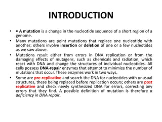
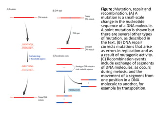
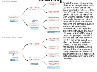
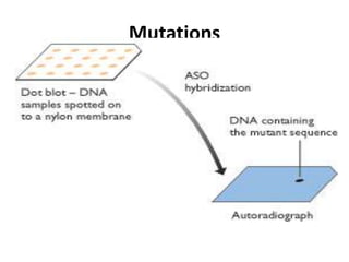
MODULE OF FOOD BIOTECHNOLOGY
UNIT of MICROBIAL GENETICS
DEPARTMENT OF BIOTECHNOLOGIES
LEVEL III FOOD BIOTECHNOLOGY
Joseph NDACYAYISENGA (M.Sc. , BIOT.)
JANUARY. 2020
Contents
• CHAPTER I: GENE
• CHAPTER II: SPONTANEOUS MUTABILITY IN BACTERIA
• CHAPTER III: BACTERIOPHAGE AS A GENETIC SYSTEM
• CHAPTER IV: GENETIC TRANSFER MECHANISMS
• CHAPTER V: MECHANISMS OF DRUG RESISTANCE IN
BACTERIA AND RECOMBINANT DNA CLONING
• CHAPTER VII: REGULATION OF GENE EXPRESSION IN
PROKARYOTES AND EUKARYOTES.
•
Definitions
• Microbial genetics is a subject area within
microbiology and genetic engineering. It studies
the genetics of very small (micro) organisms
• It is concerned with the transmission of
hereditary characters in microorganisms.
Definitions
Micro organisms include prokaryotes like bacteria,
unicellular or mycelial eukaryotes e.g., yeasts and other
fungi, and viruses, notably bacterial viruses
(bacteriophages).
Genetics is the study of what genes are, how they carry
information, how their information is expressed, and
how they are replicated and passed to subsequent
generations or other organisms.
Applications of microbial genetics in food
biotechnology
 Microbial genetics has played a unique role in
developing the fields of molecular and cell biology and
also has found applications in medicine, agriculture,
and the food and pharmaceutical industries.
 Specifically it is applied in food biotechnology in the
following ways:
Production of fermented foods.
Production of Enzymes widely applied as processing
aids in the food and beverage industry.
Applications of microbial genetics in food
biotechnology
 Production of Enzymes widely applied as processing aids in the
food and beverage industry.
 Production of food ingredients such as; flavoring agents, organic
acids, food additives and amino acids. All these ingredients are
metabolites of microorganisms during fermentation processes.
 Applicable in food testing diagnostics to underpin its safety.
 Some are used as food e.g. mycelial fungi like Mushrooms.
What is genetics?
• Study of inheritance and traits expressed in
the genetic material of the organism.
• GENOTYPE: genetic “make-up” of the
organism
• PHENOTYPE: expression of the genotype - the
organism and its traits.
Central Dogma Theory
The central dogma theory of
molecular biology is represented
by a simple pathway: DNA—
>RNA-->protein, which
demonstrates the flow of genetic
information in a living cell.
The major processes involved in
this pathway are replication,
transcription, and translation.
Central Dogma Theory
In DNA replication, the DNA
polymerase enzyme replicates all
the DNA in the nuclear genome
in a semi-conservative manner,
meaning that the double
stranded DNA is separated into
two and a template is made by
DNA polymerase.
This allows genomic material to
be duplicated so it can be evenly
partitioned between two somatic
cells (daughter cells) upon
division.
Central Dogma Theory
The process in which DNA is
copied into RNA by RNA
Polymerase is called
transcription.
Three forms of RNA are
produced
here: messenger RNA
(mRNA),
ribosomal RNA (rRNA), and t
ransfer RNA (tRNA).
Central Dogma Theory Summary
1. DNA guides the synthesis of
mRNA which in turn directs the
order in which amino acids are
assembled into proteins.
2. DNA directs its own
replication by giving rise to two
complete, identical DNA
molecules.
This replication is necessary
because each cell must inherit a
complete set of all genes in
order to carry out the cell’s life
processes.
Reverse Transcriptase
Another process in this
pathway is reverse
transcription, which
involves
copying RNA information
into
DNA using reverse
transcriptase.
Reverse Transcriptase
Recently, this processes has
been defined and may expand
the central dogma.
For example, retroviruses use
the enzyme "reverse
transcriptase" to transcribe DNA
from a RNA template.
The viral DNA then integrates into the
nucleus of the host cell. Then it is
transcribed, and further translated into
proteins.
This biological process effectively adds
another pathway to the central dogma
of molecular biology.
CHAPTER I: GENE
What is a gene and where is it located ?
Genes
• Genes are the basic physical and functional units
of heredity.
• It is a unit of inheritance.
• Each gene is located on a particular region of a
chromosome (Locus)and has a specific ordered
sequence of nucleotides (the building blocks of
DNA).
What is a locus?
• A locus describes the region of a
chromosome where a gene is located.
– 11p15.5 is the locus for the human
insulin gene. 11 is the
chromosome number,
– p indicates the short arm of the
chromosome,
– 15.5 is the number assigned to a
particular region on a
chromosome.
– When chromosomes are stained in
the lab, light and dark bands
appear, and each band is
numbered.
– The higher the number, the farther
away the band is from the
centromere.
Role of genes in development.
• Differential Regulation of Gene Expression
• Determines Cell Form and Function and the
over all shape of multicellular organisms.
What is a gene and where is it located?
1. Genetic information of an organism is stored in its
DNA .
2. On each DNA there are Segments that encode
proteins or other functional products. Such segments
are called genes.
3. Gene segments in DNA are transcribed into
messenger RNA intermediates (mRNA) / Any RNA.
4. mRNA intermediates are translated into proteins that
perform most life functions.
• DNA  RNA  Protein ….Central Dogma
variations in gene structure
Cellular and genomic organization of prokaryotes is different from that
of eukaryotes
1. Prokaryotes have no nucleus.
2. The nucleoid region in a prokaryotic cell consists of a concentrated
mass of DNA. This mass of DNA is usually one thousand times less
than what is found in a eukaryote.
3. A prokaryote may have a plasmid in addition to its major
chromosome. A plasmid is a small ring of DNA that carries accessory
genes.
Usually these genes are for antibiotic resistance!
1. Compare the organization of prokaryotic and
eukaryotic genomes.
Prokaryotic
• Usually circular
• Smaller
• Found in the nucleoid
region
• Less elaborately
structured and folded
Eukaryotic
• Complexed with a large
amount of protein to form
chromatin
• Highly extended and
tangled during interphase
• Found in the nucleus
A very simple eukaryotic gene
Eukaryotic genome organization
1. Multiple genomes: nuclear, mitochondria, chloroplasts
2. Plastid genomes resemble prokaryotic genomes
3. Multiple linear chromosomes, total size 5-10,000 MB, 5000 to
50000 genes
4. Monocistronic transcription units
5. Discontinuous coding regions (introns and exons)
Eukaryotic genome organization
(contd.)
6. Large amounts of non-coding DNA
7. Transcription and translation take place in different
compartments
8. Variety of RNAs: Coding (mRNA, rRNA, tRNA), Non-
coding (snRNA, snoRNA, microRNAs, etc).
9. Often diploid genomes and obligatory sexual
reproduction
10.Standard mechanism of recombination: meiosis
Gene – single unit of genetic function
Operon – genes transcribed in single transcript
Regulon – genes controlled by same
regulator
Modulon – genes modulated by
same stimilus
Element – plasmid, phage,
chromosome,
Genome
** order of ascending
complexity
Finding genes in eukaryotic DNA
Types of genes include
• protein-coding genes
• pseudogenes
• functional RNA genes: tRNA, rRNA and others
--snoRNA small nucleolar RNA
--snRNA small nuclear RNA
--miRNA microRNA
There are several kinds of exons:
-- noncoding
-- initial coding exons
-- internal exons
-- terminal exons
-- some single-exon genes are intronless
Prokaryotic genome organization:
Haploid circular genomes (0.5-10 Mbp, 500-10000
genes)
Genes arranged in Operons: Polycistronic transcription
units.
Environment-specific genes on plasmids and other
types of mobile genetic elements.
Usually asexual reproduction, great variety of
recombination mechanisms.
Transcription and translation take place in the same
compartment
• The organization of genes into an operon allows
for simultaneous expression of all the genes that
are located in cis (i.e., on the same contiguous
piece of DNA) in the operon
• It also allows the set of genes to undergo
horizontal gene transfer as a unit
Prokaryotic Operons
An operon
Exons vs Introns
• Eukaryotic genes have introns and exons. Exons contain
nucleotides that are translated into amino acids of
proteins. Exons are separated from one another by
intervening segments of junk DNA called introns. Introns
do not code for protein.
• They are removed when eukaryotic mRNA is processed.
Exons make up those segments of mRNA that are
spliced back together after the introns are removed; the
intron-free mRNA is used as a template to make
proteins.
Splicing
• Exons are sequences of DNA that are expressed into
protein.
• Introns are intervening sequences that are not
translated into protein
DNA
Pre-mRNA
31
1 2 3
1 32
2Spliced mRNA
3
C
C
C
Types of exons
5’
3’
Start Stop
Transcription start
Translation
StoppolyA
5’ untranslated
region
3’ untranslated
region
5’ 3’
Protein
coding
region
promoter
GT AG GT AG GT AG GT AG
Open reading frame
Gene
mRNA
Translation
Initial exon
Internal exon
Internal coding exon
Terminal exon
Exons and Coding
What’s the difference between exons and coding sequence?
Exons often are described as short segments of protein coding
sequence. This is a bit of an oversimplification. Exons are those
segments of sequence that are spliced together after the introns
have been removed from the pre-mRNA. Yes, the coding
sequence is contained in exons, but it is possible for some exons
to contain no coding sequence. Portions of exons or even entire
exons may contain sequence that is not translated into amino
acids. These are the untranslated regions or UTRs. UTRs are
found upstream and downstream of the protein-coding sequence.
Genome
• Entire genetic compliment of the organism.
• DNA - all prokaryotes and eukaryotes
• DNA or RNA - viruses
Transcription
• 1. This is the process of making a copy of a gene
(sequence of DNA that codes for a protein or
functional product)
• 2. The enzyme responsible for this process is RNA
POLYMERASE
• 3. Copies the gene in a 5’ → 3’ direction
• 4. Gene transcription begins at a site called the
PROMOTER and ends at another site called the
TERMINATOR
Events in Transcription
• Four types of RNA transcribed from DNA
– RNA primers
– mRNA
– rRNA
– tRNA
• Occurs in nucleoid of prokaryotes in nucleus of
eukaryotes.
• Three steps
– Initiation
– Elongation
– Termination
Initiation of Transcription
Elongation of the RNA Transcript
Concurrent RNA Transcription
RNA Polymerase Versus DNA
Polymerase
• RNA polymerase does not require helicase
• RNA polymerase slower than DNA polymerase
• Uracil incorporated instead of thymine
• RNA polymerase proofreading function is less
efficient than DNA polymerase (more errors)
Prokaryotic mRNA
Transcription in Eukaryotes
• RNA transcription occurs in the nucleus
• Transcription also occurs in mitochondria and
chloroplasts
• Three types of RNA polymerases
• Numerous transcription factors
• mRNA processed before translation
– Capping
– Polyadenylation
– Splicing
Eukaryotic mRNA
Transcription
(s
Another example of transcription
• 3’ GGGGGGGGGGGGGGG 5’ anti-sense
• 5’ CCCCCCCCCCCCCCC 3’ sense
Transcription (anti-sense)
5’ CCCCCCCCCCCCCCC 3’
Translation
Pro-pro-pro-pro-pro-pro-
Remember-copy the 3’ strand and by the rules of
base paring you get the sense strand sequence!
Post transcriptional processing
• This only done in eukaryotes.
• It involves:
Removal of introns,
Poly adenylation of the 3’ end and
Capping of the 5’ end.
RNA processing in eukaryotic cells
CHAPTER II: SPONTANEOUS
MUTABILITY IN BACTERIA
INTRODUCTION
• • A mutation is a change in the nucleotide sequence of a short region of a
genome.
• Many mutations are point mutations that replace one nucleotide with
another; others involve insertion or deletion of one or a few nucleotides
as we saw above.
• Mutations result either from errors in DNA replication or from the
damaging effects of mutagens, such as chemicals and radiation, which
react with DNA and change the structures of individual nucleotides. All
cells possess DNA-repair enzymes that attempt to minimize the number of
mutations that occur. These enzymes work in two ways.
• Some are pre-replicative and search the DNA for nucleotides with unusual
structures, these being replaced before replication occurs; others are post
replicative and check newly synthesized DNA for errors, correcting any
errors that they find. A possible definition of mutation is therefore a
deficiency in DNA repair.
One base pair is exchanged for another in the DNA molecule
One or more base pairs are inserted in the DNA molecule.
One or more base pairs are deleted in the DNA molecule
There is a rearrangement of sections in the DNA molecule.
There is an exchange of DNA region with another DNA molecule
(Recombination).
Some mutations harmful, some beneficial, some neutral
Changes in the DNA molecules can Cause Mutations
INTRODUCTION
• Recombination results in a restructuring of
part of a genome, for example by exchange of
segments of homologous chromosomes
during meiosis or by transposition of a mobile
element from one position to another within
a chromosome or between chromosomes.
Recombination is a cellular process which, like
other cellular processes involving DNA (e.g.
transcription and replication), is carried out
and regulated by enzymes and other proteins.
• Figure ;Mutation, repair and
recombination. (A) A
mutation is a small-scale
change in the nucleotide
sequence of a DNA molecule.
A point mutation is shown but
there are several other types
of mutation, as described in
the text. (B) DNA repair
corrects mutations that arise
as errors in replication and as
a result of mutagenic activity.
(C) Recombination events
include exchange of segments
of DNA molecules, as occurs
during meiosis, and the
movement of a segment from
one position in a DNA
molecule to another, for
example by transposition.
Mutations
• With mutations, the issues that we have to consider
are: how they arise; the effects they have on the
genome and on the organism in which the genome
resides; whether it is possible for a cell to increase its
mutation rate and induce programmed mutations
under certain circumstances
Mutations
• The causes of mutations
• Mutations arise in two ways:
• • Some mutations are spontaneous errors in
replication that evade the proofreading function
of the DNA polymerases that synthesize new
polynucleotides at the replication fork. These
mutations are called mismatches because they
are positions where the nucleotide that is
inserted into the daughter polynucleotide does
not match, by base-pairing, the nucleotide at the
corresponding position in the template DNA.
Mutations
• If the mismatch is retained in the daughter double helix
then one of the granddaughter molecules produced
during the next round of DNA replication will carry a
permanent double-stranded version of the mutation.
• • Other mutations arise because a mutagen has
reacted with the parent DNA, causing a structural
change that affects the base-pairing capability of the
altered nucleotide. Usually this alteration affects only
one strand of the parent double helix, so only one of
the daughter molecules carries the mutation, but two
of the granddaughter molecules produced during the
next round of replication will have it.
Mutations• Figure. Examples of mutations.
(A) An error in replication leads
to a mismatch in one of the
daughter double helices, in this
case a T-to-C change because
one of the As in the template
DNA was miscopied. When the
mismatched molecule is itself
replicated it gives one double
helix with the correct sequence
and one with a mutated
sequence. (B) A mutagen has
altered the structure of an A in
the lower strand of the parent
molecule, giving nucleotide X,
which does not base-pair with
the T in the other strand so, in
effect, a mismatch has been
created. When the parent
molecule is replicated, X base-
pairs with C, giving a mutated
daughter molecule. When this
daughter molecule is replicated,
both granddaughters inherit the
mutation.
Mutations
• Errors in replication are a source of point mutations
• When considered purely as a chemical reaction,
complementary base-pairing is not particularly accurate.
Nobody has yet devised a way of carrying out the template
dependent synthesis of DNA without the aid of enzymes,
but if the process could be carried out simply as a chemical
reaction in a test tube then the resulting polynucleotide
would probably have point mutations at 5 10 positions out
of every hundred. This represents an error rate of 5 10%,
which would be completely unacceptable during genome
replication. The template-dependent DNA polymerases
that carry out DNA replication must therefore increase the
accuracy of the process by several orders of magnitude.
This improvement is brought about in two ways:
Mutations
• Errors in replication are a source of point mutations
• The DNA polymerase operates a nucleotide selection
process that dramatically increases the accuracy of
template-dependent DNA synthesis.
• The accuracy of DNA synthesis is increased still further
if the DNA polymerase possesses a 3’-5’ exonuclease
activity and so is able to remove an incorrect
nucleotide that evades the base selection process and
becomes attached to the 3 end of the new
polynucleotide. This is called proofreading.
Mutations
• Mutation detection
• Rapid procedures for detecting mutations in DNA molecules.
• Any mutation can be identified by DNA sequencing but
sequencing is relatively slow and would be inappropriate for
screening a large number of samples. DNA chip technology
could also be employed, but this is not yet a widely available
option. For these reasons, a number of 'low technology'
methods have been devised. These can be divided into two
categories: mutation scanning techniques, which require no
prior information about the position of a mutation, and
mutation screening techniques, which determine whether a
specific mutation is present.
• Most scanning techniques involve analysis of the
heteroduplex formed between a single strand of the DNA
being examined and the complementary strand of a control
DNA that has the unmutated sequence:
Mutations
• Mutation detection
• Any mutation can be identified by DNA sequencing but
sequencing is relatively slow and would be inappropriate
for screening a large number of samples. DNA chip
technology could also be employed, but this is not yet a
widely available option. For these reasons, a number of
'low technology' methods have been devised. These can
be divided into two categories: mutation scanning
techniques, which require no prior information about
the position of a mutation, and mutation screening
techniques, which determine whether a specific
mutation is present.
• Most scanning techniques involve analysis of the
heteroduplex formed between a single strand of the
DNA being examined and the complementary strand of a
control DNA that has the unmutated sequence:
• If the test DNA contains a mutation then there
will be a single mismatched position in the
heteroduplex, where a base pair has not formed.
Various techniques can be used to detect
whether this mismatch is present or not:
Mutations
• Mutation detection
• Electrophoresis or high-performance liquid chromatography
(HPLC) can detect the mismatch by identifying the difference
in the mobility of the mismatched hybrid compared with the
fully base-paired one in a polyacrylamide gel or HPLC
column. This approach determines if a mismatch is present
but does not provide information on where in the test DNA
the mutation is located.
• • Cleavage of the heteroduplex at the mismatch position
followed by gel electrophoresis will locate the position of a
mismatch. If the heteroduplex stays intact then no mismatch
is present; if it is cleaved then it contains a mismatch, the
position of the mutation in the test DNA being indicated by
the sizes of the cleavage products. Cleavage is carried out by
treatment with enzymes or chemicals that cut at single-
stranded regions of mainly double-stranded DNA, or with a
single-strand-specific ribonuclease such as S1.
Mutations
• Mutation detection
• Most screening methods for detection of
specific mutations make use of the ability of
oligonucleotide hybridization to distinguish
between target DNAs whose sequences differ
at just one nucleotide position. In allele-
specific oligonucleotide (ASO) hybridization
the DNA samples are screened by probing
with an oligonucleotide that hybridizes only to
the mutant sequence:
Mutations
CHAPTER III: BACTERIOPHAGE AS
A GENETIC SYSTEM
What are Bacteriophages
Viruses that attack bacteria were
observed by Twort and d'Herelle in
1915 and 1917. They observed that
broth cultures of certain intestinal
bacteria could be dissolved by addition
of a bacteria-free filtrate obtainedfrom
sewage
Bacteriophages under Electron
Microscope
BACTRIOPHAGES
Like most viruses, bacteriophages
typically carry only the genetic
information needed for replication of
their nucleic acid and synthesis of their
protein coats.. They require precursors,
energy generation and ribosomes
supplied by their bacterial host cell.
Bacteriophage
 Bacteriophages
make up a
diverse group of
viruses, some of
which have
complex
structures,
including double-
stranded DNA.
Bacteriophage
 Also known simply as a
phage; a virus that
attacks and infects
bacteria. The infection
may or may not lead to
the death of the
bacterium, depending on
the phage and
sometimes on
conditions. Each
bacteriophage is specific
to one form of bacteria.
Composition and
Structure
 Composition
 Nucleic acid
 Genome
size
 Modified
bases
P Pr• Struc tuot(ein4
re T )
–
–
SPizreotection
HInefaedcotiron
–
Head/Capsi
d
Contractile Tail
Sheath
Tail Fibers
Base Plate
Bacteria response to bacteriophage
When bacteria are mixed with bacteriophage:
Phage entering a bacterial cell
Bacteria response to bacteriophage
When bacteria are mixed with bacteriophage:
Bacteria response to bacteriophage
• If about a billion bacteria mixed with a particular
toxin, nearly all of the bacteria are killed.
• A few will survive and give rise to colonies that
are permanently and specifically resistant to that
particular toxin
Bacteriophage showing Lytic and
lysogenic cycle
12 Dr.T .Rao M grad e Ser 16
Bacteriophages:
Virulence Factors Carried On Phage

Temperate phage can go through one of
two life cycles upon entering a host cell.
1) Lytic:
Is when growth results in lysis of the host and release
of progeny phage.
2) Lysogenic:
Is when growth results in integration of the phage DNA
into the host chromosome or stable replication as a
plasmid.
Most of the gene products of the lysogenic phage
remains dormant until it is induced to enter the lytic
cycle.
Bacteriophages:
Lysogenic Conversion

Some lysogenic phage carry genes
that can enhance the virulence of the
bacterial host.


• For example, some phage carry genes that
encode toxins.
• These genes, once integrated into the
bacterial chromosome, can cause the
once harmless bacteria to release
potent toxins that can cause disease.
Bacteriophages
•
•
•
Used for cloning foreign
genes among other
applications
Proteins and peptides are
fused to the
Capsid(surface) of the
phage
The combination of the
phage and peptide is
known as a Fusion Protein
Lytic and Lysogenic
cycle
85
Lysogenic conversion
In some interactions between lysogenic
phages and bacteria, lysogenic conversion
may occur. It is when a temperate phage
induces a change in the phenotype of the
bacteria infected that is not part of a usual
phage cycle. Changes can often involve
the external membrane of the cell by
making it impervious to other phages or
even by increasing the pathogenic
capability of the bacteria for a host.
Lytic
vs
Lysogenic
Cycle
Transduction
Transduction
Penetration:
Phage pnetrates
host cell and
injects its DNA.
Merozoites released
into bloodsteam
from liver may
infectnew red blood cells
1
Att
achment:
Phage
attaches to
host cell.
2
3
Bacteria
l cell
wall
Bacterial
chromosom
e
Capsid
Ca
S
T
Ba
Pin
Ce
P
Sh
Ta
4 Maturation:
Viral
components are
assembled into
virions.
5 Release:
Host cell lyses
and new
virions
are released.
Tail
DNA
Capsid
Tail fibers
• CHAPTER IV: GENETIC TRANSFER
MECHANISMS AND EXTRA-
CHROMOSOMAL FACTORS
Genetic transfer mechanisms in
bacteria.
• 1. Transformation
• 2. Conjugation
• 3. Transduction
Transformation: genes transferred from
one bacterium to
Another.
After cell death, some bacteria are lysed
and release cellular contents into
surrounding environment.
The recipient cell is in a physiological state
that will allow it to take up DNA.
Transformation occurs naturally among a
few organisms.
Transformation
Transformation-uptake of naked DNA
Conjugation
• Cell to cell contact
required
• Plasmid exchange
through the sex pilus
• Plasmid is called the F
factor
Conjugation
Hfr cell
Hfr x F- cell
Transduction
Transfer of genes from a
donor to a recipient by a
bacteriophage
Transduction- A defective phage is required
Extra-chromosomal factors
• These include:
 Plasmids,
Episomes,
Transposomes and
colicins
Plasmids and Plasmid Biology
Plasmids
• Small molecules of DNA that replicate
independently
• Carry information required for their own
replication, and often for one or more cellular
traits
– Not essential for normal bacterial metabolism,
growth, or reproduction
– Can confer survival advantages
Plasmids
1. Extrachromosomal DNA, usually circular-parasite?
2. Usually encode ancillary functions for in vitro growth
3. Can be essential for specific environments: virulence, antibiotics
resistance, use of unusual nutrients, production of bacteriocins
(colicins)
4. Must be a replicon - self-replicating genetic unit
5. Plasmid DNA must replicate every time host cell divides or it will be
lost
a. DNA replication
a. partitioning (making sure each progeny cells receives a plasmid)
6. High copy plasmids are usually small; low copy plasmids can be
large
7. Partitioning is strictly controlled for low copy, but loose for high copy
8. Plasmid replication requires host cell functions
9. Copy number is regulated by initiation of plasmid replication
10. Plasmids are incompatible when they cannot be stably maintained in
the same cell because they interfere with each other’s replication.
Plasmid replication
1. Plasmid replication requires host DNA replication machinery.
2. Most wild plasmids carry genes needed for transfer and copy number
control.
3. All self replication plasmids have a oriV: origin of replication
4. Some plasmids carry and oriT: origin of transfer. These plasmids will
also carry functions needed to be mobilized or mob genes.
5. Plasmid segregation is maintained by a par locus-a partition locus
that ensures each daughter cells gets on plasmid. Not all plasmids
have such sequences.
6. There are 5 main “incompatibility” groups of plasmid replication. Not
all plasmids can live with each other.
7. Agents that disrupt DNA replication destabilize or cure plasmids from
cells.
Incompatibility Groups
1. Not all plasmids can live together.
2. Plasmids that are able to coexist in the same cell do not interfere with
each other’s replication
3. A single cell can have as many group plasmids as it can tolerate and
replicate!
Partion Locus: a region on broad host range plasmids that binds to a structure on the
inner membrane of the cell to ensure proper segregation. Plasmids labeled with
fluorescent protein
-move to each daughter cell during division.
Pogliano, Joe et al. (2001) Proc. Natl. Acad. Sci. USA 98, 4486-4491
Plasmids
• Many types of plasmids
– Fertility factors
– Resistance factors
– Bacteriocin factors
– Virulence plasmids
– Cryptic plasmids
Episomes
• It is a segment of DNA in certain cells, especially bacterial cells,
that can exist either autonomously in the cytoplasm or as part of a
chromosome.
• Episome is a unit of genetic material composed of a series of genes
that sometimes has an independent existence in a host cell and at
other times is integrated into a chromosome of the cell, replicating
itself along with the chromosome.
• As autonomous units they destroy host cells, and as segments
integrated into a chromosome they multiply in cell division and are
transferred to daughter cells.
• Examples of episomes include insertion sequences and
transposons. Viruses are another example of an episome.
colicins
• Definition: Colicins are a type of bacteriocin (peptide
and protein) antibiotics released by bacteria to kill
other bacteria of the same species, in order to
provide a competitive advantage for nutrient
acquisition
• The genes that code for colicins are carried on
plasmids.
Colicins continued
• There are two general classes of colicinogenic
plasmids, large, low-copy number plasmids, and
small high copy number plasmids.
• The larger plasmids carry other genes as well as the
colicin operon. The colicin operons are generally
organized with several major genes.
Transposome,Transposons and Transposition
• Transposomes are a collection of transposons that may be
obatianed with in a cell.
• Transposons – segments of DNA that move from one location to
another in the same or different molecule
• Result is a kind of frameshift insertion (transpositions)
Transposons and Transposition
• Simplest transposons are insertion sequences
which have no more than two inverted repeats
and gene for transposase
• Complex transposons contain one or more
genes not connected with transposition (e.g.
antibiotic resistance)
Describe the effects of transposons and
retrotransposons.
• Transposons  jump and interrupt the normal functioning
may increase or decrease production of one or more proteins
- can carry a gene that can be activated when inserted
downstream from an active promoter and vice versa
• Retrotransposons  transposable elements that move within
a genome by means of an RNA intermediate, a transcript of
the retrotransposon DNA
- to insert it must be converted back to DNA by reverse
transcriptase
CHAPTER V: MECHANISMS OF DRUG
RESISTANCE IN BACTERIA AND
RECOMBINANT DNA CLONING
Antibiotics
• Antibiotics are derived primarily from three
major sources:
• -molds or fungi
• -bacteria: Streptomyces, Bacillus
• -synthetic or semisynthetic
• used internally or topically, inhibit or kill
pathogens
• work best on actively growing organisms, but
not on non-growing persisters or spores.
Types of antibiotics
• Bacteriostatic versus Bactericidal
• Static: inhibit growth
• Cidal: kill
• Cidal or static is not absolute, depending on drug
concentration, bacterial species, phase of growth of the
organism, and even the number of bacteria
• MIC (minimum inhibitory concentration): agar dilution;
broth dilution, automated antibiotic susceptibility testing
• MBC (minimum bactericidal concentration)
Antibiotic Combination
Additive: drug combination is more active
than either drug alone and the response
represents a sum of two drug effects.
Synergism: combination has a greater effect
than the sum of the two individual drug
effects.
 Antagonism: combination has less activity
than that of individual drug alone
Broad versus Narrow Spectrum
• Tetracycline: typical broad spectrum antibiotic,
active against G+ and G- bacteria,
Mycobacterium, Rickettsia, protozoan
• Penicillin: primarily G+ bacteria,
• Gentamycin: G- bacteria
• Pyrazinamide: specific for M. tuberculosis
Mechanism of Action:
Five major classes of antibiotics
Inhibition of cell wall synthesis (beta-lactams,
glycopeptides): most common.
 Inhibition of protein synthesis (amino glycoside,
chloramphenicol, tetracycline, macrolides).
 Disruption of membrane permeability (polymyxin
B for G-bacteria, gramicidin and daptomycin for
G+ bacteria).
 Inhibition of nucleic acid synthesis
(fluoroquinolones for DNA and rifampin for RNA
synthesis).
 Anti-metabolite (sulfa drugs)
Drug Resistance definition
It is a condition in which there is insensitivity to drugs that usually
cause growth inhibition or cell death at a given concentration
 People cannot be effectively treated.
 People are ill for longer.
 People are at a greater risk of dying.
 Epidemics are prolonged.
 Others are at a greater risk of infection
Causes of Resistance Problem
 Antibiotic overuse, abuse or misuse (misdiagnosis).
 Counterfeit Drugs.
 Antibiotic use in animal husbandry and food: jumping
from animals to humans; chicken contaminated with
MDR-campylobacter.
 Globalization and resistance
Antibiotic Resistance
 Natural Resistance: Bacteria may be inherently resistant to an
antibiotic. Streptomyces has some genes responsible for resistance to
its own antibiotic; or a Gram- bacteria have an outer membrane as a
permeability barrier against antibiotic (e.g., penicillin); or an organism
lacks a transport system for the antibiotic; or efflux pumps; or it lacks
the target (e.g. INH-mycolic acid synthesis) of the antibiotic.
 Acquired Resistance: Bacteria can develop resistance to antibiotics
due to (1) mutations; (2) mobile genetic elements, e.g., plasmids or
transposons carrying antibiotic resistance gene
Antibiotic Resistance Mechanisms
There are two Types of Antibiotic Resistance:
 Genetic resistance: due to chromosomal mutations or
acquisition of antibiotic resistance genes on plasmids or
transposons.
 Phenotypic resistance: due to changes in bacterial
physiological state as in stationary phase, antibiotic persisters,
dormant state
How Do Bacteria Acquire Resistance?
 Resistance due to drug selection or drug induction?
– 1950s, Joshua Lederberg devised replica plating-> demonstrating
selection of pre-existing resistant mutant- growth dependent
Spontaneous mutations
– 1988, John Cairns showed mutations arise also in nondividing or slowly
dividing cells and have some relation to the selective pressure used.
These mutations, named adaptive mutations, arise only in the presence of
a non-lethal selective pressure that favors them.
– Drug induction also plays a role, e.g., efflux
 Natural selection of spontaneous mutants in a large bacterial population:
mutation frequency to rifampicin=10-8, INH= 10-6
 Drug combination to avoid resistance: mutants resistant to both RIF and
INH occurs at 10-14
Mechanisms of Drug Resistance
• (A) Chromosomal mutations:
• 1. Reduced permeability/uptake
• 2. Enhanced efflux
• 3. Enzymatic inactivation (beta-lactamase)
• 4. Alteration of drug target
• 5. Loss of enzymes involved in drug activation (as in isoniazid
resistance-KatG, pyrazinamide resistance-PncA)
• (B) Plasmid or transposon mediated:Plasmid-mediated: 1959
Japanese found plasmid-mediated MDR (sulfonamides, streptomycin,
chloramphenicol, tetracycline) in Shigella species
• Sequential accumulation of chromosomal mutations, one at a time,
leading to MDR
Microbial genetic Questions
• What are the applications of microbial genetics?
• Explain the Role of genes in development
• How the Cellular and genomic organization of prokaryotes is different from
that of eukaryotes?
• Describe the hierarchy of gene organization
• Locate the transcription and translation and both eukaryotes and prokaryote
• What are operons, role and the components?
• What are the enzymes involved in DNA replication and their role?
• What Bacteria response to bacteriophage and what are the Proposed
mechanisms for them to survive?
• Describe the Genetic transfer mechanisms in bacteria
• Give the extra chromosomal factors in bacteria
• How plamids replicate?
• Give types of plasmids
• What are Episomes and their function?
• What are colicins and their role?
• Describe the effects of transposons and
retrotransposons
• What are the three major sources?
• What are the mechanisms of drug resistance in
bacteria?
• What the types of antibiotics and their activity?
• What the mechanism of antibiotic action?
• Explain the mechanisms of drug resistance
A. CHROMOSOMAL MUTATIONS
• 1. Reduced Permeability/Uptake
• Outer membrane porin mutations
(crossresistance): Neisseria gonorrhoeae porin
mutation cause resistance to penicillin and
tetracycline; Enterobacter aerogenes porin mutation
cause cephalosporin resistance
CHROMOSOMAL MUTATIONS
2. Increased Efflux Activity (many examples)
• Membrane bound proteins involved in extrusion of
antibiotics out of bacterial cell, energy-dependent (ATP,
proton motive force)
• Tetracyclines (first efflux mechanism): efflux proteins - TetA
to G in G- bacteria; TetK and TetL in G+ bacteria Macrolides
(Staph), ATP-dependent fluoroquinolones (pseudomonas sp.,
Staph, enterococci), streptogramins (Staph)
• Cross-resistance by efflux pump:
CHROMOSOMAL MUTATIONS cont’d
• 3. Enzymatic Inactivation
• Beta-lactamases cleave beta-lactam antibiotics and cause
resistance
• Aminoglycoside-inactivating enzymes (adding groups acetyl,
adenyl, phosphoryl to inactivate the antibiotic)
• Chloramphenicol acetyl transferase: add acetyl group to
inactivate chloramphenicol
• Streptogramin acetyl transferase: found in Staph,
Enterococci
CHROMOSOMAL MUTATIONS cont’d
• 4. Alteration of Drug Target (numerous examples)
• Penicillin-binding proteins (PBP/transpeptidase):
alteration due to mutations cause resistance to beta-
lactams commonly in G+ bacteria (e.g., methicillin
resistance in S. aureus, mecA encoding PBP2a)
• Vancomycin resistance: vancomycin prevents cross-
linking of peptidoglycan by binding to DAla-
• D-Ala dipeptide of the muramyl peptide. Most G+ bacteria
acquire vancomycin resistance by changing D-Ala-D-Ala to
D-Ala- D-lactate, which does not bind vancomycin
CHROMOSOMAL MUTATIONS cont’d
• 5. Resistance Caused by Loss of Enzymes
• Involved in Drug Activation
• The following drugs are prodrugs that need to be activated by
bacterial enzymes for activity, and mutations in the enzymes cause
inability to activate the drug, leading to resistance: e.g.
• Isoniazid (INH): KatG (catalase-peroxidase) activate INH to
produce active metabolites which then inhibit multiple targets
including mycolic acid synthesis
• Pyrazinamide (PZA): PncA (nicotinamidase/PZase) activate PZA
to active form pyrazinoic acid (POA), which targets membrane
and disrupts energy metabolism
B. TRANSFER OF RESISTANCE GENES
• Conjugation: Plasmids and Transposons:
• Plasmid-mediated: vancomycin resistance (vanA) in
Enterococcus faecium (1988)
• strA- strB streptomycin-resistance genes can be carried
on plasmid in Shigella flexneri, on transposon (Tn5393) in
pseudomonas sp
• Plasmid-mediated sulfonamide and trimethoprim
• resistance in G- bacteria: plasmids carry drug insensitive
dihydropteroate synthase or dihydrofolate reductase
Limiting Drug Resistance
• (i) Antibiotics should be used only when
• necessary
• (ii) Antibiotics can be employed such that high
• concentrations of drug is maintained over long
• periods (i.e., taking all of one's pills over the
• prescribed duration of a treatment)
• (iii) Antibiotics may be used in combination to
• prevent resistance and improve the efficacy of
• treatment
INTRODUCTION TO RECOMBINANT DNA AND
BIOTECHNOLOGY
Recombinant DNA technology is
one of the recent advances in
biotechnology, which was
developed by two scientists named
Boyer and Cohen in 1973.
Stanley N. Cohen (1935–) (top) and
Herbert Boyer (1936–) (bottom),
who constructed the first
recombinant DNA using bacterial
DNA and plasmids.
Stanley N. Cohen , who
received the Nobel Prize in
Medicine in 1986 for his
work on discoveries of
growth factors.
What is Recombinant DNA Technology?
 Recombinant DNA technology is a
technology which allows DNA to be
produced via artificial means.
 The procedure has been used to change
DNA in living organisms and may have even
more practical uses in the future.
 Recombinant DNA technology works by
taking DNA from two different sources and
combining that DNA into a single molecule.
 That alone, however, will not do much.
 Recombinant DNA technology only becomes
useful when that artificially-created DNA is
reproduced. This is known as DNA cloning.
Brief Introduction
Concept of Recombinant DNA
 Recombinant DNA is a molecule that
combines DNA from two sources .
 Creates a new combination of genetic
material
– Human gene for insulin was placed in
bacteria
– The bacteria are recombinant
organisms and produce insulin in large
quantities for diabetics
– Genetically engineered drug in 1986
 Genetic engineering is the application of
this technology to the manipulation of
genes.
 These advances were made possible by
methods for amplification of any
particular DNA segment, regardless of
source, within bacterial host cells. Or, in
the language of recombinant DNA
technology, the cloning of virtually any
DNA sequence became feasible.
 Recombinant technology begins with the isolation
of a gene of interest (target gene).
 The target gene is then inserted into the plasmid
or phage (vector) to form replicon.
 The replicon is then introduced into host cells to
cloned and either express the protein or not.
 The cloned replicon is referred to as recombinant
DNA. The procedure is called recombinant DNA
technology.
 Cloning is necessary to produce numerous copies
of the DNA since the initial supply is inadequate to
insert into host cells.
CHAPTER VII: REGULATION OF GENE
EXPRESSION IN PROKARYOTES AND
EUKARYOTES
Every cell has the same DNA and therefore
the same genes. But different genes need to be
“on” and “off” in different types of cells.
Therefore, gene expression must be regulated.
embryo
bone
liver
muscle
sperm
(The first statement on this slide is not completely true. Which of these cells
does not have exactly the same DNA as the other? Can you think of any other
examples of cells in your body that have different DNA than most of the
others?)
Gene expression must be regulated in
several different dimensions—
10 wks 14 wks 1 day
6 mos 12 mos 18 mos
In time:
At different stages of the life cycle, different genes need to be on and off.
© M. Halfon, 2007
In space:
Paddock S.W. (2001). BioTechniques 30: 756 - 761.
Each colored stripe in this fly embryo shows the expression
of a different gene or set of genes. The spatial regulation of
these genes allows the embryo to be divided up into different
regions that will give rise to the head, the internal organs, the
abdomen, etc.
and in abundance:
Clyde et al. (2003). Nature 426:849-853
Note how the gene whose expression is indicated in blue varies
in abundance from strong expression (bold arrow) to weak
(thin arrow) within its expression domain. These differences in
strength of gene expression have important functional
consequences.
• Some of the many areas in which
regulated gene expression plays a
critical role are illustrated on the
following slide.
• Gene regulation is important not
only during development but also
in mediating common variation
between individuals, diseases and
birth defects, and evolution.
Gene Regulation and Nutrition:
Development (organs, cell types)
embryo
muscle
brain
embryo
intestines
fat
liver (diseased)
With respect to nutrition, gene regulation is important to guide
the development of organs, tissues, and cell types required to
ingest, digest, and metabolize nutrients.
Genes can be regulated at many levels
RNA PROTEINDNA
TRANSCRIPTION TRANSLATION
The “Central Dogma”
Control of Gene Expression—Transcription Factors
Transcription factors (TFs) are proteins that bind to
the DNA and help to control gene expression. We
call the sequences to which they bind transcription
factor binding sites (TFBSs), which are a type of
cis-regulatory sequence.
Most transcription factors can bind to a range of similar
sequences. We call this a binding “motif.”
Wasserman, W. W. and A. Sandelin (2004). Nat Rev Genet 5(4): 276-287.
(We can represent these motifs in various ways, which
we will see in Unit 2.5)
Control of Gene Expression—Transcription Factors
Control of Gene Expression
Image adapted from Wolpert, Principles of Development
Transcription factor binding sites are found within larger
functional units of the DNA called cis-regulatory elements.
There are two main type of cis-regulatory elements: promoters,
and cis-regulatory modules (sometimes called “enhancers”).
TFBS
TFBS
transcription factor binding site (TFBS)
cis-regulatory module (CRM)
Control of Gene Expression: Promoters
Every gene has a promoter, the DNA sequence immediately
surrounding the transcription start site. The promoter is the site
where RNA polymerase and the so-called general transcription
factors bind.
Additional gene regulation takes place via the cis-regulatory
modules (CRMs), which can be located 5’ to, 3’ to, or within
introns of a gene. CRMs can be very far away from the gene
they regulate—over 50 kb—and other genes might even lie in
between!
Control of Gene Expression: CRMs
TFBS
TFBS
transcription factor binding site (TFBS)
cis-regulatory
module (CRM)
Genes are often regulated in a modular fashion—discrete cis-regulatory elements
(CRMs, “enhancers”) dictate a specific spatio-temporal expression pattern, shown
here by purple stain. A gene might have many CRMs, each responsible for a
different part of its overall expression pattern.
cis-Regulatory Modules (enhancers)
Map of 3’ regulatory region of Drosophila even skipped (Fujioka et al. 1999)
eve
Looking at cis-regulatory modules:
Reporter Genes
How can we identify and study CRMs? To do this we use a
reporter gene assay. In such an assay, we use recombinant
DNA methods to test if a DNA sequence can regulate the
expression of a gene whose expression we can easily identify
(a “reporter gene”). The jellyfish green fluorscent protein
(GFP) gene is often used, as the encoded protein emits green
light when exposed to light of the proper wavelength. We can
test for CRM activity in transfected cells in culture, or even
better, in a transgenic animal:
TFBS
TFBS
transcription factor binding site (TFBS)
cis-regulatory module (CRM)
Looking at cis-regulatory modules:
Reporter Genes
green fluorescent proteinminimal promoterCRM
muscle
e.g., from Myosin Heavy
Chain gene
green fluorescent proteinminimal promoterCRM
reporter construct
transfect cells make transgenic animal
Looking at cis-regulatory modules:
Reporter Genes
Explain the potential role that promoters and
enhancers play in transcriptional control.
Promoters
• Include the proximal control
elements
• Produces a low rate of
initiation with few RNA
transcripts
• Unless  DNA sequences
can improve the efficiency
by binding additional
transcription factors
Enhancers
• The more distant control
elements
• Bending of the DNA enables
the transcription factors
bound to enhancers to
contact proteins of the
transcription-initiation
complex at the promoter 
Compare the arrangement of coordinately controlled
genes in prokaryotes and eukaryotes.
Prokaryotic
• Prokaryotic genes that are
turned on and off together
are often clustered into
operons which are
transcribed into one mRNA
molecule and translated
together
Eukaryotic
• Eukaryotic genes coding
for enzymes of a
metabolic pathway are
often scattered over
different chromosomes
and are individually
transcribed 
Regulation of Gene Expression:
• Gene expression refers to the combined process of transcription and translation of
genetic information to a functional protein.
• Not all genes are expressed at any one time, nor are they always expressed at the
same level.
• Gene expression is tightly regulated, or controlled, so that the cell only makes the gene
products that it needs for efficient growth under its current environmental conditions.
• What’s an operon? Regulatory protein binding region and downstream gene(s).
• Regulatory proteins called repressors or activators act as off and on switches for
transcription, respectively.
• Negative regulation involves repressor proteins that respond to cell conditions so to
actively repress (prevent) RNA Polymerase from beginning transcription of the gene (or
operon) by binding onto the DNA at the operator site.
• Positive regulation of transcription also can occur. Here, environmental conditions in
the cell causes an activator protein to bind to the promoter site for a gene (or operon),
which enables RNA Polymerase to begin transcription.
Negative Regulation:
• Binding of a repressor to the operator site prevents RNA Polymerase from
transcribing the gene(s).
• A repressors ability to bind to DNA is determined by the presence or absence of
certain small molecules in the cell that bind to the repressor protein..
E.g. E. coli Lactose Catabolism (lac) Operon
However, glucose
must be absent for
transcription! Why?
When no allolactose,
repressor binds to
operator. OFF!
With allolactose, it
prevents repressor from
binding to operator. ON!
Positive Regulation:
E.g. E. coli Lactose Catabolism (lac) Operon
• E. coli growth using glucose
is more efficient than growth
on lactose.
• How is wasteful expression
of lac operon genes prevented
when glucose is present?
• lac operon needs an
activator bound to promoter
site for any chance of
transcription, regardless of the
repressor.
• The catabolic activator
protein (CAP) only binds in
the absence of glucose.
The lac Operon in Action:
• E. coli growth is slower on lactose than on glucose.
• Glucose will be used before lactose given a choice. Here the repressor does not
stop transcription; however the CAP activator does by not binding to the promoter.)
• Once glucose runs out the CAP activator binds, and the lac operon is expressed in
the presence of lactose.
• Why is there a lag in growth when glucose runs out? This is called diauxic growth.
Thank you!!

Microbial genetics notes

  • 1.
    MODULE OF FOODBIOTECHNOLOGY UNIT of MICROBIAL GENETICS DEPARTMENT OF BIOTECHNOLOGIES LEVEL III FOOD BIOTECHNOLOGY Joseph NDACYAYISENGA (M.Sc. , BIOT.) JANUARY. 2020
  • 2.
    Contents • CHAPTER I:GENE • CHAPTER II: SPONTANEOUS MUTABILITY IN BACTERIA • CHAPTER III: BACTERIOPHAGE AS A GENETIC SYSTEM • CHAPTER IV: GENETIC TRANSFER MECHANISMS • CHAPTER V: MECHANISMS OF DRUG RESISTANCE IN BACTERIA AND RECOMBINANT DNA CLONING • CHAPTER VII: REGULATION OF GENE EXPRESSION IN PROKARYOTES AND EUKARYOTES. •
  • 3.
    Definitions • Microbial geneticsis a subject area within microbiology and genetic engineering. It studies the genetics of very small (micro) organisms • It is concerned with the transmission of hereditary characters in microorganisms.
  • 4.
    Definitions Micro organisms includeprokaryotes like bacteria, unicellular or mycelial eukaryotes e.g., yeasts and other fungi, and viruses, notably bacterial viruses (bacteriophages). Genetics is the study of what genes are, how they carry information, how their information is expressed, and how they are replicated and passed to subsequent generations or other organisms.
  • 5.
    Applications of microbialgenetics in food biotechnology  Microbial genetics has played a unique role in developing the fields of molecular and cell biology and also has found applications in medicine, agriculture, and the food and pharmaceutical industries.  Specifically it is applied in food biotechnology in the following ways: Production of fermented foods. Production of Enzymes widely applied as processing aids in the food and beverage industry.
  • 6.
    Applications of microbialgenetics in food biotechnology  Production of Enzymes widely applied as processing aids in the food and beverage industry.  Production of food ingredients such as; flavoring agents, organic acids, food additives and amino acids. All these ingredients are metabolites of microorganisms during fermentation processes.  Applicable in food testing diagnostics to underpin its safety.  Some are used as food e.g. mycelial fungi like Mushrooms.
  • 7.
    What is genetics? •Study of inheritance and traits expressed in the genetic material of the organism. • GENOTYPE: genetic “make-up” of the organism • PHENOTYPE: expression of the genotype - the organism and its traits.
  • 8.
    Central Dogma Theory Thecentral dogma theory of molecular biology is represented by a simple pathway: DNA— >RNA-->protein, which demonstrates the flow of genetic information in a living cell. The major processes involved in this pathway are replication, transcription, and translation.
  • 9.
    Central Dogma Theory InDNA replication, the DNA polymerase enzyme replicates all the DNA in the nuclear genome in a semi-conservative manner, meaning that the double stranded DNA is separated into two and a template is made by DNA polymerase. This allows genomic material to be duplicated so it can be evenly partitioned between two somatic cells (daughter cells) upon division.
  • 10.
    Central Dogma Theory Theprocess in which DNA is copied into RNA by RNA Polymerase is called transcription. Three forms of RNA are produced here: messenger RNA (mRNA), ribosomal RNA (rRNA), and t ransfer RNA (tRNA).
  • 11.
    Central Dogma TheorySummary 1. DNA guides the synthesis of mRNA which in turn directs the order in which amino acids are assembled into proteins. 2. DNA directs its own replication by giving rise to two complete, identical DNA molecules. This replication is necessary because each cell must inherit a complete set of all genes in order to carry out the cell’s life processes.
  • 12.
    Reverse Transcriptase Another processin this pathway is reverse transcription, which involves copying RNA information into DNA using reverse transcriptase.
  • 13.
    Reverse Transcriptase Recently, thisprocesses has been defined and may expand the central dogma. For example, retroviruses use the enzyme "reverse transcriptase" to transcribe DNA from a RNA template. The viral DNA then integrates into the nucleus of the host cell. Then it is transcribed, and further translated into proteins. This biological process effectively adds another pathway to the central dogma of molecular biology.
  • 14.
  • 15.
    What is agene and where is it located ?
  • 16.
    Genes • Genes arethe basic physical and functional units of heredity. • It is a unit of inheritance. • Each gene is located on a particular region of a chromosome (Locus)and has a specific ordered sequence of nucleotides (the building blocks of DNA).
  • 17.
    What is alocus? • A locus describes the region of a chromosome where a gene is located. – 11p15.5 is the locus for the human insulin gene. 11 is the chromosome number, – p indicates the short arm of the chromosome, – 15.5 is the number assigned to a particular region on a chromosome. – When chromosomes are stained in the lab, light and dark bands appear, and each band is numbered. – The higher the number, the farther away the band is from the centromere.
  • 19.
    Role of genesin development. • Differential Regulation of Gene Expression • Determines Cell Form and Function and the over all shape of multicellular organisms.
  • 20.
    What is agene and where is it located? 1. Genetic information of an organism is stored in its DNA . 2. On each DNA there are Segments that encode proteins or other functional products. Such segments are called genes. 3. Gene segments in DNA are transcribed into messenger RNA intermediates (mRNA) / Any RNA. 4. mRNA intermediates are translated into proteins that perform most life functions. • DNA  RNA  Protein ….Central Dogma
  • 21.
    variations in genestructure Cellular and genomic organization of prokaryotes is different from that of eukaryotes 1. Prokaryotes have no nucleus. 2. The nucleoid region in a prokaryotic cell consists of a concentrated mass of DNA. This mass of DNA is usually one thousand times less than what is found in a eukaryote. 3. A prokaryote may have a plasmid in addition to its major chromosome. A plasmid is a small ring of DNA that carries accessory genes. Usually these genes are for antibiotic resistance!
  • 22.
    1. Compare theorganization of prokaryotic and eukaryotic genomes. Prokaryotic • Usually circular • Smaller • Found in the nucleoid region • Less elaborately structured and folded Eukaryotic • Complexed with a large amount of protein to form chromatin • Highly extended and tangled during interphase • Found in the nucleus
  • 23.
    A very simpleeukaryotic gene
  • 24.
    Eukaryotic genome organization 1.Multiple genomes: nuclear, mitochondria, chloroplasts 2. Plastid genomes resemble prokaryotic genomes 3. Multiple linear chromosomes, total size 5-10,000 MB, 5000 to 50000 genes 4. Monocistronic transcription units 5. Discontinuous coding regions (introns and exons)
  • 25.
    Eukaryotic genome organization (contd.) 6.Large amounts of non-coding DNA 7. Transcription and translation take place in different compartments 8. Variety of RNAs: Coding (mRNA, rRNA, tRNA), Non- coding (snRNA, snoRNA, microRNAs, etc). 9. Often diploid genomes and obligatory sexual reproduction 10.Standard mechanism of recombination: meiosis
  • 26.
    Gene – singleunit of genetic function Operon – genes transcribed in single transcript Regulon – genes controlled by same regulator Modulon – genes modulated by same stimilus Element – plasmid, phage, chromosome, Genome ** order of ascending complexity
  • 27.
    Finding genes ineukaryotic DNA Types of genes include • protein-coding genes • pseudogenes • functional RNA genes: tRNA, rRNA and others --snoRNA small nucleolar RNA --snRNA small nuclear RNA --miRNA microRNA There are several kinds of exons: -- noncoding -- initial coding exons -- internal exons -- terminal exons -- some single-exon genes are intronless
  • 28.
    Prokaryotic genome organization: Haploidcircular genomes (0.5-10 Mbp, 500-10000 genes) Genes arranged in Operons: Polycistronic transcription units. Environment-specific genes on plasmids and other types of mobile genetic elements. Usually asexual reproduction, great variety of recombination mechanisms. Transcription and translation take place in the same compartment
  • 29.
    • The organizationof genes into an operon allows for simultaneous expression of all the genes that are located in cis (i.e., on the same contiguous piece of DNA) in the operon • It also allows the set of genes to undergo horizontal gene transfer as a unit Prokaryotic Operons
  • 30.
  • 31.
    Exons vs Introns •Eukaryotic genes have introns and exons. Exons contain nucleotides that are translated into amino acids of proteins. Exons are separated from one another by intervening segments of junk DNA called introns. Introns do not code for protein. • They are removed when eukaryotic mRNA is processed. Exons make up those segments of mRNA that are spliced back together after the introns are removed; the intron-free mRNA is used as a template to make proteins.
  • 32.
    Splicing • Exons aresequences of DNA that are expressed into protein. • Introns are intervening sequences that are not translated into protein DNA Pre-mRNA 31 1 2 3 1 32 2Spliced mRNA 3 C C C
  • 33.
    Types of exons 5’ 3’ StartStop Transcription start Translation StoppolyA 5’ untranslated region 3’ untranslated region 5’ 3’ Protein coding region promoter GT AG GT AG GT AG GT AG Open reading frame Gene mRNA Translation Initial exon Internal exon Internal coding exon Terminal exon
  • 34.
    Exons and Coding What’sthe difference between exons and coding sequence? Exons often are described as short segments of protein coding sequence. This is a bit of an oversimplification. Exons are those segments of sequence that are spliced together after the introns have been removed from the pre-mRNA. Yes, the coding sequence is contained in exons, but it is possible for some exons to contain no coding sequence. Portions of exons or even entire exons may contain sequence that is not translated into amino acids. These are the untranslated regions or UTRs. UTRs are found upstream and downstream of the protein-coding sequence.
  • 35.
    Genome • Entire geneticcompliment of the organism. • DNA - all prokaryotes and eukaryotes • DNA or RNA - viruses
  • 36.
    Transcription • 1. Thisis the process of making a copy of a gene (sequence of DNA that codes for a protein or functional product) • 2. The enzyme responsible for this process is RNA POLYMERASE • 3. Copies the gene in a 5’ → 3’ direction • 4. Gene transcription begins at a site called the PROMOTER and ends at another site called the TERMINATOR
  • 37.
    Events in Transcription •Four types of RNA transcribed from DNA – RNA primers – mRNA – rRNA – tRNA • Occurs in nucleoid of prokaryotes in nucleus of eukaryotes. • Three steps – Initiation – Elongation – Termination
  • 38.
  • 39.
    Elongation of theRNA Transcript
  • 40.
    Elongation of theRNA Transcript
  • 41.
  • 42.
    RNA Polymerase VersusDNA Polymerase • RNA polymerase does not require helicase • RNA polymerase slower than DNA polymerase • Uracil incorporated instead of thymine • RNA polymerase proofreading function is less efficient than DNA polymerase (more errors)
  • 43.
  • 44.
    Transcription in Eukaryotes •RNA transcription occurs in the nucleus • Transcription also occurs in mitochondria and chloroplasts • Three types of RNA polymerases • Numerous transcription factors • mRNA processed before translation – Capping – Polyadenylation – Splicing
  • 45.
  • 46.
  • 47.
    Another example oftranscription • 3’ GGGGGGGGGGGGGGG 5’ anti-sense • 5’ CCCCCCCCCCCCCCC 3’ sense Transcription (anti-sense) 5’ CCCCCCCCCCCCCCC 3’ Translation Pro-pro-pro-pro-pro-pro- Remember-copy the 3’ strand and by the rules of base paring you get the sense strand sequence!
  • 48.
    Post transcriptional processing •This only done in eukaryotes. • It involves: Removal of introns, Poly adenylation of the 3’ end and Capping of the 5’ end.
  • 49.
    RNA processing ineukaryotic cells
  • 50.
  • 51.
    INTRODUCTION • • Amutation is a change in the nucleotide sequence of a short region of a genome. • Many mutations are point mutations that replace one nucleotide with another; others involve insertion or deletion of one or a few nucleotides as we saw above. • Mutations result either from errors in DNA replication or from the damaging effects of mutagens, such as chemicals and radiation, which react with DNA and change the structures of individual nucleotides. All cells possess DNA-repair enzymes that attempt to minimize the number of mutations that occur. These enzymes work in two ways. • Some are pre-replicative and search the DNA for nucleotides with unusual structures, these being replaced before replication occurs; others are post replicative and check newly synthesized DNA for errors, correcting any errors that they find. A possible definition of mutation is therefore a deficiency in DNA repair.
  • 52.
    One base pairis exchanged for another in the DNA molecule One or more base pairs are inserted in the DNA molecule. One or more base pairs are deleted in the DNA molecule There is a rearrangement of sections in the DNA molecule. There is an exchange of DNA region with another DNA molecule (Recombination). Some mutations harmful, some beneficial, some neutral Changes in the DNA molecules can Cause Mutations
  • 53.
    INTRODUCTION • Recombination resultsin a restructuring of part of a genome, for example by exchange of segments of homologous chromosomes during meiosis or by transposition of a mobile element from one position to another within a chromosome or between chromosomes. Recombination is a cellular process which, like other cellular processes involving DNA (e.g. transcription and replication), is carried out and regulated by enzymes and other proteins.
  • 54.
    • Figure ;Mutation,repair and recombination. (A) A mutation is a small-scale change in the nucleotide sequence of a DNA molecule. A point mutation is shown but there are several other types of mutation, as described in the text. (B) DNA repair corrects mutations that arise as errors in replication and as a result of mutagenic activity. (C) Recombination events include exchange of segments of DNA molecules, as occurs during meiosis, and the movement of a segment from one position in a DNA molecule to another, for example by transposition.
  • 55.
    Mutations • With mutations,the issues that we have to consider are: how they arise; the effects they have on the genome and on the organism in which the genome resides; whether it is possible for a cell to increase its mutation rate and induce programmed mutations under certain circumstances
  • 56.
    Mutations • The causesof mutations • Mutations arise in two ways: • • Some mutations are spontaneous errors in replication that evade the proofreading function of the DNA polymerases that synthesize new polynucleotides at the replication fork. These mutations are called mismatches because they are positions where the nucleotide that is inserted into the daughter polynucleotide does not match, by base-pairing, the nucleotide at the corresponding position in the template DNA.
  • 57.
    Mutations • If themismatch is retained in the daughter double helix then one of the granddaughter molecules produced during the next round of DNA replication will carry a permanent double-stranded version of the mutation. • • Other mutations arise because a mutagen has reacted with the parent DNA, causing a structural change that affects the base-pairing capability of the altered nucleotide. Usually this alteration affects only one strand of the parent double helix, so only one of the daughter molecules carries the mutation, but two of the granddaughter molecules produced during the next round of replication will have it.
  • 58.
    Mutations• Figure. Examplesof mutations. (A) An error in replication leads to a mismatch in one of the daughter double helices, in this case a T-to-C change because one of the As in the template DNA was miscopied. When the mismatched molecule is itself replicated it gives one double helix with the correct sequence and one with a mutated sequence. (B) A mutagen has altered the structure of an A in the lower strand of the parent molecule, giving nucleotide X, which does not base-pair with the T in the other strand so, in effect, a mismatch has been created. When the parent molecule is replicated, X base- pairs with C, giving a mutated daughter molecule. When this daughter molecule is replicated, both granddaughters inherit the mutation.
  • 59.
    Mutations • Errors inreplication are a source of point mutations • When considered purely as a chemical reaction, complementary base-pairing is not particularly accurate. Nobody has yet devised a way of carrying out the template dependent synthesis of DNA without the aid of enzymes, but if the process could be carried out simply as a chemical reaction in a test tube then the resulting polynucleotide would probably have point mutations at 5 10 positions out of every hundred. This represents an error rate of 5 10%, which would be completely unacceptable during genome replication. The template-dependent DNA polymerases that carry out DNA replication must therefore increase the accuracy of the process by several orders of magnitude. This improvement is brought about in two ways:
  • 60.
    Mutations • Errors inreplication are a source of point mutations • The DNA polymerase operates a nucleotide selection process that dramatically increases the accuracy of template-dependent DNA synthesis. • The accuracy of DNA synthesis is increased still further if the DNA polymerase possesses a 3’-5’ exonuclease activity and so is able to remove an incorrect nucleotide that evades the base selection process and becomes attached to the 3 end of the new polynucleotide. This is called proofreading.
  • 61.
    Mutations • Mutation detection •Rapid procedures for detecting mutations in DNA molecules. • Any mutation can be identified by DNA sequencing but sequencing is relatively slow and would be inappropriate for screening a large number of samples. DNA chip technology could also be employed, but this is not yet a widely available option. For these reasons, a number of 'low technology' methods have been devised. These can be divided into two categories: mutation scanning techniques, which require no prior information about the position of a mutation, and mutation screening techniques, which determine whether a specific mutation is present. • Most scanning techniques involve analysis of the heteroduplex formed between a single strand of the DNA being examined and the complementary strand of a control DNA that has the unmutated sequence:
  • 62.
    Mutations • Mutation detection •Any mutation can be identified by DNA sequencing but sequencing is relatively slow and would be inappropriate for screening a large number of samples. DNA chip technology could also be employed, but this is not yet a widely available option. For these reasons, a number of 'low technology' methods have been devised. These can be divided into two categories: mutation scanning techniques, which require no prior information about the position of a mutation, and mutation screening techniques, which determine whether a specific mutation is present. • Most scanning techniques involve analysis of the heteroduplex formed between a single strand of the DNA being examined and the complementary strand of a control DNA that has the unmutated sequence:
  • 63.
    • If thetest DNA contains a mutation then there will be a single mismatched position in the heteroduplex, where a base pair has not formed. Various techniques can be used to detect whether this mismatch is present or not:
  • 64.
    Mutations • Mutation detection •Electrophoresis or high-performance liquid chromatography (HPLC) can detect the mismatch by identifying the difference in the mobility of the mismatched hybrid compared with the fully base-paired one in a polyacrylamide gel or HPLC column. This approach determines if a mismatch is present but does not provide information on where in the test DNA the mutation is located. • • Cleavage of the heteroduplex at the mismatch position followed by gel electrophoresis will locate the position of a mismatch. If the heteroduplex stays intact then no mismatch is present; if it is cleaved then it contains a mismatch, the position of the mutation in the test DNA being indicated by the sizes of the cleavage products. Cleavage is carried out by treatment with enzymes or chemicals that cut at single- stranded regions of mainly double-stranded DNA, or with a single-strand-specific ribonuclease such as S1.
  • 65.
    Mutations • Mutation detection •Most screening methods for detection of specific mutations make use of the ability of oligonucleotide hybridization to distinguish between target DNAs whose sequences differ at just one nucleotide position. In allele- specific oligonucleotide (ASO) hybridization the DNA samples are screened by probing with an oligonucleotide that hybridizes only to the mutant sequence:
  • 66.
  • 67.
    CHAPTER III: BACTERIOPHAGEAS A GENETIC SYSTEM
  • 68.
    What are Bacteriophages Virusesthat attack bacteria were observed by Twort and d'Herelle in 1915 and 1917. They observed that broth cultures of certain intestinal bacteria could be dissolved by addition of a bacteria-free filtrate obtainedfrom sewage
  • 70.
  • 71.
    BACTRIOPHAGES Like most viruses,bacteriophages typically carry only the genetic information needed for replication of their nucleic acid and synthesis of their protein coats.. They require precursors, energy generation and ribosomes supplied by their bacterial host cell.
  • 72.
    Bacteriophage  Bacteriophages make upa diverse group of viruses, some of which have complex structures, including double- stranded DNA.
  • 73.
    Bacteriophage  Also knownsimply as a phage; a virus that attacks and infects bacteria. The infection may or may not lead to the death of the bacterium, depending on the phage and sometimes on conditions. Each bacteriophage is specific to one form of bacteria.
  • 74.
    Composition and Structure  Composition Nucleic acid  Genome size  Modified bases P Pr• Struc tuot(ein4 re T ) – – SPizreotection HInefaedcotiron – Head/Capsi d Contractile Tail Sheath Tail Fibers Base Plate
  • 75.
    Bacteria response tobacteriophage When bacteria are mixed with bacteriophage:
  • 77.
    Phage entering abacterial cell
  • 78.
    Bacteria response tobacteriophage When bacteria are mixed with bacteriophage:
  • 79.
    Bacteria response tobacteriophage • If about a billion bacteria mixed with a particular toxin, nearly all of the bacteria are killed. • A few will survive and give rise to colonies that are permanently and specifically resistant to that particular toxin
  • 80.
    Bacteriophage showing Lyticand lysogenic cycle 12 Dr.T .Rao M grad e Ser 16
  • 81.
    Bacteriophages: Virulence Factors CarriedOn Phage  Temperate phage can go through one of two life cycles upon entering a host cell. 1) Lytic: Is when growth results in lysis of the host and release of progeny phage. 2) Lysogenic: Is when growth results in integration of the phage DNA into the host chromosome or stable replication as a plasmid. Most of the gene products of the lysogenic phage remains dormant until it is induced to enter the lytic cycle.
  • 82.
    Bacteriophages: Lysogenic Conversion  Some lysogenicphage carry genes that can enhance the virulence of the bacterial host.   • For example, some phage carry genes that encode toxins. • These genes, once integrated into the bacterial chromosome, can cause the once harmless bacteria to release potent toxins that can cause disease.
  • 83.
    Bacteriophages • • • Used for cloningforeign genes among other applications Proteins and peptides are fused to the Capsid(surface) of the phage The combination of the phage and peptide is known as a Fusion Protein
  • 84.
  • 86.
    Lysogenic conversion In someinteractions between lysogenic phages and bacteria, lysogenic conversion may occur. It is when a temperate phage induces a change in the phenotype of the bacteria infected that is not part of a usual phage cycle. Changes can often involve the external membrane of the cell by making it impervious to other phages or even by increasing the pathogenic capability of the bacteria for a host.
  • 87.
  • 88.
  • 89.
  • 90.
    Penetration: Phage pnetrates host celland injects its DNA. Merozoites released into bloodsteam from liver may infectnew red blood cells 1 Att achment: Phage attaches to host cell. 2 3 Bacteria l cell wall Bacterial chromosom e Capsid Ca S T Ba Pin Ce P Sh Ta
  • 91.
    4 Maturation: Viral components are assembledinto virions. 5 Release: Host cell lyses and new virions are released. Tail DNA Capsid Tail fibers
  • 92.
    • CHAPTER IV:GENETIC TRANSFER MECHANISMS AND EXTRA- CHROMOSOMAL FACTORS
  • 93.
    Genetic transfer mechanismsin bacteria. • 1. Transformation • 2. Conjugation • 3. Transduction
  • 94.
    Transformation: genes transferredfrom one bacterium to Another. After cell death, some bacteria are lysed and release cellular contents into surrounding environment. The recipient cell is in a physiological state that will allow it to take up DNA. Transformation occurs naturally among a few organisms. Transformation
  • 95.
  • 96.
    Conjugation • Cell tocell contact required • Plasmid exchange through the sex pilus • Plasmid is called the F factor
  • 97.
  • 98.
  • 99.
  • 100.
    Transduction Transfer of genesfrom a donor to a recipient by a bacteriophage
  • 101.
    Transduction- A defectivephage is required
  • 102.
    Extra-chromosomal factors • Theseinclude:  Plasmids, Episomes, Transposomes and colicins
  • 103.
  • 104.
    Plasmids • Small moleculesof DNA that replicate independently • Carry information required for their own replication, and often for one or more cellular traits – Not essential for normal bacterial metabolism, growth, or reproduction – Can confer survival advantages
  • 105.
    Plasmids 1. Extrachromosomal DNA,usually circular-parasite? 2. Usually encode ancillary functions for in vitro growth 3. Can be essential for specific environments: virulence, antibiotics resistance, use of unusual nutrients, production of bacteriocins (colicins) 4. Must be a replicon - self-replicating genetic unit 5. Plasmid DNA must replicate every time host cell divides or it will be lost a. DNA replication a. partitioning (making sure each progeny cells receives a plasmid) 6. High copy plasmids are usually small; low copy plasmids can be large 7. Partitioning is strictly controlled for low copy, but loose for high copy 8. Plasmid replication requires host cell functions 9. Copy number is regulated by initiation of plasmid replication 10. Plasmids are incompatible when they cannot be stably maintained in the same cell because they interfere with each other’s replication.
  • 106.
    Plasmid replication 1. Plasmidreplication requires host DNA replication machinery. 2. Most wild plasmids carry genes needed for transfer and copy number control. 3. All self replication plasmids have a oriV: origin of replication 4. Some plasmids carry and oriT: origin of transfer. These plasmids will also carry functions needed to be mobilized or mob genes. 5. Plasmid segregation is maintained by a par locus-a partition locus that ensures each daughter cells gets on plasmid. Not all plasmids have such sequences. 6. There are 5 main “incompatibility” groups of plasmid replication. Not all plasmids can live with each other. 7. Agents that disrupt DNA replication destabilize or cure plasmids from cells.
  • 107.
    Incompatibility Groups 1. Notall plasmids can live together. 2. Plasmids that are able to coexist in the same cell do not interfere with each other’s replication 3. A single cell can have as many group plasmids as it can tolerate and replicate! Partion Locus: a region on broad host range plasmids that binds to a structure on the inner membrane of the cell to ensure proper segregation. Plasmids labeled with fluorescent protein -move to each daughter cell during division. Pogliano, Joe et al. (2001) Proc. Natl. Acad. Sci. USA 98, 4486-4491
  • 108.
    Plasmids • Many typesof plasmids – Fertility factors – Resistance factors – Bacteriocin factors – Virulence plasmids – Cryptic plasmids
  • 109.
    Episomes • It isa segment of DNA in certain cells, especially bacterial cells, that can exist either autonomously in the cytoplasm or as part of a chromosome. • Episome is a unit of genetic material composed of a series of genes that sometimes has an independent existence in a host cell and at other times is integrated into a chromosome of the cell, replicating itself along with the chromosome. • As autonomous units they destroy host cells, and as segments integrated into a chromosome they multiply in cell division and are transferred to daughter cells. • Examples of episomes include insertion sequences and transposons. Viruses are another example of an episome.
  • 110.
    colicins • Definition: Colicinsare a type of bacteriocin (peptide and protein) antibiotics released by bacteria to kill other bacteria of the same species, in order to provide a competitive advantage for nutrient acquisition • The genes that code for colicins are carried on plasmids.
  • 111.
    Colicins continued • Thereare two general classes of colicinogenic plasmids, large, low-copy number plasmids, and small high copy number plasmids. • The larger plasmids carry other genes as well as the colicin operon. The colicin operons are generally organized with several major genes.
  • 112.
    Transposome,Transposons and Transposition •Transposomes are a collection of transposons that may be obatianed with in a cell. • Transposons – segments of DNA that move from one location to another in the same or different molecule • Result is a kind of frameshift insertion (transpositions)
  • 113.
    Transposons and Transposition •Simplest transposons are insertion sequences which have no more than two inverted repeats and gene for transposase • Complex transposons contain one or more genes not connected with transposition (e.g. antibiotic resistance)
  • 114.
    Describe the effectsof transposons and retrotransposons. • Transposons  jump and interrupt the normal functioning may increase or decrease production of one or more proteins - can carry a gene that can be activated when inserted downstream from an active promoter and vice versa • Retrotransposons  transposable elements that move within a genome by means of an RNA intermediate, a transcript of the retrotransposon DNA - to insert it must be converted back to DNA by reverse transcriptase
  • 116.
    CHAPTER V: MECHANISMSOF DRUG RESISTANCE IN BACTERIA AND RECOMBINANT DNA CLONING
  • 117.
    Antibiotics • Antibiotics arederived primarily from three major sources: • -molds or fungi • -bacteria: Streptomyces, Bacillus • -synthetic or semisynthetic • used internally or topically, inhibit or kill pathogens • work best on actively growing organisms, but not on non-growing persisters or spores.
  • 118.
    Types of antibiotics •Bacteriostatic versus Bactericidal • Static: inhibit growth • Cidal: kill • Cidal or static is not absolute, depending on drug concentration, bacterial species, phase of growth of the organism, and even the number of bacteria • MIC (minimum inhibitory concentration): agar dilution; broth dilution, automated antibiotic susceptibility testing • MBC (minimum bactericidal concentration)
  • 119.
    Antibiotic Combination Additive: drugcombination is more active than either drug alone and the response represents a sum of two drug effects. Synergism: combination has a greater effect than the sum of the two individual drug effects.  Antagonism: combination has less activity than that of individual drug alone
  • 120.
    Broad versus NarrowSpectrum • Tetracycline: typical broad spectrum antibiotic, active against G+ and G- bacteria, Mycobacterium, Rickettsia, protozoan • Penicillin: primarily G+ bacteria, • Gentamycin: G- bacteria • Pyrazinamide: specific for M. tuberculosis
  • 121.
    Mechanism of Action: Fivemajor classes of antibiotics Inhibition of cell wall synthesis (beta-lactams, glycopeptides): most common.  Inhibition of protein synthesis (amino glycoside, chloramphenicol, tetracycline, macrolides).  Disruption of membrane permeability (polymyxin B for G-bacteria, gramicidin and daptomycin for G+ bacteria).  Inhibition of nucleic acid synthesis (fluoroquinolones for DNA and rifampin for RNA synthesis).  Anti-metabolite (sulfa drugs)
  • 122.
    Drug Resistance definition Itis a condition in which there is insensitivity to drugs that usually cause growth inhibition or cell death at a given concentration  People cannot be effectively treated.  People are ill for longer.  People are at a greater risk of dying.  Epidemics are prolonged.  Others are at a greater risk of infection
  • 123.
    Causes of ResistanceProblem  Antibiotic overuse, abuse or misuse (misdiagnosis).  Counterfeit Drugs.  Antibiotic use in animal husbandry and food: jumping from animals to humans; chicken contaminated with MDR-campylobacter.  Globalization and resistance
  • 124.
    Antibiotic Resistance  NaturalResistance: Bacteria may be inherently resistant to an antibiotic. Streptomyces has some genes responsible for resistance to its own antibiotic; or a Gram- bacteria have an outer membrane as a permeability barrier against antibiotic (e.g., penicillin); or an organism lacks a transport system for the antibiotic; or efflux pumps; or it lacks the target (e.g. INH-mycolic acid synthesis) of the antibiotic.  Acquired Resistance: Bacteria can develop resistance to antibiotics due to (1) mutations; (2) mobile genetic elements, e.g., plasmids or transposons carrying antibiotic resistance gene
  • 125.
    Antibiotic Resistance Mechanisms Thereare two Types of Antibiotic Resistance:  Genetic resistance: due to chromosomal mutations or acquisition of antibiotic resistance genes on plasmids or transposons.  Phenotypic resistance: due to changes in bacterial physiological state as in stationary phase, antibiotic persisters, dormant state
  • 126.
    How Do BacteriaAcquire Resistance?  Resistance due to drug selection or drug induction? – 1950s, Joshua Lederberg devised replica plating-> demonstrating selection of pre-existing resistant mutant- growth dependent Spontaneous mutations – 1988, John Cairns showed mutations arise also in nondividing or slowly dividing cells and have some relation to the selective pressure used. These mutations, named adaptive mutations, arise only in the presence of a non-lethal selective pressure that favors them. – Drug induction also plays a role, e.g., efflux  Natural selection of spontaneous mutants in a large bacterial population: mutation frequency to rifampicin=10-8, INH= 10-6  Drug combination to avoid resistance: mutants resistant to both RIF and INH occurs at 10-14
  • 127.
    Mechanisms of DrugResistance • (A) Chromosomal mutations: • 1. Reduced permeability/uptake • 2. Enhanced efflux • 3. Enzymatic inactivation (beta-lactamase) • 4. Alteration of drug target • 5. Loss of enzymes involved in drug activation (as in isoniazid resistance-KatG, pyrazinamide resistance-PncA) • (B) Plasmid or transposon mediated:Plasmid-mediated: 1959 Japanese found plasmid-mediated MDR (sulfonamides, streptomycin, chloramphenicol, tetracycline) in Shigella species • Sequential accumulation of chromosomal mutations, one at a time, leading to MDR
  • 128.
    Microbial genetic Questions •What are the applications of microbial genetics? • Explain the Role of genes in development • How the Cellular and genomic organization of prokaryotes is different from that of eukaryotes? • Describe the hierarchy of gene organization • Locate the transcription and translation and both eukaryotes and prokaryote • What are operons, role and the components? • What are the enzymes involved in DNA replication and their role? • What Bacteria response to bacteriophage and what are the Proposed mechanisms for them to survive? • Describe the Genetic transfer mechanisms in bacteria • Give the extra chromosomal factors in bacteria • How plamids replicate? • Give types of plasmids
  • 129.
    • What areEpisomes and their function? • What are colicins and their role? • Describe the effects of transposons and retrotransposons • What are the three major sources? • What are the mechanisms of drug resistance in bacteria? • What the types of antibiotics and their activity? • What the mechanism of antibiotic action? • Explain the mechanisms of drug resistance
  • 130.
    A. CHROMOSOMAL MUTATIONS •1. Reduced Permeability/Uptake • Outer membrane porin mutations (crossresistance): Neisseria gonorrhoeae porin mutation cause resistance to penicillin and tetracycline; Enterobacter aerogenes porin mutation cause cephalosporin resistance
  • 131.
    CHROMOSOMAL MUTATIONS 2. IncreasedEfflux Activity (many examples) • Membrane bound proteins involved in extrusion of antibiotics out of bacterial cell, energy-dependent (ATP, proton motive force) • Tetracyclines (first efflux mechanism): efflux proteins - TetA to G in G- bacteria; TetK and TetL in G+ bacteria Macrolides (Staph), ATP-dependent fluoroquinolones (pseudomonas sp., Staph, enterococci), streptogramins (Staph) • Cross-resistance by efflux pump:
  • 132.
    CHROMOSOMAL MUTATIONS cont’d •3. Enzymatic Inactivation • Beta-lactamases cleave beta-lactam antibiotics and cause resistance • Aminoglycoside-inactivating enzymes (adding groups acetyl, adenyl, phosphoryl to inactivate the antibiotic) • Chloramphenicol acetyl transferase: add acetyl group to inactivate chloramphenicol • Streptogramin acetyl transferase: found in Staph, Enterococci
  • 133.
    CHROMOSOMAL MUTATIONS cont’d •4. Alteration of Drug Target (numerous examples) • Penicillin-binding proteins (PBP/transpeptidase): alteration due to mutations cause resistance to beta- lactams commonly in G+ bacteria (e.g., methicillin resistance in S. aureus, mecA encoding PBP2a) • Vancomycin resistance: vancomycin prevents cross- linking of peptidoglycan by binding to DAla- • D-Ala dipeptide of the muramyl peptide. Most G+ bacteria acquire vancomycin resistance by changing D-Ala-D-Ala to D-Ala- D-lactate, which does not bind vancomycin
  • 134.
    CHROMOSOMAL MUTATIONS cont’d •5. Resistance Caused by Loss of Enzymes • Involved in Drug Activation • The following drugs are prodrugs that need to be activated by bacterial enzymes for activity, and mutations in the enzymes cause inability to activate the drug, leading to resistance: e.g. • Isoniazid (INH): KatG (catalase-peroxidase) activate INH to produce active metabolites which then inhibit multiple targets including mycolic acid synthesis • Pyrazinamide (PZA): PncA (nicotinamidase/PZase) activate PZA to active form pyrazinoic acid (POA), which targets membrane and disrupts energy metabolism
  • 135.
    B. TRANSFER OFRESISTANCE GENES • Conjugation: Plasmids and Transposons: • Plasmid-mediated: vancomycin resistance (vanA) in Enterococcus faecium (1988) • strA- strB streptomycin-resistance genes can be carried on plasmid in Shigella flexneri, on transposon (Tn5393) in pseudomonas sp • Plasmid-mediated sulfonamide and trimethoprim • resistance in G- bacteria: plasmids carry drug insensitive dihydropteroate synthase or dihydrofolate reductase
  • 136.
    Limiting Drug Resistance •(i) Antibiotics should be used only when • necessary • (ii) Antibiotics can be employed such that high • concentrations of drug is maintained over long • periods (i.e., taking all of one's pills over the • prescribed duration of a treatment) • (iii) Antibiotics may be used in combination to • prevent resistance and improve the efficacy of • treatment
  • 137.
    INTRODUCTION TO RECOMBINANTDNA AND BIOTECHNOLOGY Recombinant DNA technology is one of the recent advances in biotechnology, which was developed by two scientists named Boyer and Cohen in 1973.
  • 138.
    Stanley N. Cohen(1935–) (top) and Herbert Boyer (1936–) (bottom), who constructed the first recombinant DNA using bacterial DNA and plasmids. Stanley N. Cohen , who received the Nobel Prize in Medicine in 1986 for his work on discoveries of growth factors.
  • 139.
    What is RecombinantDNA Technology?  Recombinant DNA technology is a technology which allows DNA to be produced via artificial means.  The procedure has been used to change DNA in living organisms and may have even more practical uses in the future.
  • 140.
     Recombinant DNAtechnology works by taking DNA from two different sources and combining that DNA into a single molecule.  That alone, however, will not do much.  Recombinant DNA technology only becomes useful when that artificially-created DNA is reproduced. This is known as DNA cloning.
  • 141.
  • 142.
    Concept of RecombinantDNA  Recombinant DNA is a molecule that combines DNA from two sources .  Creates a new combination of genetic material – Human gene for insulin was placed in bacteria – The bacteria are recombinant organisms and produce insulin in large quantities for diabetics – Genetically engineered drug in 1986
  • 143.
     Genetic engineeringis the application of this technology to the manipulation of genes.  These advances were made possible by methods for amplification of any particular DNA segment, regardless of source, within bacterial host cells. Or, in the language of recombinant DNA technology, the cloning of virtually any DNA sequence became feasible.
  • 144.
     Recombinant technologybegins with the isolation of a gene of interest (target gene).  The target gene is then inserted into the plasmid or phage (vector) to form replicon.  The replicon is then introduced into host cells to cloned and either express the protein or not.  The cloned replicon is referred to as recombinant DNA. The procedure is called recombinant DNA technology.  Cloning is necessary to produce numerous copies of the DNA since the initial supply is inadequate to insert into host cells.
  • 145.
    CHAPTER VII: REGULATIONOF GENE EXPRESSION IN PROKARYOTES AND EUKARYOTES
  • 146.
    Every cell hasthe same DNA and therefore the same genes. But different genes need to be “on” and “off” in different types of cells. Therefore, gene expression must be regulated. embryo bone liver muscle sperm (The first statement on this slide is not completely true. Which of these cells does not have exactly the same DNA as the other? Can you think of any other examples of cells in your body that have different DNA than most of the others?)
  • 147.
    Gene expression mustbe regulated in several different dimensions— 10 wks 14 wks 1 day 6 mos 12 mos 18 mos In time: At different stages of the life cycle, different genes need to be on and off. © M. Halfon, 2007
  • 148.
    In space: Paddock S.W.(2001). BioTechniques 30: 756 - 761. Each colored stripe in this fly embryo shows the expression of a different gene or set of genes. The spatial regulation of these genes allows the embryo to be divided up into different regions that will give rise to the head, the internal organs, the abdomen, etc.
  • 149.
    and in abundance: Clydeet al. (2003). Nature 426:849-853 Note how the gene whose expression is indicated in blue varies in abundance from strong expression (bold arrow) to weak (thin arrow) within its expression domain. These differences in strength of gene expression have important functional consequences.
  • 150.
    • Some ofthe many areas in which regulated gene expression plays a critical role are illustrated on the following slide. • Gene regulation is important not only during development but also in mediating common variation between individuals, diseases and birth defects, and evolution.
  • 151.
    Gene Regulation andNutrition: Development (organs, cell types) embryo muscle brain embryo intestines fat liver (diseased) With respect to nutrition, gene regulation is important to guide the development of organs, tissues, and cell types required to ingest, digest, and metabolize nutrients.
  • 152.
    Genes can beregulated at many levels RNA PROTEINDNA TRANSCRIPTION TRANSLATION The “Central Dogma”
  • 153.
    Control of GeneExpression—Transcription Factors Transcription factors (TFs) are proteins that bind to the DNA and help to control gene expression. We call the sequences to which they bind transcription factor binding sites (TFBSs), which are a type of cis-regulatory sequence.
  • 154.
    Most transcription factorscan bind to a range of similar sequences. We call this a binding “motif.” Wasserman, W. W. and A. Sandelin (2004). Nat Rev Genet 5(4): 276-287. (We can represent these motifs in various ways, which we will see in Unit 2.5) Control of Gene Expression—Transcription Factors
  • 155.
    Control of GeneExpression Image adapted from Wolpert, Principles of Development Transcription factor binding sites are found within larger functional units of the DNA called cis-regulatory elements. There are two main type of cis-regulatory elements: promoters, and cis-regulatory modules (sometimes called “enhancers”). TFBS TFBS transcription factor binding site (TFBS) cis-regulatory module (CRM)
  • 156.
    Control of GeneExpression: Promoters Every gene has a promoter, the DNA sequence immediately surrounding the transcription start site. The promoter is the site where RNA polymerase and the so-called general transcription factors bind.
  • 157.
    Additional gene regulationtakes place via the cis-regulatory modules (CRMs), which can be located 5’ to, 3’ to, or within introns of a gene. CRMs can be very far away from the gene they regulate—over 50 kb—and other genes might even lie in between! Control of Gene Expression: CRMs TFBS TFBS transcription factor binding site (TFBS) cis-regulatory module (CRM)
  • 158.
    Genes are oftenregulated in a modular fashion—discrete cis-regulatory elements (CRMs, “enhancers”) dictate a specific spatio-temporal expression pattern, shown here by purple stain. A gene might have many CRMs, each responsible for a different part of its overall expression pattern. cis-Regulatory Modules (enhancers) Map of 3’ regulatory region of Drosophila even skipped (Fujioka et al. 1999) eve
  • 159.
    Looking at cis-regulatorymodules: Reporter Genes How can we identify and study CRMs? To do this we use a reporter gene assay. In such an assay, we use recombinant DNA methods to test if a DNA sequence can regulate the expression of a gene whose expression we can easily identify (a “reporter gene”). The jellyfish green fluorscent protein (GFP) gene is often used, as the encoded protein emits green light when exposed to light of the proper wavelength. We can test for CRM activity in transfected cells in culture, or even better, in a transgenic animal:
  • 160.
    TFBS TFBS transcription factor bindingsite (TFBS) cis-regulatory module (CRM) Looking at cis-regulatory modules: Reporter Genes green fluorescent proteinminimal promoterCRM muscle e.g., from Myosin Heavy Chain gene
  • 161.
    green fluorescent proteinminimalpromoterCRM reporter construct transfect cells make transgenic animal Looking at cis-regulatory modules: Reporter Genes
  • 162.
    Explain the potentialrole that promoters and enhancers play in transcriptional control. Promoters • Include the proximal control elements • Produces a low rate of initiation with few RNA transcripts • Unless  DNA sequences can improve the efficiency by binding additional transcription factors Enhancers • The more distant control elements • Bending of the DNA enables the transcription factors bound to enhancers to contact proteins of the transcription-initiation complex at the promoter 
  • 165.
    Compare the arrangementof coordinately controlled genes in prokaryotes and eukaryotes. Prokaryotic • Prokaryotic genes that are turned on and off together are often clustered into operons which are transcribed into one mRNA molecule and translated together Eukaryotic • Eukaryotic genes coding for enzymes of a metabolic pathway are often scattered over different chromosomes and are individually transcribed 
  • 167.
    Regulation of GeneExpression: • Gene expression refers to the combined process of transcription and translation of genetic information to a functional protein. • Not all genes are expressed at any one time, nor are they always expressed at the same level. • Gene expression is tightly regulated, or controlled, so that the cell only makes the gene products that it needs for efficient growth under its current environmental conditions. • What’s an operon? Regulatory protein binding region and downstream gene(s). • Regulatory proteins called repressors or activators act as off and on switches for transcription, respectively. • Negative regulation involves repressor proteins that respond to cell conditions so to actively repress (prevent) RNA Polymerase from beginning transcription of the gene (or operon) by binding onto the DNA at the operator site. • Positive regulation of transcription also can occur. Here, environmental conditions in the cell causes an activator protein to bind to the promoter site for a gene (or operon), which enables RNA Polymerase to begin transcription.
  • 168.
    Negative Regulation: • Bindingof a repressor to the operator site prevents RNA Polymerase from transcribing the gene(s). • A repressors ability to bind to DNA is determined by the presence or absence of certain small molecules in the cell that bind to the repressor protein..
  • 169.
    E.g. E. coliLactose Catabolism (lac) Operon However, glucose must be absent for transcription! Why? When no allolactose, repressor binds to operator. OFF! With allolactose, it prevents repressor from binding to operator. ON!
  • 170.
    Positive Regulation: E.g. E.coli Lactose Catabolism (lac) Operon • E. coli growth using glucose is more efficient than growth on lactose. • How is wasteful expression of lac operon genes prevented when glucose is present? • lac operon needs an activator bound to promoter site for any chance of transcription, regardless of the repressor. • The catabolic activator protein (CAP) only binds in the absence of glucose.
  • 171.
    The lac Operonin Action: • E. coli growth is slower on lactose than on glucose. • Glucose will be used before lactose given a choice. Here the repressor does not stop transcription; however the CAP activator does by not binding to the promoter.) • Once glucose runs out the CAP activator binds, and the lac operon is expressed in the presence of lactose. • Why is there a lag in growth when glucose runs out? This is called diauxic growth.
  • 172.