![Antigen-Antibody Binding
All immunochemical methods are based on a highly specific and sensitive reaction between an antigen and
an antibody. Antigen is a substance that induces the production of antibodies [ie, proteins from the class of
immunoglobulins (MW about 150 kDa) that are produced in the immune system of any vertebrate or
human as a result of a defense reaction (immunity) to this foreign substance]. Antibodies are a large family
of glycoproteins that share key structural and functional properties. Functionally, they can be characterized
by their ability to bind both to antigens and specialized cells or proteins of the immune system.
Immunochemical techniques
Immunochemical techniques are based on a reaction of antigen with antibody, or more exactly, on a
reaction of an antigenic determinants with the binding site of the antibody. The antibodies used are
produced by various ways.
1. Precipitation methods
Quantitative precipitin curve
Measurement of antigen-antibody complex formation has proved extremely useful for analysing many
constituents of body fluids. A wide variety of immunochemical methods has been developed based on the
fundamental principle of quantitative precipitin curve described by Heidelberger and Kendall in 1935. A
soluble antigen possessing multiple antigenic determinants reacts with the corresponding antibody and the
resulting antigen-antibody complex precipitates out of solution. The precipitin curve describes the
relationship between the antigen concentration and the amount of precipitate for a constant quantity of
antibody. This immunoprecipitin curve forms the basis of many immunochemical assays that can be performed in a
solution as well as in a gel.
Precipitation methods in gel
As a support matrix, agar or agarose gels are used most often. In single immunodiffusion only one
component (i.e. antigen or antibody) diffuses from the place of sample application, while the other reaction
partner is dispersed evenly in the gel. If both components of the immunochemical reaction diffuse in the
gel against each other from places of their application, the technique is called double immunodiffusion. In
the area of antigen antibody reaction a precipitation zone appears as line, crescent or circle.
Single radial immunodiffusion
Single radial immunodiffusion represents a simple method without requirements for expensive instruments.
The antigen is applied into wells that are cut in the agarose gel containing dispersed corresponding
monospecific antibody. The agarose plate is incubated at room temperature for 48-72 hours depending on
the specific protein in question. The antigen from the sample diffuses out from the wells into the agarose
where the antibody concentration is constant. In a distance from the well where antigen concentration is
equivalent to the antibody concentration (i.e., zone of equivalence is reached), the complex antigen-
antibody precipitates and appears as a strong white ring around the well (see fig. 3). The square of the ring
diameter is directly proportional to the antigen concentration. The protocol involves several steps:](data:image/gif;base64,R0lGODlhAQABAIAAAAAAAP///yH5BAEAAAAALAAAAAABAAEAAAIBRAA7)
Recommended
More Related Content
What's hot
What's hot (20)
Similar to Biochemistry Immunochemical techniques part 1.pdf
Similar to Biochemistry Immunochemical techniques part 1.pdf (20)
More from RinaDas9
More from RinaDas9 (17)
Recently uploaded
Recently uploaded (20)
Biochemistry Immunochemical techniques part 1.pdf
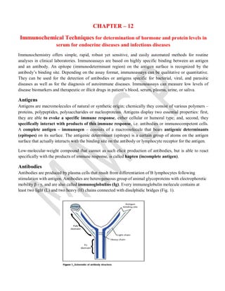
- 1. CHAPTER – 12 Immunochemical Techniques for determination of hormone and protein levels in serum for endocrine diseases and infectious diseases Immunochemistry offers simple, rapid, robust yet sensitive, and easily automated methods for routine analyses in clinical laboratories. Immunoassays are based on highly specific binding between an antigen and an antibody. An epitope (immunodeterminant region) on the antigen surface is recognized by the antibody’s binding site. Depending on the assay format, immunoassays can be qualitative or quantitative. They can be used for the detection of antibodies or antigens specific for bacterial, viral, and parasitic diseases as well as for the diagnosis of autoimmune diseases. Immunoassays can measure low levels of disease biomarkers and therapeutic or illicit drugs in patient’s blood, serum, plasma, urine, or saliva. Antigens Antigens are macromolecules of natural or synthetic origin; chemically they consist of various polymers – proteins, polypeptides, polysaccharides or nucleoproteins. Antigens display two essential properties: first, they are able to evoke a specific immune response, either cellular or humoral type; and, second, they specifically interact with products of this immune response, i.e. antibodies or immunocompetent cells. A complete antigen – immunogen – consists of a macromolecule that bears antigenic determinants (epitopes) on its surface. The antigenic determinant (epitope) is a certain group of atoms on the antigen surface that actually interacts with the binding site on the antibody or lymphocyte receptor for the antigen. Low-molecular-weight compound that cannot as such elicit production of antibodies, but is able to react specifically with the products of immune response, is called hapten (incomplete antigen). Antibodies Antibodies are produced by plasma cells that result from differentiation of B lymphocytes following stimulation with antigen. Antibodies are heterogeneous group of animal glycoproteins with electrophoretic mobility β - γ, and are also called immunoglobulins (Ig). Every immunoglobulin molecule contains at least two light (L) and two heavy (H) chains connected with disulphidic bridges (Fig. 1).
- 2. Antigen-Antibody Binding All immunochemical methods are based on a highly specific and sensitive reaction between an antigen and an antibody. Antigen is a substance that induces the production of antibodies [ie, proteins from the class of immunoglobulins (MW about 150 kDa) that are produced in the immune system of any vertebrate or human as a result of a defense reaction (immunity) to this foreign substance]. Antibodies are a large family of glycoproteins that share key structural and functional properties. Functionally, they can be characterized by their ability to bind both to antigens and specialized cells or proteins of the immune system. Immunochemical techniques Immunochemical techniques are based on a reaction of antigen with antibody, or more exactly, on a reaction of an antigenic determinants with the binding site of the antibody. The antibodies used are produced by various ways. 1. Precipitation methods Quantitative precipitin curve Measurement of antigen-antibody complex formation has proved extremely useful for analysing many constituents of body fluids. A wide variety of immunochemical methods has been developed based on the fundamental principle of quantitative precipitin curve described by Heidelberger and Kendall in 1935. A soluble antigen possessing multiple antigenic determinants reacts with the corresponding antibody and the resulting antigen-antibody complex precipitates out of solution. The precipitin curve describes the relationship between the antigen concentration and the amount of precipitate for a constant quantity of antibody. This immunoprecipitin curve forms the basis of many immunochemical assays that can be performed in a solution as well as in a gel. Precipitation methods in gel As a support matrix, agar or agarose gels are used most often. In single immunodiffusion only one component (i.e. antigen or antibody) diffuses from the place of sample application, while the other reaction partner is dispersed evenly in the gel. If both components of the immunochemical reaction diffuse in the gel against each other from places of their application, the technique is called double immunodiffusion. In the area of antigen antibody reaction a precipitation zone appears as line, crescent or circle. Single radial immunodiffusion Single radial immunodiffusion represents a simple method without requirements for expensive instruments. The antigen is applied into wells that are cut in the agarose gel containing dispersed corresponding monospecific antibody. The agarose plate is incubated at room temperature for 48-72 hours depending on the specific protein in question. The antigen from the sample diffuses out from the wells into the agarose where the antibody concentration is constant. In a distance from the well where antigen concentration is equivalent to the antibody concentration (i.e., zone of equivalence is reached), the complex antigen- antibody precipitates and appears as a strong white ring around the well (see fig. 3). The square of the ring diameter is directly proportional to the antigen concentration. The protocol involves several steps:
- 3. • Boiled agarose is cooled to 50°C and monospecific serum is added to it. The gel is poured onto a glass plate or a Petri dish. • After the agarose has solidified, round-shaped wells (starts) are cut out. Samples and standards are applied to the wells. • The plate is incubated in a wet chamber. The antigen diffuses radially to the gel and reacts with antibody. • Then, the plate is stained in order to visualise the precipitate rings, and dried. • Diameters of the rings are measured and squared values are calculated. Calibration curve is plotted using standards. Finally, concentration of antigen in samples is read from the plot. The sensitivity of this technique is about 10-200 mg/L, and therefore it is suitable for estimation of most serum proteins (e.g. immunoglobulin IgG, IgM, IgA, prealbumin, transferrin, α2-macroglobulin, ceruloplasmin). This technique has been, however, currently largely replaced with immunoturbidimetry or nephelometry. Fig 3: Double immunodiffusion In double immunodiffusion reaction, the antigen and the antibody diffuse towards each other. It is based on wells punched into the agarose gel in a rosette pattern that are filled with antigen or antibody solutions, respectively. Both antigen and antibody molecules are allowed to diffuse radially into the gel surrounding the wells; and where the antigen and specific reactive antibody meet, a precipitin line forms. If antiserum to several possible antigens is placed into the central well and the outer wells are filled by different antigens, precipitin lines of various shapes can arise. For instance, if two antigenic mixtures are applied into two adjacent wells, the following patterns of precipitin lines can be observed, in dependence on the relationship between the two antigenic mixtures (see fig. 4): a reaction of identity – if two identical antigens are applied into two adjacent wells, the precipitin bands form a continuous arc. a reaction of non-identity – the precipitin bands form lines that intersect. a reaction of partial identity – it is characterised by a formation of a spur. The common hooked precipitation line arises from the reaction of the common antigenic determinants on both antigens with the antibody. The spur means that the second antigen lacks an epitope present in the first antigen that is recognised by one of the antibodies in the antiserum.
- 4. Fig 4: Double immunodiffusion is a qualitative technique suitable for the identification of antigens and the estimation of their mutual immunochemical relationships if the specific antisera are available. On the other hand, this method may be used for characterisation of antibodies using known purified antigens. Immunoelectrophoresis Immunoelectrophoresis is a qualitative method that combines protein electrophoresis with immunodiffusion. Fig 5:
- 5. It is performed in two steps. The first one involves the separation of antigens according to their charges/size in an electrical field. In the second step, a suitable antiserum (polyspecific or monospecific) is applied to grooves running parallel to the electrophoresis migration zone. The separated antigens and antibodies are allowed to diffuse into the gel towards one another. The precipitation line is formed in the area when the antigen with the reacting antibody meets (Fig. 5). Immunofixation Immunofixation is a method used for the detection and isotyping of monoclonal immunoglobulins in serum, urine and cerebrospinal fluid. Similarly to immunoelectrophoresis, immunofixation is carried out in two stages. In the first one, serum proteins are separated by electrophoresis. In the second step, the monoclonal immunoglobulins are identified by means of immunoprecipitation with specific antibodies. The typical procedure of immunofixation is following: The aliquots of an identical patient serum are applied into six indicated origins in an agarose gel. All the specimens are separated by electrophoresis. At the end of electrophoresis, the gel is covered with the template, in which the areas of the electrophoresis migration zones are cut out. A fixative solution is applied on the first track to denaturate and immobilise the serum protein and to create an electrophoresis reference pattern. The monospecific antibodies, directed against constant regions of IgG, IgM, and IgA heavy chains, and light chains κ and λ, are added to each of the following tracks. The specific antibodies diffuse into the gel. The antigen antibody complexes are formed in the zone where appropriate antibody and antigen meet. The immune complexes are retained in the porous structure of gel and form a marked band, while all the nonprecipited components are subsequently removed by washing. Finally, the gel is stained for protein in order to visualise the formed immunoprecipitates. Use of appropriately diluted sample is the critical step. It follows from the immunoprecipitin curve that excess of either antigen or antibody leads to decay of the immunocomplex. Thus, if both over-diluted or over- concentrated sample is examined, a false negative result may be obtained. In clinical practice, immunofixation is employed especially for detection of monoclonal immunoglobulins. Immunofixation is easier to perform, more sensitive, and its evaluation is less complicated.The clinical conditions associated with monoclonal immnunoglubulins - monoclonal gammapathies – include a wide spectrum from a benign form to the malignant diseases. Precipitation methods in solution The precipitate, which shapes in an agarose gel, can also form in a solution. Two techniques are used to quantitate this precipitate: immunoturbidimetry and immunonephelometry. Turbidimetry and nephelometry: When a diluted antigen solution is combined with a solution of the corresponding antibody, the formation of small aggregates (immunoprecipitates) results in turbidity (cloudy appearance) of the solution. Two approaches can be used to quantify this turbidity: Turbidimetry is based on the measurement of intensity of light transmission; i.e., light that passes through the cuvette is measured. Such measurement can be performed on a conventional spectrophotometer. In
- 6. contrast, nephelometry measures directly the intensity of scattered light. Nephelometry requires a special apparatus – the nephelometer, which uses laser as the light source (Fig. 6). The conventional way of checking whether the measured absorbance lies within the antibody excess or antigen excess zone of precipitin curve is to repeat the measurement with a higher dilution of the sample. With a new sophisticated equipment we can measure the rate of turbidity formation, which is directly proportional to the concentration of antigen. Sensitivity of these techniques can be increased by binding of antibody or antigen onto latex particles, which considerably increase light scatter due to immunocomplex formation. The immunoprecipitation techniques in solution are much faster than radial immunodiffusion, but also more expensive. Fig 6: