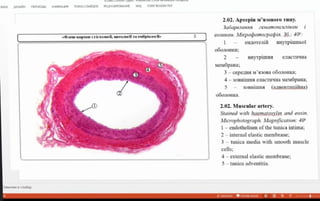
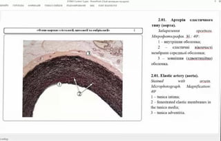
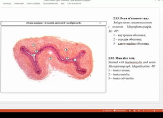
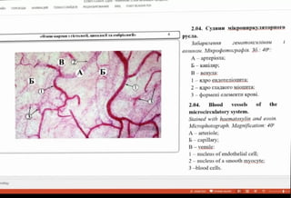
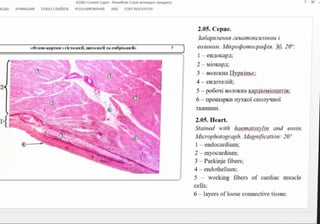
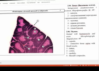
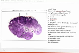
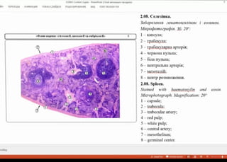
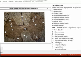
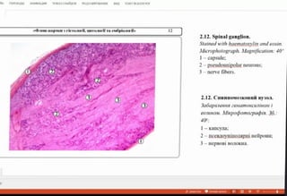
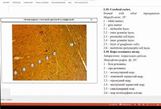
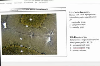
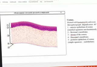
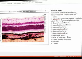
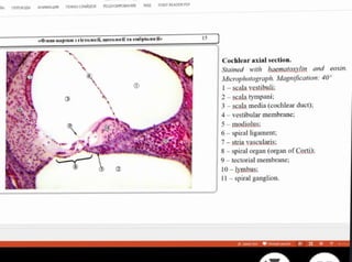
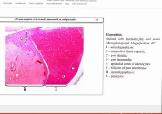
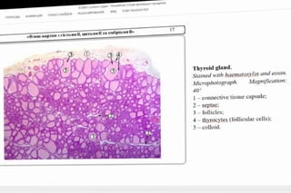
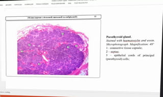
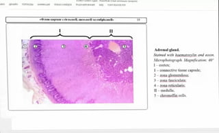
1. The document provides microscopic images and descriptions of various tissues and organs stained with hematoxylin and eosin.
2. The tissues and organs include arteries, veins, smooth muscle, heart, thymus, lymph node, spleen, spinal cord, brain, eye, ear, pituitary gland, thyroid gland, and parathyroid gland.
3. For each image, labels are provided identifying key cellular structures and components visible at 40x magnification.