SlideShare a Scribd company logo
1 of 40
THE ORBIT
Dr. Hadi munib
Oral and maxillofacial surgery resident
The Orbital Region
 The orbits are a pair of bony cavities that contain the eyeballs; their associated muscles,
nerves, vessels, and fat; and most of the lacrimal apparatus.
 The orbital opening is guarded by the eyelids.
 Eyelids; two thin, movable folds that protect the eye from injury and excessive light by their
closure.
 The upper eyelid is larger and more mobile than the lower, and they meet each other at the
medial and lateral angles.
 The palpebral fissure is the elliptical opening between the eyelids and is the entrance into
the conjunctival sac.
 When the eye is closed, the upper eyelid completely covers the cornea of the eye.
 When the eye is open and looking straight ahead, the upper lid just covers the upper margin
of the cornea.
 The lower lid lies just below the cornea when the eye is open and rises only slightly when the
eye is closed
The Orbital Region
 The superficial surface of the eyelids is covered by skin, and the deep surface is covered by a
mucous membrane called the conjunctiva.
 The eyelashes are short, curved hairs on the free edges of the eyelids, arranged in double or
triple rows at the mucocutaneous junction.
 The sebaceous glands (glands of Zeis) open directly into the eyelash follicles.
 The ciliary glands (glands of Moll) are modified sweat glands that open separately between
adjacent lashes.
 The tarsal glands are long, modified sebaceous glands that pour their oily secretion onto the
margin of the lid; their openings lie behind the eyelashes.
 This oily material prevents the overflow of tears and helps make the closed eyelids airtight.
 The more rounded medial angle is separated from the eyeball by a small space, the lacus
lacrimalis, in the center of which is a small, reddish yellow elevation, the caruncula
Lacrimalis.
 A reddish semilunar fold, called the plica semilunaris, lies on the lateral side of the caruncle.
The Orbital Region
 Near the medial angle of the eye a small elevation, the papilla lacrimalis, is present.
 On the summit of the papilla is a small hole, the punctum lacrimale, which leads into the
canaliculus lacrimalis.
 The papilla lacrimalis projects into the lacus, and the punctum and canaliculus carry
tears down into the nose
 The conjunctiva is a thin mucous membrane that lines the eyelids and is reflected at the
superior and inferior fornices onto the anterior surface of the eyeball.
 Its epithelium is continuous with that of the cornea.
 The upper lateral part of the superior fornix is pierced by the ducts of the lacrimal gland.
 The conjunctiva thus forms a potential space, the conjunctival sac, which is open at the
palpebral fissure.
 Beneath the eyelid is a groove, the subtarsal sulcus, which runs close to and parallel with
the margin of the lid
The Orbital Region
 The sulcus tends to trap small foreign particles introduced into the conjunctival sac and is
thus clinically important.
 The framework of the eyelids is formed by a fibrous sheet, the orbital septum.
 This is attached to the periosteum at the orbital margins.
 The orbital septum is thickened at the margins of the lids to form the superior and inferior
tarsal plates.
 The lateral ends of the plates are attached by a band, the lateral palpebral ligament, to a
bony tubercle just within the orbital margin.
 The medial ends of the plates are attached by a band, the medial palpebral ligament, to the
crest of the lacrimal bone.
 The tarsal glands are embedded in the posterior surface of the tarsal plates.
 The superficial surface of the tarsal plates and the orbital septum are covered by the
palpebral fibers of the orbicularis oculi muscle.
 The aponeurosis of insertion of the levator palpebrae superioris muscle pierces the orbital
septum to reach the anterior surface of the superior tarsal plate and the skin
Movements of the Eyelid
 The position of the eyelids at rest depends on the tone of the orbicularis oculi and the
levator palpebrae superioris muscles and the position of the eyeball.
 The eyelids are closed by the contraction of the orbicularis oculi and the relaxation of
the levator palpebrae superioris muscles.
 The eye is opened by the levator palpebrae superioris raising the upper lid.
 On looking upward, the levator palpebrae superioris contracts, and the upper lid moves
with the eyeball.
 On looking downward, both lids move, the upper lid continues to cover the upper part
of the cornea, and the lower lid is pulled downward slightly by the conjunctiva, which is
attached to the sclera and the lower lid.
Lacrimal Apparatus
 Lacrimal Gland
 Consists of a large orbital part and a small palpebral part, which are continuous with
each other around the lateral edge of the aponeurosis of the levator palpebrae
superioris.
 It is situated above the eyeball in the anterior and upper part of the orbit posterior to
the orbital septum.
 The gland opens into the lateral part of the superior fornix of the conjunctiva by 12
ducts.
 The parasympathetic secretomotor nerve supply is derived from the lacrimal nucleus of
the facial nerve.
 The preganglionic fibers reach the pterygopalatine ganglion (sphenopalatine ganglion)
via the nervus intermedius and its great petrosal branch and via the nerve of the
pterygoid canal.
 The postganglionic fibers leave the ganglion and join the maxillary nerve.
 They then pass into its zygomatic branch and the zygomaticotemporal nerve.
Lacrimal Apparatus
 The sympathetic postganglionic nerve supply is from the internal carotid plexus and travels in the
deep petrosal nerve, the nerve of the pterygoid canal, the maxillary nerve, the zygomatic nerve,
the zygomaticotemporal nerve, and finally the lacrimal nerve.
 Lacrimal Ducts
 The tears circulate across the cornea and accumulate in the lacus lacrimalis.
 From here, the tears enter the canaliculi lacrimales through the puncta lacrimalis.
 The canaliculi lacrimales pass medially and open into the lacrimal sac which lies in the lacrimal
groove behind the medial palpebral ligament and is the upper blind end of the nasolacrimal duct.
 The nasolacrimal duct is about 0.5 in. (1.3 cm) long and emerges from the lower end of the
lacrimal sac.
 The duct descends downward, backward, and laterally in a bony canal and opens into the inferior
meatus of the nose.
 The opening is guarded by a fold of mucous membrane known as the lacrimal fold.
 This prevents air from being forced up the duct into the lacrimal sac on blowing the nose.
The Orbit
 A pyramidal cavity with its base anterior and its apex posterior.
 The orbital margin is formed above by the frontal bone.
 Roof: Formed by the orbital plate of the frontal bone, which separates the orbital cavity
from the anterior cranial fossa and the frontal lobe of the cerebral hemisphere
 Lateral wall: Formed by the zygomatic bone and the greater wing of the sphenoid.
 Floor: Formed by the orbital plate of the maxilla, which separates the orbital cavity from
the maxillary sinus
 Medial wall: Formed from before backward by the frontal process of the maxilla, the
lacrimal bone, the orbital plate of the ethmoid (which separates the orbital cavity from the
ethmoid sinuses), and the body of the sphenoid
Openings into the Orbital Cavity
 Orbital opening: Lies anteriorly, About one sixth of the eye is exposed; the remainder is
protected by the walls of the orbit.
 Supraorbital notch (Foramen): The supraorbital notch is situated on the superior orbital margin.
It transmits the supraorbital nerve and blood vessels.
 Infraorbital groove and canal: Situated on the floor of the orbit in the orbital plate of the
maxilla; they transmit the infraorbital nerve (a continuation of the maxillary nerve) and blood
vessels.
 Nasolacrimal canal: Located anteriorly on the medial wall; it communicates with the inferior
meatus of the nose; It transmits the nasolacrimal duct.
 Inferior orbital fissure: Located posteriorly between the maxilla and the greater wing of the
sphenoid; it communicates with the pterygopalatine fossa. It transmits the maxillary nerve and
its zygomatic branch, the inferior ophthalmic vein, and sympathetic nerves.
 Superior orbital fissure: Located posteriorly between the greater and lesser wings of the
sphenoid; it communicates with the middle cranial fossa. It transmits the lacrimal nerve, the
frontal nerve, the trochlear nerve, the oculomotor nerve (upper and lower divisions), the
abducent nerve, the nasociliary nerve, and the superior ophthalmic vein.
Openings into the Orbital Cavity
 Optic canal: Located posteriorly in the lesser wing of the sphenoid; it communicates
with the middle cranial fossa. It transmits the optic nerve and the ophthalmic artery
Orbital Fascia
 The periosteum of the bones that form the walls of the orbit.
 It is loosely attached to the bones and is continuous through the foramina and
fissures with the periosteum covering the outer surfaces of the bones.
 The muscle of Müller, or orbitalis muscle, is a thin layer of smooth muscle that
bridges the inferior orbital fissure.
 It is supplied by sympathetic nerves, and its function is unknown.
Nerves of the Orbit – Optic Nerve
 Enters the orbit from the middle cranial fossa by passing through the optic canal.
 Accompanied by the ophthalmic artery, which lies on its lower lateral side.
 The nerve is surrounded by sheaths of pia mater, arachnoid mater, and dura mater.
 It runs forward and laterally within the cone of the recti muscles and pierces the
sclera at a point medial to the posterior pole of the eyeball.
 The meninges fuse with the sclera so that the subarachnoid space with its contained
cerebrospinal fluid extends forward from the middle cranial fossa, around the optic
nerve, and through the optic canal, as far as the eyeball.
 A rise in pressure of the cerebrospinal fluid within the cranial cavity therefore is
transmitted to the back of the eyeball.
Nerves of the Orbit – Lacrimal Nerve
 The lacrimal nerve arises from the ophthalmic division of the trigeminal nerve.
 It enters the orbit through the upper part of the superior orbital fissure and passes
forward along the upper border of the lateral rectus muscle
 It is joined by a branch of the zygomaticotemporal nerve, which later leaves it to
enter the lacrimal gland (parasympathetic secretomotor fibers).
 The lacrimal nerve ends by supplying the skin of the lateral part of the upper lid.
 Trochlear Nerve
 Enters the orbit through the upper part of the superior orbital fissure.
 It runs forward and supplies the superior oblique muscle
Nerves of the Orbit – Frontal Nerve
 Frontal Nerve
 Arises from the ophthalmic division of the trigeminal nerve.
 It enters the orbit through the upper part of the superior orbital fissure and passes
forward on the upper surface of the levator palpebrae superioris beneath the roof of
the orbit.
 It divides into the supratrochlear and supraorbital nerves that wind around the upper
margin of the orbital cavity to supply the skin of the forehead; the supraorbital nerve
also supplies the mucous membrane of the frontal air sinus.
Nerves of the Orbit – Oculomotor Nerve
 The superior ramus of the oculomotor nerve enters the orbit through the lower part
of the superior orbital fissure
 It supplies the superior rectus muscle, then pierces it, and supplies the levator
palpebrae superioris muscle.
 The inferior ramus of the oculomotor nerve enters the orbit in a similar manner and
supplies the inferior rectus, the medial rectus, and the inferior oblique muscles.
 The nerve to the inferior oblique gives off a branch that passes to the ciliary
ganglion and carries parasympathetic fibers to the sphincter pupillae and the ciliary
muscle.
Nerves of the Orbit - Nasociliary Nerve
 Arises from the ophthalmic division of the trigeminal nerve.
 It enters the orbit through the lower part of the superior orbital fissure.
 It crosses above the optic nerve, runs forward along the upper margin of the medial
rectus muscle, and ends by dividing into the anterior ethmoidal and infratrochlear
nerves
Branches of the Nasociliary Nerve
 Communicating branch to the ciliary ganglion is a sensory nerve; from the eyeball pass
to the ciliary ganglion via the short ciliary nerves, pass through the ganglion without
interruption, and then join the nasociliary nerve by means of the communicating
branch.
 Long ciliary nerves; two or three in number, arise from the nasociliary nerve as it
crosses the optic nerve. They contain sympathetic fibers for the dilator pupillae
muscle.
The nerves pass forward with the short ciliary nerves and pierce the sclera of the
eyeball.
They continue forward between the sclera and the choroid to reach the iris.
 The posterior ethmoidal nerve supplies the ethmoidal and sphenoidal air sinuses
 The infratrochlear nerve passes forward below the pulley of the superior oblique
muscle and supplies the skin of the medial part of the upper eyelid and the adjacent
part of the nose
Branches of the Nasociliary Nerve
 Anterior ethmoidal nerve passes through the anterior ethmoidal foramen and enters
the anterior cranial fossa on the upper surface of the cribriform plate of the ethmoid
 It enters the nasal cavity through a slitlike opening alongside the crista galli.
 After supplying an area of mucous membrane, it appears on the face as the external
nasal branch at the lower border of the nasal bone, and supplies the skin of the nose
down as far as the tip.
Nerves of the Orbit – Abducent Nerve
 Enters the orbit through the lower part of the superior orbital fissure.
 It supplies the lateral rectus muscle.
 Ciliary Ganglion
 A parasympathetic ganglion about the size of a pinhead and situated in the posterior
part of the orbit.
 It receives its preganglionic parasympathetic fibers from the oculomotor nerve via the
nerve to the inferior oblique.
 The postganglionic fibers leave the ganglion in the short ciliary nerves, which enter the
back of the eyeball and supply the sphincter pupillae and the ciliary muscle.
 A number of sympathetic fibers pass from the internal carotid plexus into the orbit and
run through the ganglion without interruption.
Blood Vessels and Lymph Vessels of the Orbit
 Ophthalmic Artery
 A branch of the internal carotid artery after that vessel emerges from the cavernous
sinus
 It enters the orbit through the optic canal with the optic nerve.
 It runs forward and crosses the optic nerve to reach the medial wall of the orbit.
 It gives off numerous branches, which accompany the nerves in the orbital cavity.
 Branches of the Ophthalmic Artery
 Central artery of the retina is a small branch that pierces the meningeal sheaths of the
optic nerve to gain entrance to the nerve; It runs in the substance of the optic nerve and
enters the eyeball at the center of the optic disc. Here, it divides into branches, which
may be studied in a patient through an ophthalmoscope. The branches are end
arteries.
Ophthalmic Artery Branches
 The muscular branches
 The ciliary arteries can be divided into anterior and posterior groups. The former
group enters the eyeball near the corneoscleral junction; the latter group enters
near the optic nerve.
 The lacrimal artery to the lacrimal gland
 The supratrochlear and supraorbital arteries are distributed to the skin of the
forehead
Blood Vessels and Lymph Vessels of the Orbit
 Ophthalmic Veins
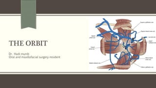
 Superior ophthalmic vein communicates in front with the facial vein.
 The inferior ophthalmic vein communicates through the inferior orbital fissure with
the pterygoid venous plexus.
 Both veins pass backward through the superior orbital fissure and drain into the
cavernous sinus.
 Lymph Vessels; No lymph vessels or nodes are present in the orbital cavity.
References
Chapter 11; The Head and Neck (Pages; 549 – 556)
THANK YOU!

More Related Content

What's hot

Anatomy of orbit ophthalm
Anatomy of orbit ophthalmAnatomy of orbit ophthalm
Anatomy of orbit ophthalmDrArino John
 
Anatomy of the orbit
Anatomy of the orbitAnatomy of the orbit
Anatomy of the orbitPriyanka Raj
 
anatomy of The orbit (2)
anatomy of  The orbit (2)anatomy of  The orbit (2)
anatomy of The orbit (2)ddert
 
Anatomy of orbit
Anatomy of orbitAnatomy of orbit
Anatomy of orbitveeru1984
 
Bony orbit and its contents
Bony orbit and its contentsBony orbit and its contents
Bony orbit and its contentsmgmcri1234
 
Surgical anatomy of orbit 1 /certified fixed orthodontic courses by Indian de...
Surgical anatomy of orbit 1 /certified fixed orthodontic courses by Indian de...Surgical anatomy of orbit 1 /certified fixed orthodontic courses by Indian de...
Surgical anatomy of orbit 1 /certified fixed orthodontic courses by Indian de...Indian dental academy
 
Gross anatomy of the head and neck
Gross anatomy of the head and neckGross anatomy of the head and neck
Gross anatomy of the head and neckLucidante1
 
Anatomy of human orbit
Anatomy of human orbitAnatomy of human orbit
Anatomy of human orbitrakshyabasnet1
 
Lateral skull base anatomy and applied science by Dr, bomkar bam
Lateral skull base anatomy and applied science by Dr, bomkar bamLateral skull base anatomy and applied science by Dr, bomkar bam
Lateral skull base anatomy and applied science by Dr, bomkar bamBomkar Bam
 
Anatomy of the orbit
Anatomy of the orbitAnatomy of the orbit
Anatomy of the orbitMohamed Ezz
 
Anatomy of orbit paresh varsat
Anatomy of orbit paresh varsatAnatomy of orbit paresh varsat
Anatomy of orbit paresh varsatDrParesh Varsat
 
“BASE OF SKULL: ANATOMY and APPLIED ASPECT”
“BASE OF SKULL: ANATOMY and APPLIED ASPECT”“BASE OF SKULL: ANATOMY and APPLIED ASPECT”
“BASE OF SKULL: ANATOMY and APPLIED ASPECT”RAKESHTALUKDAR2
 

What's hot (20)

Anatomy of orbit ophthalm
Anatomy of orbit ophthalmAnatomy of orbit ophthalm
Anatomy of orbit ophthalm
 
Anatomy of the orbit
Anatomy of the orbitAnatomy of the orbit
Anatomy of the orbit
 
anatomy of The orbit (2)
anatomy of  The orbit (2)anatomy of  The orbit (2)
anatomy of The orbit (2)
 
The Orbit
The OrbitThe Orbit
The Orbit
 
anatomy of orbital
anatomy of orbital anatomy of orbital
anatomy of orbital
 
Anatomy of orbit
Anatomy of orbitAnatomy of orbit
Anatomy of orbit
 
The Orbit
The OrbitThe Orbit
The Orbit
 
Bony orbit and its contents
Bony orbit and its contentsBony orbit and its contents
Bony orbit and its contents
 
Orbit anatomy
Orbit anatomyOrbit anatomy
Orbit anatomy
 
Surgical anatomy of orbit 1 /certified fixed orthodontic courses by Indian de...
Surgical anatomy of orbit 1 /certified fixed orthodontic courses by Indian de...Surgical anatomy of orbit 1 /certified fixed orthodontic courses by Indian de...
Surgical anatomy of orbit 1 /certified fixed orthodontic courses by Indian de...
 
Gross anatomy of the head and neck
Gross anatomy of the head and neckGross anatomy of the head and neck
Gross anatomy of the head and neck
 
Anatomy of human orbit
Anatomy of human orbitAnatomy of human orbit
Anatomy of human orbit
 
Lateral skull base anatomy and applied science by Dr, bomkar bam
Lateral skull base anatomy and applied science by Dr, bomkar bamLateral skull base anatomy and applied science by Dr, bomkar bam
Lateral skull base anatomy and applied science by Dr, bomkar bam
 
Anatomy of the orbit
Anatomy of the orbitAnatomy of the orbit
Anatomy of the orbit
 
Sphenoid bone (osteology)
Sphenoid bone (osteology)Sphenoid bone (osteology)
Sphenoid bone (osteology)
 
Anatomy of orbit
Anatomy of orbitAnatomy of orbit
Anatomy of orbit
 
Orbital anatomy
Orbital anatomyOrbital anatomy
Orbital anatomy
 
Anatomy of orbital cavity
Anatomy of orbital cavityAnatomy of orbital cavity
Anatomy of orbital cavity
 
Anatomy of orbit paresh varsat
Anatomy of orbit paresh varsatAnatomy of orbit paresh varsat
Anatomy of orbit paresh varsat
 
“BASE OF SKULL: ANATOMY and APPLIED ASPECT”
“BASE OF SKULL: ANATOMY and APPLIED ASPECT”“BASE OF SKULL: ANATOMY and APPLIED ASPECT”
“BASE OF SKULL: ANATOMY and APPLIED ASPECT”
 

Similar to The Orbit

2 The orbital region part 1.pdf
2 The orbital region  part 1.pdf2 The orbital region  part 1.pdf
2 The orbital region part 1.pdfDr Rafid Al-Temimi
 
anatomy and physiology of lacrimal apparatus ppt
anatomy and physiology of lacrimal apparatus  pptanatomy and physiology of lacrimal apparatus  ppt
anatomy and physiology of lacrimal apparatus pptRohit Rao
 
Anatomy of the Eye (whole).pptx
Anatomy of the Eye (whole).pptxAnatomy of the Eye (whole).pptx
Anatomy of the Eye (whole).pptxdratulkranand
 
Anatomy of the lacrimal apparatus sivateja
Anatomy of the lacrimal apparatus sivatejaAnatomy of the lacrimal apparatus sivateja
Anatomy of the lacrimal apparatus sivatejaSivateja Challa
 
Anatomy of the lacrimal apparatus
Anatomy of the lacrimal apparatusAnatomy of the lacrimal apparatus
Anatomy of the lacrimal apparatusSSSIHMS-PG
 
Anatomyoflacrimalapparatus 160313074220
Anatomyoflacrimalapparatus 160313074220Anatomyoflacrimalapparatus 160313074220
Anatomyoflacrimalapparatus 160313074220Ankur gaur
 
Orbit. anatomy power point presentation
Orbit. anatomy power point presentationOrbit. anatomy power point presentation
Orbit. anatomy power point presentationesalama
 
Facial region of Head by Adiev and Shaikh Masood.pptx
Facial region of Head by Adiev and Shaikh Masood.pptxFacial region of Head by Adiev and Shaikh Masood.pptx
Facial region of Head by Adiev and Shaikh Masood.pptxshahajipawale0
 
Eye and orbit revision ppt
Eye and orbit revision pptEye and orbit revision ppt
Eye and orbit revision pptA K
 
Anatomy and physiology of eyelids
Anatomy and physiology of eyelidsAnatomy and physiology of eyelids
Anatomy and physiology of eyelidsKanwal Perveen
 

Similar to The Orbit (20)

Eye by meher
Eye by meherEye by meher
Eye by meher
 
Orbital region
Orbital regionOrbital region
Orbital region
 
Orbit anatomy
Orbit anatomyOrbit anatomy
Orbit anatomy
 
2 The orbital region part 1.pdf
2 The orbital region  part 1.pdf2 The orbital region  part 1.pdf
2 The orbital region part 1.pdf
 
Anatomy of eye
Anatomy of eye Anatomy of eye
Anatomy of eye
 
anatomy and physiology of lacrimal apparatus ppt
anatomy and physiology of lacrimal apparatus  pptanatomy and physiology of lacrimal apparatus  ppt
anatomy and physiology of lacrimal apparatus ppt
 
Lacrimal Apparatus.pptx
Lacrimal Apparatus.pptxLacrimal Apparatus.pptx
Lacrimal Apparatus.pptx
 
Anatomy of the Eye (whole).pptx
Anatomy of the Eye (whole).pptxAnatomy of the Eye (whole).pptx
Anatomy of the Eye (whole).pptx
 
Anatomy of the lacrimal apparatus sivateja
Anatomy of the lacrimal apparatus sivatejaAnatomy of the lacrimal apparatus sivateja
Anatomy of the lacrimal apparatus sivateja
 
Anatomy of the lacrimal apparatus
Anatomy of the lacrimal apparatusAnatomy of the lacrimal apparatus
Anatomy of the lacrimal apparatus
 
anatomy And Physiology of lacrimal secretions
anatomy And Physiology of lacrimal secretions anatomy And Physiology of lacrimal secretions
anatomy And Physiology of lacrimal secretions
 
Orbit Anatomy
Orbit AnatomyOrbit Anatomy
Orbit Anatomy
 
Anatomyoflacrimalapparatus 160313074220
Anatomyoflacrimalapparatus 160313074220Anatomyoflacrimalapparatus 160313074220
Anatomyoflacrimalapparatus 160313074220
 
Anatomy of lacrimal apparatus
Anatomy of lacrimal apparatusAnatomy of lacrimal apparatus
Anatomy of lacrimal apparatus
 
Structure of eye
Structure of eyeStructure of eye
Structure of eye
 
Pns
PnsPns
Pns
 
Orbit. anatomy power point presentation
Orbit. anatomy power point presentationOrbit. anatomy power point presentation
Orbit. anatomy power point presentation
 
Facial region of Head by Adiev and Shaikh Masood.pptx
Facial region of Head by Adiev and Shaikh Masood.pptxFacial region of Head by Adiev and Shaikh Masood.pptx
Facial region of Head by Adiev and Shaikh Masood.pptx
 
Eye and orbit revision ppt
Eye and orbit revision pptEye and orbit revision ppt
Eye and orbit revision ppt
 
Anatomy and physiology of eyelids
Anatomy and physiology of eyelidsAnatomy and physiology of eyelids
Anatomy and physiology of eyelids
 

More from Hadi Munib

Principles of Management of Odontogenic Infections.pptx
Principles of Management of Odontogenic Infections.pptxPrinciples of Management of Odontogenic Infections.pptx
Principles of Management of Odontogenic Infections.pptxHadi Munib
 
Medication-Related Osteonecrosis of the jaws (MRONJ).pptx
Medication-Related Osteonecrosis of the jaws (MRONJ).pptxMedication-Related Osteonecrosis of the jaws (MRONJ).pptx
Medication-Related Osteonecrosis of the jaws (MRONJ).pptxHadi Munib
 
Airway and Anesthetic Management of the Traumatized Patient.pptx
Airway and Anesthetic Management of the Traumatized Patient.pptxAirway and Anesthetic Management of the Traumatized Patient.pptx
Airway and Anesthetic Management of the Traumatized Patient.pptxHadi Munib
 
Initial Management of the Trauma Patient II.pptx
Initial Management of the Trauma Patient II.pptxInitial Management of the Trauma Patient II.pptx
Initial Management of the Trauma Patient II.pptxHadi Munib
 
Initial Management of the Trauma Patient.pptx
Initial Management of the Trauma Patient.pptxInitial Management of the Trauma Patient.pptx
Initial Management of the Trauma Patient.pptxHadi Munib
 
Suturing and Wound Closure
Suturing and Wound ClosureSuturing and Wound Closure
Suturing and Wound ClosureHadi Munib
 
Post Operative Complications
Post Operative Complications  Post Operative Complications
Post Operative Complications Hadi Munib
 
Surgical Tubes used in General Surgery
Surgical Tubes used in General SurgerySurgical Tubes used in General Surgery
Surgical Tubes used in General SurgeryHadi Munib
 
Medical Management and Perioperative Assessment of Renal, Hepatic and Hematol...
Medical Management and Perioperative Assessment of Renal, Hepatic and Hematol...Medical Management and Perioperative Assessment of Renal, Hepatic and Hematol...
Medical Management and Perioperative Assessment of Renal, Hepatic and Hematol...Hadi Munib
 
Medical Management and Perioperative Assessment of Cardiovascular Diseases
Medical Management and Perioperative Assessment of Cardiovascular DiseasesMedical Management and Perioperative Assessment of Cardiovascular Diseases
Medical Management and Perioperative Assessment of Cardiovascular DiseasesHadi Munib
 
Medical Management and Perioperative Assessment of Respiratory Diseases
Medical Management and Perioperative Assessment of Respiratory DiseasesMedical Management and Perioperative Assessment of Respiratory Diseases
Medical Management and Perioperative Assessment of Respiratory DiseasesHadi Munib
 
Immunodeficiency Syndrome
Immunodeficiency SyndromeImmunodeficiency Syndrome
Immunodeficiency SyndromeHadi Munib
 
Basic Features of Autoimmune Diseases
Basic Features of Autoimmune DiseasesBasic Features of Autoimmune Diseases
Basic Features of Autoimmune DiseasesHadi Munib
 
Basic Principles of Hypersensitivity Reactions
Basic Principles of Hypersensitivity ReactionsBasic Principles of Hypersensitivity Reactions
Basic Principles of Hypersensitivity ReactionsHadi Munib
 
Basic Principles of the Immune System
Basic Principles of the Immune SystemBasic Principles of the Immune System
Basic Principles of the Immune SystemHadi Munib
 
Hemodynamic Disorders
Hemodynamic DisordersHemodynamic Disorders
Hemodynamic DisordersHadi Munib
 
Basic Features of the Cell
Basic Features of the CellBasic Features of the Cell
Basic Features of the CellHadi Munib
 
Basic Features of Inflammation and Repair
Basic Features of Inflammation and RepairBasic Features of Inflammation and Repair
Basic Features of Inflammation and RepairHadi Munib
 

More from Hadi Munib (20)

Principles of Management of Odontogenic Infections.pptx
Principles of Management of Odontogenic Infections.pptxPrinciples of Management of Odontogenic Infections.pptx
Principles of Management of Odontogenic Infections.pptx
 
Medication-Related Osteonecrosis of the jaws (MRONJ).pptx
Medication-Related Osteonecrosis of the jaws (MRONJ).pptxMedication-Related Osteonecrosis of the jaws (MRONJ).pptx
Medication-Related Osteonecrosis of the jaws (MRONJ).pptx
 
Airway and Anesthetic Management of the Traumatized Patient.pptx
Airway and Anesthetic Management of the Traumatized Patient.pptxAirway and Anesthetic Management of the Traumatized Patient.pptx
Airway and Anesthetic Management of the Traumatized Patient.pptx
 
Initial Management of the Trauma Patient II.pptx
Initial Management of the Trauma Patient II.pptxInitial Management of the Trauma Patient II.pptx
Initial Management of the Trauma Patient II.pptx
 
Initial Management of the Trauma Patient.pptx
Initial Management of the Trauma Patient.pptxInitial Management of the Trauma Patient.pptx
Initial Management of the Trauma Patient.pptx
 
Burn Injuries
Burn InjuriesBurn Injuries
Burn Injuries
 
Suturing and Wound Closure
Suturing and Wound ClosureSuturing and Wound Closure
Suturing and Wound Closure
 
Post Operative Complications
Post Operative Complications  Post Operative Complications
Post Operative Complications
 
Surgical Tubes used in General Surgery
Surgical Tubes used in General SurgerySurgical Tubes used in General Surgery
Surgical Tubes used in General Surgery
 
Medical Management and Perioperative Assessment of Renal, Hepatic and Hematol...
Medical Management and Perioperative Assessment of Renal, Hepatic and Hematol...Medical Management and Perioperative Assessment of Renal, Hepatic and Hematol...
Medical Management and Perioperative Assessment of Renal, Hepatic and Hematol...
 
Medical Management and Perioperative Assessment of Cardiovascular Diseases
Medical Management and Perioperative Assessment of Cardiovascular DiseasesMedical Management and Perioperative Assessment of Cardiovascular Diseases
Medical Management and Perioperative Assessment of Cardiovascular Diseases
 
Medical Management and Perioperative Assessment of Respiratory Diseases
Medical Management and Perioperative Assessment of Respiratory DiseasesMedical Management and Perioperative Assessment of Respiratory Diseases
Medical Management and Perioperative Assessment of Respiratory Diseases
 
Wound Healing
Wound HealingWound Healing
Wound Healing
 
Immunodeficiency Syndrome
Immunodeficiency SyndromeImmunodeficiency Syndrome
Immunodeficiency Syndrome
 
Basic Features of Autoimmune Diseases
Basic Features of Autoimmune DiseasesBasic Features of Autoimmune Diseases
Basic Features of Autoimmune Diseases
 
Basic Principles of Hypersensitivity Reactions
Basic Principles of Hypersensitivity ReactionsBasic Principles of Hypersensitivity Reactions
Basic Principles of Hypersensitivity Reactions
 
Basic Principles of the Immune System
Basic Principles of the Immune SystemBasic Principles of the Immune System
Basic Principles of the Immune System
 
Hemodynamic Disorders
Hemodynamic DisordersHemodynamic Disorders
Hemodynamic Disorders
 
Basic Features of the Cell
Basic Features of the CellBasic Features of the Cell
Basic Features of the Cell
 
Basic Features of Inflammation and Repair
Basic Features of Inflammation and RepairBasic Features of Inflammation and Repair
Basic Features of Inflammation and Repair
 

Recently uploaded

Low Rate Call Girls Pune Esha 9907093804 Short 1500 Night 6000 Best call girl...
Low Rate Call Girls Pune Esha 9907093804 Short 1500 Night 6000 Best call girl...Low Rate Call Girls Pune Esha 9907093804 Short 1500 Night 6000 Best call girl...
Low Rate Call Girls Pune Esha 9907093804 Short 1500 Night 6000 Best call girl...Miss joya
 
Call Girls Colaba Mumbai ❤️ 9920874524 👈 Cash on Delivery
Call Girls Colaba Mumbai ❤️ 9920874524 👈 Cash on DeliveryCall Girls Colaba Mumbai ❤️ 9920874524 👈 Cash on Delivery
Call Girls Colaba Mumbai ❤️ 9920874524 👈 Cash on Deliverynehamumbai
 
Russian Call Girls in Pune Tanvi 9907093804 Short 1500 Night 6000 Best call g...
Russian Call Girls in Pune Tanvi 9907093804 Short 1500 Night 6000 Best call g...Russian Call Girls in Pune Tanvi 9907093804 Short 1500 Night 6000 Best call g...
Russian Call Girls in Pune Tanvi 9907093804 Short 1500 Night 6000 Best call g...Miss joya
 
College Call Girls Pune Mira 9907093804 Short 1500 Night 6000 Best call girls...
College Call Girls Pune Mira 9907093804 Short 1500 Night 6000 Best call girls...College Call Girls Pune Mira 9907093804 Short 1500 Night 6000 Best call girls...
College Call Girls Pune Mira 9907093804 Short 1500 Night 6000 Best call girls...Miss joya
 
Russian Call Girls Chickpet - 7001305949 Booking and charges genuine rate for...
Russian Call Girls Chickpet - 7001305949 Booking and charges genuine rate for...Russian Call Girls Chickpet - 7001305949 Booking and charges genuine rate for...
Russian Call Girls Chickpet - 7001305949 Booking and charges genuine rate for...narwatsonia7
 
Call Girls Service in Bommanahalli - 7001305949 with real photos and phone nu...
Call Girls Service in Bommanahalli - 7001305949 with real photos and phone nu...Call Girls Service in Bommanahalli - 7001305949 with real photos and phone nu...
Call Girls Service in Bommanahalli - 7001305949 with real photos and phone nu...narwatsonia7
 
Call Girls Chennai Megha 9907093804 Independent Call Girls Service Chennai
Call Girls Chennai Megha 9907093804 Independent Call Girls Service ChennaiCall Girls Chennai Megha 9907093804 Independent Call Girls Service Chennai
Call Girls Chennai Megha 9907093804 Independent Call Girls Service ChennaiNehru place Escorts
 
Call Girls Service Jaipur Grishma WhatsApp ❤8445551418 VIP Call Girls Jaipur
Call Girls Service Jaipur Grishma WhatsApp ❤8445551418 VIP Call Girls JaipurCall Girls Service Jaipur Grishma WhatsApp ❤8445551418 VIP Call Girls Jaipur
Call Girls Service Jaipur Grishma WhatsApp ❤8445551418 VIP Call Girls Jaipurparulsinha
 
Russian Call Girls in Chennai Pallavi 9907093804 Independent Call Girls Servi...
Russian Call Girls in Chennai Pallavi 9907093804 Independent Call Girls Servi...Russian Call Girls in Chennai Pallavi 9907093804 Independent Call Girls Servi...
Russian Call Girls in Chennai Pallavi 9907093804 Independent Call Girls Servi...Nehru place Escorts
 
Aspirin presentation slides by Dr. Rewas Ali
Aspirin presentation slides by Dr. Rewas AliAspirin presentation slides by Dr. Rewas Ali
Aspirin presentation slides by Dr. Rewas AliRewAs ALI
 
CALL ON ➥9907093804 🔝 Call Girls Hadapsar ( Pune) Girls Service
CALL ON ➥9907093804 🔝 Call Girls Hadapsar ( Pune)  Girls ServiceCALL ON ➥9907093804 🔝 Call Girls Hadapsar ( Pune)  Girls Service
CALL ON ➥9907093804 🔝 Call Girls Hadapsar ( Pune) Girls ServiceMiss joya
 
Vip Call Girls Anna Salai Chennai 👉 8250192130 ❣️💯 Top Class Girls Available
Vip Call Girls Anna Salai Chennai 👉 8250192130 ❣️💯 Top Class Girls AvailableVip Call Girls Anna Salai Chennai 👉 8250192130 ❣️💯 Top Class Girls Available
Vip Call Girls Anna Salai Chennai 👉 8250192130 ❣️💯 Top Class Girls AvailableNehru place Escorts
 
Call Girls Service In Shyam Nagar Whatsapp 8445551418 Independent Escort Service
Call Girls Service In Shyam Nagar Whatsapp 8445551418 Independent Escort ServiceCall Girls Service In Shyam Nagar Whatsapp 8445551418 Independent Escort Service
Call Girls Service In Shyam Nagar Whatsapp 8445551418 Independent Escort Serviceparulsinha
 
Call Girl Bangalore Nandini 7001305949 Independent Escort Service Bangalore
Call Girl Bangalore Nandini 7001305949 Independent Escort Service BangaloreCall Girl Bangalore Nandini 7001305949 Independent Escort Service Bangalore
Call Girl Bangalore Nandini 7001305949 Independent Escort Service Bangalorenarwatsonia7
 
Russian Call Girl Brookfield - 7001305949 Escorts Service 50% Off with Cash O...
Russian Call Girl Brookfield - 7001305949 Escorts Service 50% Off with Cash O...Russian Call Girl Brookfield - 7001305949 Escorts Service 50% Off with Cash O...
Russian Call Girl Brookfield - 7001305949 Escorts Service 50% Off with Cash O...narwatsonia7
 
Call Girls In Andheri East Call 9920874524 Book Hot And Sexy Girls
Call Girls In Andheri East Call 9920874524 Book Hot And Sexy GirlsCall Girls In Andheri East Call 9920874524 Book Hot And Sexy Girls
Call Girls In Andheri East Call 9920874524 Book Hot And Sexy Girlsnehamumbai
 
VIP Call Girls Tirunelveli Aaradhya 8250192130 Independent Escort Service Tir...
VIP Call Girls Tirunelveli Aaradhya 8250192130 Independent Escort Service Tir...VIP Call Girls Tirunelveli Aaradhya 8250192130 Independent Escort Service Tir...
VIP Call Girls Tirunelveli Aaradhya 8250192130 Independent Escort Service Tir...narwatsonia7
 
Housewife Call Girls Hoskote | 7001305949 At Low Cost Cash Payment Booking
Housewife Call Girls Hoskote | 7001305949 At Low Cost Cash Payment BookingHousewife Call Girls Hoskote | 7001305949 At Low Cost Cash Payment Booking
Housewife Call Girls Hoskote | 7001305949 At Low Cost Cash Payment Bookingnarwatsonia7
 
Call Girls Yelahanka Just Call 7001305949 Top Class Call Girl Service Available
Call Girls Yelahanka Just Call 7001305949 Top Class Call Girl Service AvailableCall Girls Yelahanka Just Call 7001305949 Top Class Call Girl Service Available
Call Girls Yelahanka Just Call 7001305949 Top Class Call Girl Service Availablenarwatsonia7
 

Recently uploaded (20)

Low Rate Call Girls Pune Esha 9907093804 Short 1500 Night 6000 Best call girl...
Low Rate Call Girls Pune Esha 9907093804 Short 1500 Night 6000 Best call girl...Low Rate Call Girls Pune Esha 9907093804 Short 1500 Night 6000 Best call girl...
Low Rate Call Girls Pune Esha 9907093804 Short 1500 Night 6000 Best call girl...
 
Call Girls Colaba Mumbai ❤️ 9920874524 👈 Cash on Delivery
Call Girls Colaba Mumbai ❤️ 9920874524 👈 Cash on DeliveryCall Girls Colaba Mumbai ❤️ 9920874524 👈 Cash on Delivery
Call Girls Colaba Mumbai ❤️ 9920874524 👈 Cash on Delivery
 
Russian Call Girls in Delhi Tanvi ➡️ 9711199012 💋📞 Independent Escort Service...
Russian Call Girls in Delhi Tanvi ➡️ 9711199012 💋📞 Independent Escort Service...Russian Call Girls in Delhi Tanvi ➡️ 9711199012 💋📞 Independent Escort Service...
Russian Call Girls in Delhi Tanvi ➡️ 9711199012 💋📞 Independent Escort Service...
 
Russian Call Girls in Pune Tanvi 9907093804 Short 1500 Night 6000 Best call g...
Russian Call Girls in Pune Tanvi 9907093804 Short 1500 Night 6000 Best call g...Russian Call Girls in Pune Tanvi 9907093804 Short 1500 Night 6000 Best call g...
Russian Call Girls in Pune Tanvi 9907093804 Short 1500 Night 6000 Best call g...
 
College Call Girls Pune Mira 9907093804 Short 1500 Night 6000 Best call girls...
College Call Girls Pune Mira 9907093804 Short 1500 Night 6000 Best call girls...College Call Girls Pune Mira 9907093804 Short 1500 Night 6000 Best call girls...
College Call Girls Pune Mira 9907093804 Short 1500 Night 6000 Best call girls...
 
Russian Call Girls Chickpet - 7001305949 Booking and charges genuine rate for...
Russian Call Girls Chickpet - 7001305949 Booking and charges genuine rate for...Russian Call Girls Chickpet - 7001305949 Booking and charges genuine rate for...
Russian Call Girls Chickpet - 7001305949 Booking and charges genuine rate for...
 
Call Girls Service in Bommanahalli - 7001305949 with real photos and phone nu...
Call Girls Service in Bommanahalli - 7001305949 with real photos and phone nu...Call Girls Service in Bommanahalli - 7001305949 with real photos and phone nu...
Call Girls Service in Bommanahalli - 7001305949 with real photos and phone nu...
 
Call Girls Chennai Megha 9907093804 Independent Call Girls Service Chennai
Call Girls Chennai Megha 9907093804 Independent Call Girls Service ChennaiCall Girls Chennai Megha 9907093804 Independent Call Girls Service Chennai
Call Girls Chennai Megha 9907093804 Independent Call Girls Service Chennai
 
Call Girls Service Jaipur Grishma WhatsApp ❤8445551418 VIP Call Girls Jaipur
Call Girls Service Jaipur Grishma WhatsApp ❤8445551418 VIP Call Girls JaipurCall Girls Service Jaipur Grishma WhatsApp ❤8445551418 VIP Call Girls Jaipur
Call Girls Service Jaipur Grishma WhatsApp ❤8445551418 VIP Call Girls Jaipur
 
Russian Call Girls in Chennai Pallavi 9907093804 Independent Call Girls Servi...
Russian Call Girls in Chennai Pallavi 9907093804 Independent Call Girls Servi...Russian Call Girls in Chennai Pallavi 9907093804 Independent Call Girls Servi...
Russian Call Girls in Chennai Pallavi 9907093804 Independent Call Girls Servi...
 
Aspirin presentation slides by Dr. Rewas Ali
Aspirin presentation slides by Dr. Rewas AliAspirin presentation slides by Dr. Rewas Ali
Aspirin presentation slides by Dr. Rewas Ali
 
CALL ON ➥9907093804 🔝 Call Girls Hadapsar ( Pune) Girls Service
CALL ON ➥9907093804 🔝 Call Girls Hadapsar ( Pune)  Girls ServiceCALL ON ➥9907093804 🔝 Call Girls Hadapsar ( Pune)  Girls Service
CALL ON ➥9907093804 🔝 Call Girls Hadapsar ( Pune) Girls Service
 
Vip Call Girls Anna Salai Chennai 👉 8250192130 ❣️💯 Top Class Girls Available
Vip Call Girls Anna Salai Chennai 👉 8250192130 ❣️💯 Top Class Girls AvailableVip Call Girls Anna Salai Chennai 👉 8250192130 ❣️💯 Top Class Girls Available
Vip Call Girls Anna Salai Chennai 👉 8250192130 ❣️💯 Top Class Girls Available
 
Call Girls Service In Shyam Nagar Whatsapp 8445551418 Independent Escort Service
Call Girls Service In Shyam Nagar Whatsapp 8445551418 Independent Escort ServiceCall Girls Service In Shyam Nagar Whatsapp 8445551418 Independent Escort Service
Call Girls Service In Shyam Nagar Whatsapp 8445551418 Independent Escort Service
 
Call Girl Bangalore Nandini 7001305949 Independent Escort Service Bangalore
Call Girl Bangalore Nandini 7001305949 Independent Escort Service BangaloreCall Girl Bangalore Nandini 7001305949 Independent Escort Service Bangalore
Call Girl Bangalore Nandini 7001305949 Independent Escort Service Bangalore
 
Russian Call Girl Brookfield - 7001305949 Escorts Service 50% Off with Cash O...
Russian Call Girl Brookfield - 7001305949 Escorts Service 50% Off with Cash O...Russian Call Girl Brookfield - 7001305949 Escorts Service 50% Off with Cash O...
Russian Call Girl Brookfield - 7001305949 Escorts Service 50% Off with Cash O...
 
Call Girls In Andheri East Call 9920874524 Book Hot And Sexy Girls
Call Girls In Andheri East Call 9920874524 Book Hot And Sexy GirlsCall Girls In Andheri East Call 9920874524 Book Hot And Sexy Girls
Call Girls In Andheri East Call 9920874524 Book Hot And Sexy Girls
 
VIP Call Girls Tirunelveli Aaradhya 8250192130 Independent Escort Service Tir...
VIP Call Girls Tirunelveli Aaradhya 8250192130 Independent Escort Service Tir...VIP Call Girls Tirunelveli Aaradhya 8250192130 Independent Escort Service Tir...
VIP Call Girls Tirunelveli Aaradhya 8250192130 Independent Escort Service Tir...
 
Housewife Call Girls Hoskote | 7001305949 At Low Cost Cash Payment Booking
Housewife Call Girls Hoskote | 7001305949 At Low Cost Cash Payment BookingHousewife Call Girls Hoskote | 7001305949 At Low Cost Cash Payment Booking
Housewife Call Girls Hoskote | 7001305949 At Low Cost Cash Payment Booking
 
Call Girls Yelahanka Just Call 7001305949 Top Class Call Girl Service Available
Call Girls Yelahanka Just Call 7001305949 Top Class Call Girl Service AvailableCall Girls Yelahanka Just Call 7001305949 Top Class Call Girl Service Available
Call Girls Yelahanka Just Call 7001305949 Top Class Call Girl Service Available
 

The Orbit

  • 1. THE ORBIT Dr. Hadi munib Oral and maxillofacial surgery resident
  • 2. The Orbital Region  The orbits are a pair of bony cavities that contain the eyeballs; their associated muscles, nerves, vessels, and fat; and most of the lacrimal apparatus.  The orbital opening is guarded by the eyelids.  Eyelids; two thin, movable folds that protect the eye from injury and excessive light by their closure.  The upper eyelid is larger and more mobile than the lower, and they meet each other at the medial and lateral angles.  The palpebral fissure is the elliptical opening between the eyelids and is the entrance into the conjunctival sac.  When the eye is closed, the upper eyelid completely covers the cornea of the eye.  When the eye is open and looking straight ahead, the upper lid just covers the upper margin of the cornea.  The lower lid lies just below the cornea when the eye is open and rises only slightly when the eye is closed
  • 3. The Orbital Region  The superficial surface of the eyelids is covered by skin, and the deep surface is covered by a mucous membrane called the conjunctiva.  The eyelashes are short, curved hairs on the free edges of the eyelids, arranged in double or triple rows at the mucocutaneous junction.  The sebaceous glands (glands of Zeis) open directly into the eyelash follicles.  The ciliary glands (glands of Moll) are modified sweat glands that open separately between adjacent lashes.  The tarsal glands are long, modified sebaceous glands that pour their oily secretion onto the margin of the lid; their openings lie behind the eyelashes.  This oily material prevents the overflow of tears and helps make the closed eyelids airtight.  The more rounded medial angle is separated from the eyeball by a small space, the lacus lacrimalis, in the center of which is a small, reddish yellow elevation, the caruncula Lacrimalis.  A reddish semilunar fold, called the plica semilunaris, lies on the lateral side of the caruncle.
  • 4. The Orbital Region  Near the medial angle of the eye a small elevation, the papilla lacrimalis, is present.  On the summit of the papilla is a small hole, the punctum lacrimale, which leads into the canaliculus lacrimalis.  The papilla lacrimalis projects into the lacus, and the punctum and canaliculus carry tears down into the nose  The conjunctiva is a thin mucous membrane that lines the eyelids and is reflected at the superior and inferior fornices onto the anterior surface of the eyeball.  Its epithelium is continuous with that of the cornea.  The upper lateral part of the superior fornix is pierced by the ducts of the lacrimal gland.  The conjunctiva thus forms a potential space, the conjunctival sac, which is open at the palpebral fissure.  Beneath the eyelid is a groove, the subtarsal sulcus, which runs close to and parallel with the margin of the lid
  • 5. The Orbital Region  The sulcus tends to trap small foreign particles introduced into the conjunctival sac and is thus clinically important.  The framework of the eyelids is formed by a fibrous sheet, the orbital septum.  This is attached to the periosteum at the orbital margins.  The orbital septum is thickened at the margins of the lids to form the superior and inferior tarsal plates.  The lateral ends of the plates are attached by a band, the lateral palpebral ligament, to a bony tubercle just within the orbital margin.  The medial ends of the plates are attached by a band, the medial palpebral ligament, to the crest of the lacrimal bone.  The tarsal glands are embedded in the posterior surface of the tarsal plates.  The superficial surface of the tarsal plates and the orbital septum are covered by the palpebral fibers of the orbicularis oculi muscle.  The aponeurosis of insertion of the levator palpebrae superioris muscle pierces the orbital septum to reach the anterior surface of the superior tarsal plate and the skin
  • 6.
  • 7.
  • 8.
  • 9. Movements of the Eyelid  The position of the eyelids at rest depends on the tone of the orbicularis oculi and the levator palpebrae superioris muscles and the position of the eyeball.  The eyelids are closed by the contraction of the orbicularis oculi and the relaxation of the levator palpebrae superioris muscles.  The eye is opened by the levator palpebrae superioris raising the upper lid.  On looking upward, the levator palpebrae superioris contracts, and the upper lid moves with the eyeball.  On looking downward, both lids move, the upper lid continues to cover the upper part of the cornea, and the lower lid is pulled downward slightly by the conjunctiva, which is attached to the sclera and the lower lid.
  • 10.
  • 11.
  • 12. Lacrimal Apparatus  Lacrimal Gland  Consists of a large orbital part and a small palpebral part, which are continuous with each other around the lateral edge of the aponeurosis of the levator palpebrae superioris.  It is situated above the eyeball in the anterior and upper part of the orbit posterior to the orbital septum.  The gland opens into the lateral part of the superior fornix of the conjunctiva by 12 ducts.  The parasympathetic secretomotor nerve supply is derived from the lacrimal nucleus of the facial nerve.  The preganglionic fibers reach the pterygopalatine ganglion (sphenopalatine ganglion) via the nervus intermedius and its great petrosal branch and via the nerve of the pterygoid canal.  The postganglionic fibers leave the ganglion and join the maxillary nerve.  They then pass into its zygomatic branch and the zygomaticotemporal nerve.
  • 13. Lacrimal Apparatus  The sympathetic postganglionic nerve supply is from the internal carotid plexus and travels in the deep petrosal nerve, the nerve of the pterygoid canal, the maxillary nerve, the zygomatic nerve, the zygomaticotemporal nerve, and finally the lacrimal nerve.  Lacrimal Ducts  The tears circulate across the cornea and accumulate in the lacus lacrimalis.  From here, the tears enter the canaliculi lacrimales through the puncta lacrimalis.  The canaliculi lacrimales pass medially and open into the lacrimal sac which lies in the lacrimal groove behind the medial palpebral ligament and is the upper blind end of the nasolacrimal duct.  The nasolacrimal duct is about 0.5 in. (1.3 cm) long and emerges from the lower end of the lacrimal sac.  The duct descends downward, backward, and laterally in a bony canal and opens into the inferior meatus of the nose.  The opening is guarded by a fold of mucous membrane known as the lacrimal fold.  This prevents air from being forced up the duct into the lacrimal sac on blowing the nose.
  • 14. The Orbit  A pyramidal cavity with its base anterior and its apex posterior.  The orbital margin is formed above by the frontal bone.  Roof: Formed by the orbital plate of the frontal bone, which separates the orbital cavity from the anterior cranial fossa and the frontal lobe of the cerebral hemisphere  Lateral wall: Formed by the zygomatic bone and the greater wing of the sphenoid.  Floor: Formed by the orbital plate of the maxilla, which separates the orbital cavity from the maxillary sinus  Medial wall: Formed from before backward by the frontal process of the maxilla, the lacrimal bone, the orbital plate of the ethmoid (which separates the orbital cavity from the ethmoid sinuses), and the body of the sphenoid
  • 15.
  • 16.
  • 17. Openings into the Orbital Cavity  Orbital opening: Lies anteriorly, About one sixth of the eye is exposed; the remainder is protected by the walls of the orbit.  Supraorbital notch (Foramen): The supraorbital notch is situated on the superior orbital margin. It transmits the supraorbital nerve and blood vessels.  Infraorbital groove and canal: Situated on the floor of the orbit in the orbital plate of the maxilla; they transmit the infraorbital nerve (a continuation of the maxillary nerve) and blood vessels.  Nasolacrimal canal: Located anteriorly on the medial wall; it communicates with the inferior meatus of the nose; It transmits the nasolacrimal duct.  Inferior orbital fissure: Located posteriorly between the maxilla and the greater wing of the sphenoid; it communicates with the pterygopalatine fossa. It transmits the maxillary nerve and its zygomatic branch, the inferior ophthalmic vein, and sympathetic nerves.  Superior orbital fissure: Located posteriorly between the greater and lesser wings of the sphenoid; it communicates with the middle cranial fossa. It transmits the lacrimal nerve, the frontal nerve, the trochlear nerve, the oculomotor nerve (upper and lower divisions), the abducent nerve, the nasociliary nerve, and the superior ophthalmic vein.
  • 18. Openings into the Orbital Cavity  Optic canal: Located posteriorly in the lesser wing of the sphenoid; it communicates with the middle cranial fossa. It transmits the optic nerve and the ophthalmic artery
  • 19.
  • 20. Orbital Fascia  The periosteum of the bones that form the walls of the orbit.  It is loosely attached to the bones and is continuous through the foramina and fissures with the periosteum covering the outer surfaces of the bones.  The muscle of Müller, or orbitalis muscle, is a thin layer of smooth muscle that bridges the inferior orbital fissure.  It is supplied by sympathetic nerves, and its function is unknown.
  • 21. Nerves of the Orbit – Optic Nerve  Enters the orbit from the middle cranial fossa by passing through the optic canal.  Accompanied by the ophthalmic artery, which lies on its lower lateral side.  The nerve is surrounded by sheaths of pia mater, arachnoid mater, and dura mater.  It runs forward and laterally within the cone of the recti muscles and pierces the sclera at a point medial to the posterior pole of the eyeball.  The meninges fuse with the sclera so that the subarachnoid space with its contained cerebrospinal fluid extends forward from the middle cranial fossa, around the optic nerve, and through the optic canal, as far as the eyeball.  A rise in pressure of the cerebrospinal fluid within the cranial cavity therefore is transmitted to the back of the eyeball.
  • 22. Nerves of the Orbit – Lacrimal Nerve  The lacrimal nerve arises from the ophthalmic division of the trigeminal nerve.  It enters the orbit through the upper part of the superior orbital fissure and passes forward along the upper border of the lateral rectus muscle  It is joined by a branch of the zygomaticotemporal nerve, which later leaves it to enter the lacrimal gland (parasympathetic secretomotor fibers).  The lacrimal nerve ends by supplying the skin of the lateral part of the upper lid.  Trochlear Nerve  Enters the orbit through the upper part of the superior orbital fissure.  It runs forward and supplies the superior oblique muscle
  • 23. Nerves of the Orbit – Frontal Nerve  Frontal Nerve  Arises from the ophthalmic division of the trigeminal nerve.  It enters the orbit through the upper part of the superior orbital fissure and passes forward on the upper surface of the levator palpebrae superioris beneath the roof of the orbit.  It divides into the supratrochlear and supraorbital nerves that wind around the upper margin of the orbital cavity to supply the skin of the forehead; the supraorbital nerve also supplies the mucous membrane of the frontal air sinus.
  • 24. Nerves of the Orbit – Oculomotor Nerve  The superior ramus of the oculomotor nerve enters the orbit through the lower part of the superior orbital fissure  It supplies the superior rectus muscle, then pierces it, and supplies the levator palpebrae superioris muscle.  The inferior ramus of the oculomotor nerve enters the orbit in a similar manner and supplies the inferior rectus, the medial rectus, and the inferior oblique muscles.  The nerve to the inferior oblique gives off a branch that passes to the ciliary ganglion and carries parasympathetic fibers to the sphincter pupillae and the ciliary muscle.
  • 25. Nerves of the Orbit - Nasociliary Nerve  Arises from the ophthalmic division of the trigeminal nerve.  It enters the orbit through the lower part of the superior orbital fissure.  It crosses above the optic nerve, runs forward along the upper margin of the medial rectus muscle, and ends by dividing into the anterior ethmoidal and infratrochlear nerves
  • 26. Branches of the Nasociliary Nerve  Communicating branch to the ciliary ganglion is a sensory nerve; from the eyeball pass to the ciliary ganglion via the short ciliary nerves, pass through the ganglion without interruption, and then join the nasociliary nerve by means of the communicating branch.  Long ciliary nerves; two or three in number, arise from the nasociliary nerve as it crosses the optic nerve. They contain sympathetic fibers for the dilator pupillae muscle. The nerves pass forward with the short ciliary nerves and pierce the sclera of the eyeball. They continue forward between the sclera and the choroid to reach the iris.  The posterior ethmoidal nerve supplies the ethmoidal and sphenoidal air sinuses  The infratrochlear nerve passes forward below the pulley of the superior oblique muscle and supplies the skin of the medial part of the upper eyelid and the adjacent part of the nose
  • 27. Branches of the Nasociliary Nerve  Anterior ethmoidal nerve passes through the anterior ethmoidal foramen and enters the anterior cranial fossa on the upper surface of the cribriform plate of the ethmoid  It enters the nasal cavity through a slitlike opening alongside the crista galli.  After supplying an area of mucous membrane, it appears on the face as the external nasal branch at the lower border of the nasal bone, and supplies the skin of the nose down as far as the tip.
  • 28. Nerves of the Orbit – Abducent Nerve  Enters the orbit through the lower part of the superior orbital fissure.  It supplies the lateral rectus muscle.  Ciliary Ganglion  A parasympathetic ganglion about the size of a pinhead and situated in the posterior part of the orbit.  It receives its preganglionic parasympathetic fibers from the oculomotor nerve via the nerve to the inferior oblique.  The postganglionic fibers leave the ganglion in the short ciliary nerves, which enter the back of the eyeball and supply the sphincter pupillae and the ciliary muscle.  A number of sympathetic fibers pass from the internal carotid plexus into the orbit and run through the ganglion without interruption.
  • 29.
  • 30. Blood Vessels and Lymph Vessels of the Orbit  Ophthalmic Artery  A branch of the internal carotid artery after that vessel emerges from the cavernous sinus  It enters the orbit through the optic canal with the optic nerve.  It runs forward and crosses the optic nerve to reach the medial wall of the orbit.  It gives off numerous branches, which accompany the nerves in the orbital cavity.  Branches of the Ophthalmic Artery  Central artery of the retina is a small branch that pierces the meningeal sheaths of the optic nerve to gain entrance to the nerve; It runs in the substance of the optic nerve and enters the eyeball at the center of the optic disc. Here, it divides into branches, which may be studied in a patient through an ophthalmoscope. The branches are end arteries.
  • 31. Ophthalmic Artery Branches  The muscular branches  The ciliary arteries can be divided into anterior and posterior groups. The former group enters the eyeball near the corneoscleral junction; the latter group enters near the optic nerve.  The lacrimal artery to the lacrimal gland  The supratrochlear and supraorbital arteries are distributed to the skin of the forehead
  • 32. Blood Vessels and Lymph Vessels of the Orbit  Ophthalmic Veins  Superior ophthalmic vein communicates in front with the facial vein.  The inferior ophthalmic vein communicates through the inferior orbital fissure with the pterygoid venous plexus.  Both veins pass backward through the superior orbital fissure and drain into the cavernous sinus.  Lymph Vessels; No lymph vessels or nodes are present in the orbital cavity.
  • 33.
  • 34.
  • 35.
  • 36.
  • 37.
  • 38.
  • 39. References Chapter 11; The Head and Neck (Pages; 549 – 556)

Editor's Notes

  1. NOTE: To change the image on this slide, select the picture and delete it. Then click the Pictures icon in the placeholder to insert your own image.