
Recommended
Recommended
More Related Content
What's hot
What's hot (20)
Similar to Airway and Anesthetic Management of the Traumatized Patient.pptx
Similar to Airway and Anesthetic Management of the Traumatized Patient.pptx (20)
More from Hadi Munib
More from Hadi Munib (20)
Recently uploaded
Recently uploaded (20)
Airway and Anesthetic Management of the Traumatized Patient.pptx
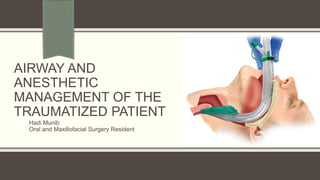
- 1. AIRWAY AND ANESTHETIC MANAGEMENT OF THE TRAUMATIZED PATIENT Hadi Munib Oral and Maxillofacial Surgery Resident
- 2. Outline Importance of Airway in maxillofacial Trauma Initial Airway Assessment History Physical Examination Anesthetic Strategies References
- 3. Importance of Airway in Maxillofacial Trauma The maxillofacial trauma patient can present with impending airway compromise. Patent airway is mandatory. Maxillofacial injuries can prevent the trauma surgeon from securing an airway. An airway obstruction can be due to: Foreign body Mandibular injury or maxillary injury Tracheal injury, laryngeal injury, or tongue injury Which can be compounded by hemorrhage or edema resulting from injuries to these or associated structures.
- 4. Importance of Airway in Maxillofacial Trauma Great care must be taken to avoid excessive movement of the cervical spine. The patient’s neck should not be rotated, hyper-flexed, or hyperextended. In order to minimize iatrogenic injury, the patient’s neck should be immobilized until a detailed cervical spine examination and complete cervical spine radiographic series are performed and a cervical spine injury has been ruled out. A common error in trauma management during the primary survey involves delay in securing the patient’s airway. Maxillofacial Injuries may complicate airway management: compromise airway integrity, resulting in poor exchange or obstruction, and the soft tissue or skeletal injury may make providing respiratory support difficult.
- 5. Importance of Airway in maxillofacial Trauma Gruen et al studied 2594 trauma mortality patients in order to identify patterns of errors contributing to inpatient deaths. They found that failure to intubate, secure or protect the airway was the most common factor related to patient mortality, responsible for 16% of inpatient deaths.
- 6. Initial Airway Assessment The airway should be assessed to ascertain patency. The well-established approach to the ABCs (airway, breathing, circulation) is the standard approach. Is the patient able to communicate verbally? Is the patient breathing spontaneously? Does the patient have adequate oxygenation? A negative response to any of these questions will require emergent intervention to secure adequate oxygenation and ventilation. The inability to obtain control of the airway can result in irreversible brain damage due to hypoxia in as little as 4 minutes. Severe head-injury patients with a Glasgow Coma Scale (GCS) score of 8 or less usually require the placement of a definitive airway.
- 7. Initial Airway Assessment Airway patency does not ensure adequate ventilation. Ventilation requires adequate gas exchange with adequate oxygenation with normal function of the lungs, diaphragm, and chest wall. Chest auscultation and percussion can help in assessing the patency of oxygenation; Auscultation is the most important technique in assessing air flow through the tracheobronchial tree. Breath sounds may be decreased when air flow is decreased (due to muscular weakness) or when the transmission of sound is poor (due to pleural effusion or pneumothorax). The chest wall should be visually inspected to rule out any chest injuries. Conditions such as flail chest or massive hemothorax, as well as tension or open pneumothorax, can compromise ventilation and oxygenation and need immediate attention.
- 8. Initial Airway Assessment Every trauma patient should receive supplemental oxygen. Maintaining good oxygen saturation and preventing hypercarbia are key factors. A pulse oximeter is essential in assessing adequate hemoglobin saturation. Potential for a false initial assessment for a patient who may have both normal appearing skin tone and oxygen saturation secondary to carbon monoxide exposure. Capnography could be helpful in monitoring the concentration or partial pressure of carbon dioxide (CO2). It is usually presented as a graph of expiratory CO2 plotted against time. The capnogram is a direct monitor of the inhaled and exhaled concentration or partial pressure of CO2 and is actually an indirect monitor of the CO2 partial pressure in the arterial blood.
- 10. Initial Airway Assessment Vital signs are an important part of the assessment. Hypotension is usually indicative of hypovolemia. Hemorrhage is the most common cause of shock after injury. Almost all trauma patients have an element of hypovolemia, which is indicative of hemorrhagic shock. Cardiogenic shock due to myocardial dysfunction can occur in trauma patients, especially if they sustain blunt cardiac injury, cardiac tamponade, or myocardial infarction. Skin color could be an important tool in assessing perfusion and oxygenation.
- 11. Initial Airway Assessment Capillary refill; the rate at which blood refills empty capillaries; Capillary refill is usually 1.5 to 2 seconds. Prolonged capillary refill suggests diminished localized blood flow or dehydration.
- 12. Airway Assessment Identify the patient with a potentially difficult airway. History and Physical Examination A patient’s history should inquire as to difficulties with airway management during previous anesthetics, presence of obstructive sleep apnea (OSA), and previous surgeries on the airway. page 12
- 13. Airway Assessment The physical examination focuses on several potentially contributing anatomic abnormalities, which have been shown to correlate well with increasing difficulty with intubation: Modified Mallampati test; based on the ability to visualize the uvula and facial pillars with the mouth open wide, tongue maximally protruded, neck extended forward into the sniffing position, while the patient phonates page 13
- 14. page 14
- 15. Airway Assessment Thyromental distance; a measurement from the mandibular menton to the prominence of the thyroid cartilage, with the neck fully extended. The distance should be at least 6 cm or approximately three ordinary fingerbreadths. A short thyromental distance equates with an anterior larynx that is at a more acute angle and also results in less space for the tongue to be compressed into by the laryngoscope blade page 15
- 17. Airway Assessment Mandibular retrognathia. Interincisal opening less than 3 cm (reduced temporomandibular joint [TMJ] mobility, trismus, scarring, fibrosis). Short neck. Neck circumference greater than 17 cm. Enlarged tonsils (grade 3–4). Limited neck flexion or head extension (e.g., rheumatoid arthritis, ankylosing spondylitis). Prominent upper incisors. Abnormal oropharyngeal/neck masses. Congenital, developmental, or acquired facial deformities (e.g., craniofacial syndromes, burns to the head and neck). page 17
- 18. page 18
- 19. Airway Assessment Five independent predictors have been associated with impossible mask ventilation: 1. Neck radiation changes 2. Male sex 3. OSA 4. Mallampati 3 or 4 5. Presence of a beard. Certain features such as: obesity, snoring, and lack of teeth were reported to potentially increase the difficulty of mask ventilation but could be overcome using various maneuvers and were not found to be predictors of impossible mask ventilation page 19
- 20. LEMON Assessment
- 21. History In many acute trauma patients, history cannot be obtained from the patient. Pre-hospital personnel and family members may be helpful in providing some of this relevant information. The AMPLE history is a quick mnemonic to use in evaluating the trauma patient: A: Allergies M: Medications (currently used) P: Pregnancy/Past illnesses L: Last meal E: Events/Environment related to the injury
- 22. History A patient who is on a beta blocker can have a “normal” heart rate even in hypovolemic shock. Patients on anticoagulants are prone to severe bleeding and hemorrhage. With many elderly patients on anticoagulants, a neurologic hemorrhage must be ruled out in this patient population. There is the potential for airway compromise associated with an increased risk of aspiration, decreased functional residual volume, and increased difficulty in intubating the airway. There are hemodynamic changes that impact fluid management, as well as the pharmacokinetics and pharmacodynamics of anesthetic and emergency agents
- 23. History The mechanism of injury can provide the trauma surgeon with pertinent information about the type of injuries sustained. Blunt injuries usually result from motor vehicle accidents (MVAs), falls, and occupation- related injuries. Important prehospital information to obtain from MVA patients includes seat belt usage, direction and type of impact, steering wheel deformation, and automobile damage. Rear impact in an MVA will probably result in cervical spine injury and/or soft tissue injury to the neck. The extent of injury depends on the region of the body affected, the path of the penetrating object, and the organs in proximity to the injury, as well as the velocity of the missile.
- 24. Six Specific Situations Associated with Maxillofacial Trauma in which these situations might affect the Airway 1. Posteroinferior displacement of a fractured maxilla parallel to the inclined plane of the skull base may block the nasopharyngeal airway. 2. A bilateral fracture in the mandible (comminuted in anterior mandible/ Bilateral Parasympheseal) 3. Fractured or exfoliated teeth, bone fragments, vomitus and blood as well as foreign bodies – dentures, debris, shrapnel etc. 4. Hemorrhage, either from distinct vessels in open wounds or severe nasal bleeding from complex blood supply of the nose, may also contribute to airway obstruction. 5. Soft tissue swelling and edema resulting from trauma to the head and neck may cause delayed airway compromise. 6. Trauma to the larynx and trachea may cause swelling and displacement of structures, such as the epiglottis, arytenoid cartilages and vocal cords, thereby increasing the risk of cervical airway obstruction.
- 26. Physical Examination Airway patency can affect the ability to ventilate and intubate the patient. Part of a detailed trauma examination is to recognize the problem and manage it appropriately. Airway compromise may be partial or complete. Signs such as stridor, dyspnea, tachypnea, and dysphagia may be indicative of airway obstruction. Tachypnea could be an early sign of airway or ventilatory compromise. The patient with an altered level of consciousness is at high risk of airway compromise and will require a secure airway.
- 27. Physical Examination Soft tissue: A detailed soft tissue examination must be performed. Significant tissue avulsions or injuries may result in an inability to secure a mask and provide ventilatory support. Avulsion Laceration Abrasion Puncture Contusion
- 30. Physical Examination Hemorrhage: can lead to airway irritation, aspiration, and laryngospasm. Pressure packing may be needed to control the bleeding. If the bleeding does not stop, surgical exploration or interventional radiology might be indicated (especially in neck trauma). Hemorrhage can result in an expanding hematoma that can compromise airway integrity. Securing an airway either via intubation or tracheotomy may be required
- 33. Physical Examination Skeleton (a) Mandibular fractures are common in trauma patients. Bilateral mandibular fractures can lead to a flail mandible; upper airway obstruction due to posterior movement of the tongue. Lying supine can further aggravate the obstruction. Airway adjuncts (e.g., nasopharyngeal airway) may assist in supporting and maintaining airway patency.
- 35. Physical Examination (b) Trauma to the midface can result in injuries to the nasopharynx and oropharynx. These fractures could be associated with hemorrhage, dislodged teeth, or unstable segments (maxilla or midface), any of which could lead to airway compromise. Posterior and downward displacement of a Le Fort fracture can result in an open bite and difficulty with mask ventilation. Displacement of a Le Fort II/III fracture can result in obstruction of the nasopharynx
- 37. Physical Examination (c) Nasal fractures can lead to posterior bleeding and aspiration and thus to nasopharyngeal obstruction. Nasal intubation is relatively contraindicated in midface fractures because of the risk of the endotracheal tube entering either the cranium or the orbit. A hematoma in the oropharynx or nasopharynx can lead to partial or complete airway obstruction. A nasal septal hematoma can lead to nasopharyngeal obstruction. An urgent surgical airway may be needed if endotracheal intubation is not possible.
- 38. Physical Examination Laryngeal trauma: Laryngeal trauma is associated with the triad of hoarseness, subcutaneous emphysema, and a palpable fracture. Patients with arytenoid injury are at higher risk of aspiration. Some of the symptoms of arytenoid injury include hoarseness, dysphagia, sore throat, dyspnea, vocal fatigue, loss of vocal control, and cough. A flexible endoscopically guided intubation may be helpful in laryngeal trauma. Early diagnosis of tooth aspiration in the multi-trauma patient can be difficult because aspiration is rarely considered in the absence of an acute clinical presentation. Accounting for teeth is an important aspect of the maxillofacial exam.
- 40. Injuries Affecting Airway: Cervical Spine Injury Cervical spine injury must be presumed in any patient with maxillofacial or head trauma. The incidence of cervical injury in the presence of facial fractures has been reported to be 1% to 6%. Cervical spine injuries tend to occur in patients between 15 and 45 years of age and are seen more commonly in males (7:3). The most common level of fracture is C2 Dislocations occur most commonly at the C5/6 and C6/7 levels. The exclusion of cervical spine injuries, or “clearance,” requires the exclusion of both bony and ligamentous injuries.
- 41. They suggest injuries to upper cervical spine segments are associated with lower third facial fractures, principally mandibular fractures Injuries to the lower cervical spine segments are associated with middle third facial fractures. Our study lends some support to this model in that three of the four cases of CSI associated with isolated mandibular fractures occurred at C2 and the remaining case at C1
- 43. Examination of the neck includes inspection, palpation and auscultation. The standard three-view plain film series for a c-spine evaluation includes lateral, anteroposterior, and open-mouth view. The lateral cervical spine film must include the base of the occiput and the top of the first thoracic vertebra. The lateral view alone is inadequate and will miss up to 15% of cervical spine injuries. The lower cervical spine may be difficult to examine, and caudal traction on the arms should be used to improve visualization. If the lower cervical spine is not visualized, a computed tomography (CT) scan of the region is indicated Injuries Affecting Airway: Cervical Spine Injury
- 44. Thin cut (2-mm) axial CT scanning of specific bone windows, with sagittal and coronal reconstruction, should be used to evaluate abnormal, suspicious, or poorly visualized areas on plain radiology. With technically adequate studies and experienced interpretation, the combination of plain radiology and directed CT scanning provides a false-negative rate of less than 0.1%. Magnetic resonance imaging (MRI) is the gold standard for clearing the cervical spine in a clinically suspicious or unresponsive trauma patient. In most trauma centers, cervical fixation is usually maintained until the patient is awake and can respond to command. Injuries Affecting Airway: Cervical Spine Injury
- 45. Rigid cervical collars (by themselves) limit rotation and lateral movement only to 50% of normal and flexion and extension only to 30% of normal and do not provide adequate stabilization. Complete dissection of the spinal cord above C3 will cause apnea and death, unless the patient receives immediate intubation and ventilatory support. For lesions between C3 and C5, the degree of respiratory failure is variable. The vital capacity can be reduced to 15% to 20% of normal. These patients are at risk of increasing diaphragmatic fatigue due to slowly progressive ascending injury resulting from cord swelling and edema. This usually leads to decompensation and retention of secretions on day 3 to 4 post injury. Intubation and ventilation is needed to prevent respiratory failure. Injuries Affecting Airway: Cervical Spine Injury
- 46. Patients with high cervical spine lesions have increased bronchial secretions, possibly due to altered neuronal control of mucous glands. Injuries at C5 or below spare the phrenic nerve and diaphragmatic breathing. Expiratory reserve may be impaired secondary to accessory muscle paresis Injuries Affecting Airway: Cervical Spine Injury
- 47. The decision to intubate depends on several factors, Loss of innervation of the diaphragm (e.g., phrenic nerve injury) Fatigue of muscles of respiration Failure to clear secretions Failure to maintain a patent airway History of aspiration Presence of other injuries (e.g., head, maxillofacial, and/ or chest injuries) Premorbid conditions, especially respiratory disease (chronic obstructive pulmonary disease) and/or obesity Injuries Affecting Airway: Cervical Spine Injury
- 48. Injuries to the cervical airway may result from blunt or penetrating trauma. The incidence is usually rare. Blunt trauma to the airway either from a direct blow to the cervical airway or from severe flexion/extension injuries may result in a thyroid or a cricoid cartilage fracture. Injuries Affecting Airway: Cervical Airway Injury
- 49. Injuries Affecting the Airway: Thermal and Inhalation Injury Suspected in any patient presenting with a history of exposure to fire or smoke. CO Toxicity does not cause direct injury to the lungs. Its toxic effect results from a displacement of oxygen from hemoglobin-binding sites, thereby decreasing the oxygen-carrying capacity of the blood. This causes a shift of the oxyhemoglobin dissociation curve to the left; tissue hypoxia. CO reacts with myoglobin to further impair oxygen uptake by decreasing facilitated diffusion of oxygen into muscle. This leads to impaired tissue oxygen availability and delivery. Cyanide causes tissue asphyxiation through the inhibition of intracellular cytochrome oxidase.
- 50. It blocks the final step in oxidative phosphorylation and prevents mitochondrial oxygen use. The organs most affected to cellular hypoxia include the central nervous system and the heart Inhalation injury can occur with or without evidence of burns. Burns on the face and singed eyebrows and/or facial hair are indicative of smoke inhalation. Patients with respiratory injury present with many symptoms, including minor eye irritation, cough, dyspnea, hyperventilation, tachypnea, rales, wheezing, and facial burns. This can lead to acute respiratory failure. These symptoms usually present within 6 to 24 hours post injury Injuries Affecting the Airway: Thermal and Inhalation Injury
- 51. Pulse oximetry is an accurate technique to monitor oxygen Saturation; it is falsely elevated by CO. Arterial oxygen tension (partial pressure arterial oxygen [PaO2]) does not accurately reflect the degree of CO poisoning or cellular hypoxia. The PaO2 level reflects the oxygen dissolved in blood that is not altered by the hemoglobin-bound CO. PaO2 level within the reference range may lead to serious underestimation of the decrement in tissue oxygen delivery and the degree of hypoxia at the cellular level that occurs when CO blocks the delivery of oxygen to the tissues; dissolved oxygen makes up only a small fraction of arterial oxygen content Arterial blood gas (ABG) measurements are still useful Injuries Affecting the Airway: Thermal and Inhalation Injury
- 52. Cyanide levels correlate closely with the level of exposure and toxicity; These levels may not be readily available. Indications of Cyanide Poisoning: Persistent neurologic dysfunction Unresponsive to use of supplemental oxygen Cardiac dysfunction Severe lactic acidosis In a setting consistent with potential cyanide exposure, empiric therapy should be started Injuries Affecting the Airway: Thermal and Inhalation Injury
- 55. Both direct laryngoscopy and fibreoptic bronchoscopy are both diagnostic and therapeutic. Visualization of erythema, edema, ulceration, and soot deposition make bronchoscopy useful in evaluating the extent of injury to the tracheobronchial tree. Fiberoptic bronchoscopy can facilitate endotracheal tube placement. Injuries Affecting the Airway: Thermal and Inhalation Injury
- 56. Post-Operative Period The patient with a difficult airway is also at high risk for complications in the post- operative period. Following surgery, mucous membranes are edematous, soft tissue is swollen and the air pathway may be compressed. Neck expandability is relatively low and even a small hemorrhage in the region could result in airway compromise. It was found that complications arose throughout the perioperative period: 67% upon induction, 15% during surgery, 12% at extubation 5% during recovery.
- 57. Post-Operative Period In intubated maxillofacial trauma patients, extubation should be deferred until normal anatomy is restored or at least until the edema subsides. During extubation the patient should be monitored closely and the care providers should be prepared for the possibility of re-intubation. In a case of tracheotomy tube, the patient may be awakened and allowed to breathe spontaneously through the tracheostomy tube for a few days, providing a safer recovery.
- 58. Indications of Definitive Airway in Maxillofacial Injury
- 59. STRATEGIES OF GENERAL ANESTHESIA IN THE TRAUMA PATIENT
- 60. Triage To classify patients into various categories, identifying the urgency of treatment. Traumatized patients are divided into two major categories: Those patients who require airway control as part of the resuscitation: Usually apneic or have total or near-total airway obstruction. They are identified during the primary survey and will require establishment of a patent airway. The mouth must be inspected for the presence of any foreign material. Then administration of positive pressure ventilation by mask with 100% oxygen is performed.
- 61. Triage If ventilation is unsuccessful, placement of an oropharyngeal or nasopharyngeal airway may be used to facilitate mask ventilation. The oropharyngeal airway can induce gagging and coughing; may lead to increased intracranial or intraocular pressure and/or vomiting. The nasopharyngeal airway can lead to epistaxis, and care must be used when using it in a patient with a base-of skull fracture. If the patient cannot be effectively ventilated, laryngoscopy and intubation must be attempted. An LMA (laryngeal mask airway) or a Combitube (double-lumen airway) may be considered in these cases.
- 65. Triage Those patients who require semi-elective airway control at some time during hospitalization for surgical management of the maxillofacial injury. Usually require elective intubation or, in selected situations, a tracheostomy. Intubation or a surgical airway in this situation: Provides a patent airway Protects against aspiration of secretions Allows positive pressure ventilation Decreases anatomic dead space Provides a potential emergency route for drug administration.
- 66. Basic Principles of Management - Prophylaxis Against Aspiration Trauma patients are always considered to have full stomach. This is due to multiple reasons including: Ingestion of food or liquids before injury Swallowed blood from oral or nasal injury Delayed gastric emptying Administration of liquid contrast medium
- 67. Various actions are considered to prevent aspiration: Administer non-particulate antacid prior to induction Cricoid pressure/Sellick maneuver applied continuously during airway management Rapid-sequence induction (RSI) Avoidance of ventilation between administration of medication and intubation Awake intubation with or without topical or anesthetic blocks to the upper airway Basic Principles of Management - Prophylaxis Against Aspiration
- 69. Induction of General Anesthesia There is no ideal anesthetic drug for trauma patients with hemorrhagic shock. The key to safe anesthetic management is to administer small incremental doses of whichever agents are selected. Titration of the general anesthetic is a key factor in preventing complications. Some of the induction agents are propofol, etomidate, ketamine, and dexmedetomidine.
- 70. Propofol An ideal general anesthetic agent used in induction. It has a rapid onset of action as well as fast recovery from its sedative-hypnotic effects. Propofol has antiemetic effects that may be beneficial, especially for a patient who may have maxilla-mandibular fixation. Has some unwanted side effects in trauma patients. It may potentiate profound hypotension due to its direct myocardial depression and decrease in systemic vascular resistance. These effects can be potentiated in the elderly or cardiac patient and can be exacerbated in the hypovolemic trauma patient
- 71. Etomidate Widely used in the trauma population because of its cardiovascular stability relative to other induction agents. Etomidate provides advantages for induction of anesthesia in the setting of hemorrhagic shock. Its pharmacodynamics and pharmacokinetics are minimally altered in contrast to other anesthetic drugs. Etomidate may still produce profound hypotension due to its inhibition of catecholamine release. It can cause adrenocortical suppression. Adrenal suppression was found to last 6 to 8 hours in patients following a single induction dose of etomidate, and more than 24 hours following etomidate infusion. The impact of single-dose etomidate on outcomes in critically ill patients remains unclear.
- 72. Ketamine Ketamine can be used for induction in trauma patients. It is most suitable in children who are hemodynamically unstable or hypovolemic. Ketamine is also helpful in asthmatic patients because of its broncho-dilating properties. It can result in myocardial depression. In normal patients, the effects of catecholamine release usually will mask cardiac depression and result in tachycardia and hypertension. In patients with severe hemorrhagic shock, the cardiac depression may be unmasked. This will lead to cardiovascular collapse.
- 73. Dexmedetomidine An agonist of Alpha-2-adrenergic receptors in certain parts of the brain. It is indicated for sedation of critically ill or trauma patients in an intensive care unit setting. Dexmedetomidine may also be used to provide sedation for an awake intubation. Clinically it provide sedation and analgesia. It does so with minimal respiratory compromise. Dexmedetomidine is administered via a slow bolus supplemented with a continuous infusion. It is not administered with incremental boluses because of concerns about peripheral Alpha-2-receptor stimulation, with resulting hypotension and bradycardia. Intravenous infusion is commonly initiated with a 1 mcg/kg loading dose, administered over 10 minutes, followed by a maintenance infusion of 0.2 to 1.0 mcg/kg/hour.
- 74. Succinylcholine Remains the neuromuscular blocker with the fastest onset (30 seconds) and shortest duration of action It is the agent of choice for RSI. It is especially indicated in trauma patients presenting for an emergency procedure with a potentially full stomach or who have recently eaten. Succinylcholine may be contraindicated in selected trauma patients: Burn Patients; After the first 24 hours due to raised Pottassium Crush Injury: Risk of Hyperkalemia Ocular Injury: due to increased IOP, within 1 minute of induction, peak at 2-4 minutes and subsides at 6 minutes Intracranial Injury: ICP
- 75. Rocuronium Rocuronium can be also used safely during RSI (1 mg/kg), with an onset of relaxation of 60 seconds. At this dose the duration of action will be 1 to 2 hours. Sugammadex is an agent for reversing the neuromuscular blockade caused by a non- depolarizing agent such as rocuronium. Its main advantage is reversal of neuromuscular blockade without relying on the inhibition of acetylcholinesterase. Its administration is associated with much greater cardiovascular and autonomic stability than the traditional reversal agents.
- 76. Advanced Airway Technique Two-handed mask ventilation: Bag-mask ventilation is the cornerstone of basic airway management. When a second person is available to provide ventilations by compressing the bag, a two-hand technique can be used. The surgeon creates opposing semicircles with the thumb and index finger of each hand to form a ring around the mask connector and hold the mask on the patient’s face. Then, the mandible can be lifted with the remaining digits
- 77. C-E
- 80. Advanced Airway Technique Combitube (double-lumen) A blind insertion airway device (BIAD) used in the prehospital and emergency setting. It is ideal to provide an airway for the trauma patient in respiratory distress. The simplicity of placement is a major advantage over endotracheal intubation. This technique has some serious but rare complications, including: Aspiration Esophageal perforation Facial nerve dysfunction
- 82. ( A ) right-sided Robertshaw double-lumen tube ( B ) left-sided Rusch double-lumen tracheostomy tube.
- 83. Advanced Airway Technique LMA: The laryngeal mask airway (LMA) is an easy way to ventilate a trauma patient. It is a supraglottic airway device and is a good alternative to bag-valve-mask ventilation (freeing the hands of the provider with the benefit of less gastric distention). The LMA is actually placed into the back of the mouth following the curve of the tongue. The rate of complications is rare (0.15%). These complications include: Aspiration of gastric contents, local irritation, pressure-induced lesions, facial nerve palsies, mild sympathetic response, obstruction, and laryngospasm.
- 84. LMA
- 85. LMA
- 86. Laryngoscope
- 88. Advanced Airway Technique The laryngoscope is a useful technique to visualize the vocal cords and the glottis.
- 89. Advanced Airway Technique Videolaryngoscope: The failure of direct laryngoscopy to provide an adequate view for tracheal intubation led to the development of videolaryngoscopy. The first camera screen laryngoscope was developed by Berall in 1998. Currently, there are several manufacturers of video laryngoscopes. These laryngoscopes employ a variety of features, such as a monitor on the handle and/or channels, to assist in guiding the endotracheal tube into the trachea. The superior performance of video laryngoscopy in airway management where cervical spine injury is suspected.
- 92. Advanced Airway Technique Retrograde intubation is another alternative to secure an airway in the trauma patient. Under local anesthesia, a cannula is inserted through the cricothyroid membrane into the trachea, and a guide wire is then passed through the needle upward through the vocal cords into the oral cavity. The wire is then used to guide an ET tube through the vocal cords.
- 97. Surgical Airway Emergency airway management in the trauma patient often involves a combination of factors that increase the technical difficulty of intubation and increase patient risk. These factors include: Dynamically deteriorating clinical situation in the unstable patient Respiratory and ventilatory compromise Impaired ventilation and oxygenation Full stomach (increased risk of regurgitation, vomiting, aspiration) Increased secretions, blood, vomitus, and potential for foreign body Distorted anatomy If intubation is not possible, a surgical airway is advised.
- 98. Tracheostomy
- 99. Tracheostomy A surgical procedure which consists of making an incision on the anterior aspect of the neck and opening a direct airway through an incision in the trachea The resulting stoma can serve independently as an airway or as a site for tracheostomy tube to be inserted This tube allows a person to breathe without the use of the nose or mouth. There are four main indications: Emergency airway access Airway access for prolonged mechanical ventilation Functional or mechanical upper airway obstruction Decreased/incompetent clearance of tracheobronchial secretions
- 100. Tracheostomy There are no absolute contraindications to tracheostomy except for active cellulitis of the anterior neck skin
- 101. An outer cannula (top item) with inflatable cuff (top right), an inner cannula (center item) and an obturator (bottom item)
- 103. Early Complications Bleeding Pneumothorax Subcutaneous Emphysema Injury to Recurrent Laryngeal Nerve Blocked Tracheostomy Tube Accidental Displacement of Tracheostomy Tube Infection
- 104. Bleeding Most likely cause of fatality after a tracheostomy. Injury to the anterior jugular venous system is a typical source of bleeding, which if encountered is ligated and divided It usually occurs due to a Tracheoarterial Fistula, an abnormal connection between the trachea and nearby blood vessels, and most commonly manifests between 3 days to 6 weeks after the procedure is done. Fistulas can result from incorrectly positioned equipment, high cuff pressures causing pressure sores or mucosal damage, a low surgical trachea site, repetitive neck movement, radiotherapy, or prolonged intubation. Use of the bronchoscope can help with proper placement of instruments and better visualization of anatomical structures. Bleeding can also occur from frequent or improper suctioning; Use of a red rubber suction may reduce any trauma from suctioning.
- 105. Pneumothorax Collapsed Lung; Partial or complete 17% incidence; Increased incidence in children possibly due to the higher position of the pleural dome Pneumothorax is a potentially life threatening complication and can occur from direct injury to the pleura or the cupola of the lung (especially in children) or from high negative inspiratory pressures of patients who are awake and distressed on mechanical ventilation. Patients with chronic obstructive pulmonary disorder (COPD) are at particularly high risk because intrinsic air-trapping causes their chest to be barrel shaped, and the apex of the lung is much higher in the chest cavity. Chest X-ray
- 106. Subcutaneous emphysema The result of air or gas trapped under the skin, typically seen in the chest walls and neck regions. It can develop rapidly, and may be the first clinical sign of a pneumothorax. It can result from trauma during the tracheotomy procedure or tracheal intubation. The most common sign is swelling around the neck and chest pain. Other signs and symptoms include tender sore throat, aching neck, difficulty in swallowing, breathlessness, wheezing, and distension. Subcutaneous emphysema has a characteristic crackling feel when palpating the skin, which is termed subcutaneous crepitation
- 107. Recurrent Laryngeal Nerve Injury Damage to the recurrent laryngeal nerve resulting in vocal cord paralysis, is also a potential immediate complication of tracheostomy. Damage can cause hoarse or breathy vocal quality, dysphagia and aspiration pneumonia. If midline is properly maintained during dissection, the nerves should not be encountered, except for cases where significant misplacement of the tracheostomy tube has occurred. Tracheostomy itself may be needed when bilateral recurrent laryngeal nerve injury has occurred during another procedure (ie., total thyroidectomy). Bilateral recurrent laryngeal nerve injury results in the vocal folds paralyzed medially, causing difficulty breathing past the closed vocal folds, requiring emergent intubation or tracheostomy.
- 108. Blocked Tracheostomy Tube A blocked tracheostomy tube is typically secondary to a mucus plug or blood clot. This can be prevented with proper suctioning of the airway, adequate humidification, and frequent changing or cleaning of the inner cannula when working with a double lumen tube. The use of dual cannula tubes lessens the potential of mucus plugging because the inner cannula can be removed for cleaning while the outer cannula safely maintains patency of the fresh tract. Inner cannulas should be cleaned daily if non-disposable, or changed daily if disposable in order to avoid mucous plugging. Partial blockage is indicated by resistance to the passage of a suction catheter over the first 10 cm or when the inside of the tracheostomy tube feels roughened by accumulated, dried secretions. A completely blocked tube is indicated by the inability to pass a suction catheter down the tracheostomy tube.
- 109. Signs of Blocked Tracheostomy Tube
- 110. Late Complications Tracheal Stenosis Tracheomalacia Tracheoesophegeal Fistula Tracheoinnominate Fistula Pneumonia
- 111. Tracheal Stenosis An abnormal narrowing of the trachea that can develop from prolonged intubation causing scarring due to pressure injury. It can also occur due to high cuff pressures from either the tracheostomy tube or the endotracheal tube cuff. Tracheal stenosis commonly develops at the stoma site or at the level of the cuff. When stenosis occurs at the stoma, it is usually due to bacterial infection and chondritis. Granulation tissue can develop at the stoma site and may obstruct the airway at the stoma site. The tissue can cause difficulty with replacing the tracheostomy Tube Diagnosis of tracheal stenosis is often delayed; Approximately 3 to 12% of all patients will develop clinically significant stenosis that will require intervention; Extra-long Tracheostomy Tube
- 112. Tracheomalacia Weakening of the tracheal wall. In adults, this is usually secondary to trauma of some kind to the tracheal wall, including recurrent infections, where the tube and/or cuff rubs against it, or with over inflation of the cuff. Signs and symptoms of tracheomalacia include: shortness of breath, hoarse voice, chronic cough, difficulty swallowing, recurring respiratory tract infections, and, during mechanical ventilation there is often the presence of a cuff leak, or the need to over- inflate the cuff in order to maintain control of mechanical ventilation If tracheomalacia is suspected, bronchoscopy or a dynamic CT scan of the trachea can reveal excessive expiratory collapse of the trachea. Treatment options depend on the severity and include insertion of a longer tracheostomy tube to bypass the region of trachomalacia or double cuffed tracheostomy tubes.
- 113. Care of Tracheostomy Caring for a tracheotomy mostly includes Suctioning to prevent occlusions Replacing supplies, such as replacement of the inner cannula and/or suction devices. Risks include hypoxia and so suctioning is limited to 10 to 20 seconds at a time and the patient is hyper-oxygenated just before and after suctioning. Risks also include atelectasis, or collapsing lung tissue from high suction pressure, and so pressure is limited to 80–120 mm Hg. Risks also include tissue damage. The suction catheter is inserted no more than 1 cm past the length of the tube to avoid contact with trachea tissue.
- 114. Tracheostomy Horizontal skin incisions are cosmetically superior to vertical incisions.
- 116. Emergency Cricothyrotomy Emergency cricothyrotomy is one of the last-resort and potentially life-saving procedures for maxillofacial trauma patients who cannot be intubated or ventilated. A surgical airway has been established by making an incision through the skin and the cricothyroid membrane into the tracheal lumen, through which an endotracheal tube is inserted. In recent years, a number of commercial kits that include all of the necessary equipment to establish a surgical airway have reached the market. The surgeon must be familiar with the kit used at his or her trauma center. The tube can stay in place for up to 3 days. In surgical cricothyrotomy, a scalpel is used to make a horizontal incision over the lower half of the cricothyroid membrane. The only absolute contraindication to surgical cricothyrotomy is age.
- 117. Cricothyrotomy Some general indications for this procedure include: Inability to intubate Inability to ventilate Inability to maintain SpO2 >90% Severe traumatic injury that prevents oral or nasal tracheal intubation
- 118. Cricothyrotomy Contraindications Inability to identify landmarks (cricothyroid membrane) Underlying anatomical abnormality such as a tumor or severe goiter Tracheal transection Acute laryngeal disease due to infection or trauma Small children under 12 years old (a 10–14 gauge catheter over the needle may be used) In patients below this cutoff age, needle cricothyrotomy is indicated because infants and children younger than 12 years usually have a smaller cricothyroid membrane as well as a more funnel-shaped, rostral, and compliant larynx
- 121. Cricothyrotomy
- 124. References Fonseca; Volume 2; Airway and Anesthetic Management of the Traumatized Patient Peterson’s Principles of Oral and Maxillofacial Surgery; Chapter 4 Outpatient Anesthesia Complications of Tracheostomy; https://www.tracheostomyeducation.com/blog/complications-of- tracheostomy/#:~:text=termed%20subcutaneous%20crepitation.- ,Injury%20to%20the%20Recurrent%20Laryngeal%20Nerve,quality%2C%20dysphagia %20and%20aspiration%20pneumonia. Maxillofacial Trauma Patient: Coping with Difficult Airway
- 125. References
- 126. THANK YOU!
Editor's Notes
- NOTE: To change the image on this slide, select the picture and delete it. Then click the Pictures icon in the placeholder to insert your own image.