SlideShare a Scribd company logo
1 of 17
ANATOMY OF
THE EYE AND
ORBIT
REVISION
WHAT BONES MAKE
UP THE BOUNDARIES
OF THE ORBIT?
Wall Bones
Superior • Orbital plate of frontal
bone
• LW sphenoid
Inferior • Orbital surface of
maxilla
• Palatine bone
• Orbital surface of
zygomatic
Medial • Ethmoid (orbital plate)
• LW sphenoid
• Frontal bone
• Lacrimal bone
• Frontal process of
maxilla
Lateral (thickest) • Zygomatic
• GW sphenoid
ORBIT SURROUNDED
BY PARANASAL AIR-
FILLED SINUSES
 Air filled sinuses located within
the bones of the skull and face
 Functions:
 Lightening weight of the head
 Humidifying and heating inhaled
air
 Increasing resonance of speech
 Protect vital structures in injury
 Superior to the orbit- frontal
sinus
 Inferiorly- maxillary sinus
 Medially- ethmoid sinus
THE EYEBALL
 Has 3 layers:
 Fibrous outer layer:
 Sclera: dense, white, avascular, continuous with the cornea
 Vascular middle layer
 Choroid: pigmented, vascular, continuous with ciliary body and iris
 Ciliary body = muscular thickening that provides attachment for the lens.
Secretes aq humor
 Iris = on anterior surface of the lens. It is a thin contractile diaphragm
 Inner retina layer
 Light sensitive
 Sensory neural layer
THE PUPIL
 Two involuntary muscles control
the size of the pupil:
 Dilator pupillae- sympathetic
innervation (makes the pupil
larger)
 Sphincter pupillae-
parasympathetic
ACCOMMODATION
 Ciliary m. control the shape of the lens- which
enables the eye to focus (accommodation)
 To focus on near objects:
 Ciliary m. contract through parasympathetic
innervation
 Release tension on the zonal fibers
 Lens becomes rounder/ convex- increased
curvature shifts focal point closer to the eye
MUSCLES OF THE EYELID
 Levator palpebra superioris m.
 Only in the UPPER eyelid
 Raises the eyelid
 Innervation via CN3
 Inserts into the tarsus
 In companion with the LPS m. is a collection of SM
fibers = superior tarsal m.
 Raises the eyelid
 Innervation via the sympathetic fibers from superior
cervical ganglion
Thus, CN3 lesion/ sympathetic
lesion leads to ptosis – drooping
eyelid
MUSCLES OF THE EYELID
 Orbicularis Oris m.
 A sphincter muscle located in the upper and lower eyelids
 Palpebral part of the muscle close the eyelids during
blinking and sleep
 Orbital part: voluntary action- screws the eyes tightly shut
for protection
 Innervated by the zygomatic and temporal branches of CN7
 Damage to CN7  paralysis  eyelids don’t close fully +
eversion of the lower eyelid  drying + damage to cornea
 Also assists in pumping tears into nasolacrimal duct system
EXTRA-OCULAR MUSCLES
Muscle Attachements Actions Innervation
LPS From LW sphenoid to the superior tarsal
plate of upper eyelid
Elevates upper eyelid CN3
Superior rectus Superior part of the common tendinous
ring to the superior and anterior sclera
Elevation
Adduction
Medial rotation
CN3
Inferior rectus Inferior common tendinous ring to the
inferior and anterior sclera
Depression
Adduction
Lateral rotation
CN3
Medical rectus Medical part of common tendinous ring to
the anteromedial sclera
Adducts eyeball CN3
Lateral rectus Lateral part of the common tendinous ring
to the anterolateral sclera
Abducts CN4
Superior oblique Body of the sphenoid, attaches to the
sclera posterior to superior rectus
Depresses
Abducts
Medially rotates
CN4
Inferior oblique Anterior aspect of the orbital floor to the
sclera of the eye, posterior to lateral rectus
Elevates
Abducts
Laterally rotates
CN3
Located in the orbit, but extrinsic to the eyeball.
They act to control movements of the eyeball and
superior eyelid.
EYELIDS, CONJUNCTIVA
AND TEARS PROTECT THE
EYE
 Eyelids:
 Dense connective tissue
called Tarsus
 Embedded in tarsus = tarsal
glands
 Sebaceous secretions
which prevent tears from
evaporating
 Conjunctiva:
 Provides protection and
lubrication
 Contains blood vessels to combat
infection
 Consists of an epithelial layer
composed of stratified squamous
and stratified columnar epithelium
 Non-keratinized with
interspersed goblet cells
 Within the epithelial layer there
are BVs, fibrous tissue,
lymphatics, melanocytes, T/B
cells, Langerhan's cells,
accessory lacrimal glands
Note: the suspensory ligament is hammock
shaped thickening, enclosing the inferior
rectus m. it supports the eyeball
TEARS
Tears/ lacrimal fluid from lacrimal gland
produced by parasympathetic impulses from
CN7
Secreted through lacrimal ducts to surface of
conjunctiva
When eyelids blink, it pushes tears medially
over the cornea
Fluid containing foreign material pushes to
medial angle of the eye
Drains through lacrimal canaliculi into the
lacrimal sac
Nasolacrimal duct conveys the fluid to the
nasal cavity
Function of tears:
 Lubricate – basal tears
 Removing irritants – reflex
tears
 Aiding immune system
PARASYMPATHETIC
AUTONOMIC
INNERVATION OF THE
EYE
 Parasympathetic preganglionic
fibers for the eye are axons of cells
in the accessory oculomotor
nucleus.
 They run in the CN3 and exit in
the motor root of the ciliary
ganglion
 The axons of the ciliary root
ganglion are postganglionic
parasympathetic fibers which
reach the eye in the short ciliary
nerves and are distributed to the
sphincter pupillae, ciliary m. and
BVs
SYMPATHETIC
AUTONOMIC
INNERVATION OF THE
EYE
 Some of the filaments passing
through the superior orbital
fissure form the sympathetic root
of the ciliary ganglion
 Some pass through the ganglion
as short ciliary nerves.
 Other filaments join the
ophthalmic nerve or its nasociliary
branch and reach the eye in long
ciliary nerves that supply the
dilator pupillae
 Both long and short ciliary nerves
also contain afferents to the
cornea, iris and choroid
SYMPATHETIC
AUTONOMIC
INNERVATION OF THE
EYE
 Some of the fibers in the short
ciliary nerves pass through a
communicating ramus from the
ciliary ganglion to the
nasociliary nerve-
 This ramus is called the sensory
root of the ciliary ganglion
 Involved in reflex actions
 Other sympathetic fibers from
the internal carotid plexus reach
the eye through the ophthalmic
periarterial plexus
SUPERIOR ORBITAL
FISSURE AND OPTIC
CANAL
 ”What structures run through the
foramina of the orbit?”
 Superior orbital fissure: CN3,
4, 6 and sensory innervation to
the eye
 Optic canal: CN2 and
opothalmic a. and optic n.
BLOOD SUPPLY TO
THE ORBIT
 Ophthalmic a:
 First branch of the internal carotid,
runs anteriorly through the optic
canal along with optic n.
(inferolaterally)
 Branches supply all structures in
the orbit in addition to the nose,
face, meninges
 Central retinal a. is the first branch
of the ophthalmic a. which runs in
the dura mater inferior to the optic
n.
 Supplies the internal retinal layers
 Second branch is the lacrimal a.
which runs along the superior
edge of the lateral rectus m.
 Supplies the lacrimal gland, eyelids
and conjunctiva
VENOUS DRAINAGE
OF THE ORBIT
 Ophthalmic v.
 Superior and inferior subdivisions-
pass through the superior orbital
fissure and enter the cavernous
sinus
 Central retinal v.
 Drains blood from the capillaries
of the retina into the superior
ophthalmic v. or the cavernous
sinus directly

More Related Content

What's hot (20)

Embryology of eye
Embryology of eyeEmbryology of eye
Embryology of eye
 
Histology of eye
Histology of eyeHistology of eye
Histology of eye
 
Anatomy of the eyelids
Anatomy of the eyelidsAnatomy of the eyelids
Anatomy of the eyelids
 
Anatomy of eye
Anatomy of eyeAnatomy of eye
Anatomy of eye
 
Anatomy of human orbit
Anatomy of human orbitAnatomy of human orbit
Anatomy of human orbit
 
Anatomy of optic nerve and its clinical significance
Anatomy of optic nerve and its clinical significanceAnatomy of optic nerve and its clinical significance
Anatomy of optic nerve and its clinical significance
 
Cavernous sinus
Cavernous sinusCavernous sinus
Cavernous sinus
 
Anatomy of Optic Nerve
Anatomy of Optic NerveAnatomy of Optic Nerve
Anatomy of Optic Nerve
 
ORBIT Anatomy
ORBIT AnatomyORBIT Anatomy
ORBIT Anatomy
 
Anatomy of the orbit (nerves and vessels)
Anatomy of the orbit (nerves and vessels)Anatomy of the orbit (nerves and vessels)
Anatomy of the orbit (nerves and vessels)
 
Orbital blood supply
Orbital blood supplyOrbital blood supply
Orbital blood supply
 
Anatomy of the lacrimal apparatus
Anatomy of the lacrimal apparatusAnatomy of the lacrimal apparatus
Anatomy of the lacrimal apparatus
 
Anatomy of lacrimal apparatus
Anatomy of lacrimal apparatusAnatomy of lacrimal apparatus
Anatomy of lacrimal apparatus
 
Ophthalmic artery
Ophthalmic arteryOphthalmic artery
Ophthalmic artery
 
Anatomy of retina
Anatomy of retinaAnatomy of retina
Anatomy of retina
 
Anatomy of the eye
Anatomy of the eyeAnatomy of the eye
Anatomy of the eye
 
BLOOD AND NERVE SUPPLY OF EYE
BLOOD AND NERVE SUPPLY OF EYE BLOOD AND NERVE SUPPLY OF EYE
BLOOD AND NERVE SUPPLY OF EYE
 
Lacrimal apparatus & nose
Lacrimal apparatus & noseLacrimal apparatus & nose
Lacrimal apparatus & nose
 
Anatomy of the eyelids.
Anatomy of the eyelids.Anatomy of the eyelids.
Anatomy of the eyelids.
 
#ANATOMY OF UVEA (IRIS, CILLIARY BODY, CHOROID)
#ANATOMY OF UVEA (IRIS, CILLIARY BODY, CHOROID) #ANATOMY OF UVEA (IRIS, CILLIARY BODY, CHOROID)
#ANATOMY OF UVEA (IRIS, CILLIARY BODY, CHOROID)
 

Similar to Anatomy of the Eye and Orbit: Bones, Muscles, Blood Supply

Similar to Anatomy of the Eye and Orbit: Bones, Muscles, Blood Supply (20)

Orbital region
Orbital regionOrbital region
Orbital region
 
ANATOMY_OF_THE_EYE_AND_ORBITS.ppt
ANATOMY_OF_THE_EYE_AND_ORBITS.pptANATOMY_OF_THE_EYE_AND_ORBITS.ppt
ANATOMY_OF_THE_EYE_AND_ORBITS.ppt
 
Sensory organs
Sensory organsSensory organs
Sensory organs
 
Eye by meher
Eye by meherEye by meher
Eye by meher
 
Sensory organs
Sensory organsSensory organs
Sensory organs
 
Anatomy of eye
Anatomy of eye Anatomy of eye
Anatomy of eye
 
Anatomy and physiology of eyelids
Anatomy and physiology of eyelidsAnatomy and physiology of eyelids
Anatomy and physiology of eyelids
 
Contents of orbit
Contents of orbitContents of orbit
Contents of orbit
 
eyelid
eyelideyelid
eyelid
 
The Orbit
The OrbitThe Orbit
The Orbit
 
Anatomy of the eye for studentss
Anatomy of the eye for studentssAnatomy of the eye for studentss
Anatomy of the eye for studentss
 
Unit 4; Special Senses, Educational Platform.pptx
Unit 4; Special Senses, Educational Platform.pptxUnit 4; Special Senses, Educational Platform.pptx
Unit 4; Special Senses, Educational Platform.pptx
 
Anatomy of Eyelids & Its Clinical Correlations
Anatomy of Eyelids & Its Clinical CorrelationsAnatomy of Eyelids & Its Clinical Correlations
Anatomy of Eyelids & Its Clinical Correlations
 
د احمد اسامه هاشم عيون ophthalmology
د احمد اسامه هاشم عيون ophthalmologyد احمد اسامه هاشم عيون ophthalmology
د احمد اسامه هاشم عيون ophthalmology
 
Anatomy of eye
Anatomy of eyeAnatomy of eye
Anatomy of eye
 
anatomy and physiology of lacrimal apparatus ppt
anatomy and physiology of lacrimal apparatus  pptanatomy and physiology of lacrimal apparatus  ppt
anatomy and physiology of lacrimal apparatus ppt
 
Eyeball 2013
Eyeball 2013Eyeball 2013
Eyeball 2013
 
Anatomy of external as well internal eye
Anatomy of external as well internal eye Anatomy of external as well internal eye
Anatomy of external as well internal eye
 
anatomy.ppt
anatomy.pptanatomy.ppt
anatomy.ppt
 
Eyelid Anatomy-2.pptx
Eyelid Anatomy-2.pptxEyelid Anatomy-2.pptx
Eyelid Anatomy-2.pptx
 

Recently uploaded

History Class XII Ch. 3 Kinship, Caste and Class (1).pptx
History Class XII Ch. 3 Kinship, Caste and Class (1).pptxHistory Class XII Ch. 3 Kinship, Caste and Class (1).pptx
History Class XII Ch. 3 Kinship, Caste and Class (1).pptxsocialsciencegdgrohi
 
Employee wellbeing at the workplace.pptx
Employee wellbeing at the workplace.pptxEmployee wellbeing at the workplace.pptx
Employee wellbeing at the workplace.pptxNirmalaLoungPoorunde1
 
BASLIQ CURRENT LOOKBOOK LOOKBOOK(1) (1).pdf
BASLIQ CURRENT LOOKBOOK  LOOKBOOK(1) (1).pdfBASLIQ CURRENT LOOKBOOK  LOOKBOOK(1) (1).pdf
BASLIQ CURRENT LOOKBOOK LOOKBOOK(1) (1).pdfSoniaTolstoy
 
How to Make a Pirate ship Primary Education.pptx
How to Make a Pirate ship Primary Education.pptxHow to Make a Pirate ship Primary Education.pptx
How to Make a Pirate ship Primary Education.pptxmanuelaromero2013
 
Introduction to AI in Higher Education_draft.pptx
Introduction to AI in Higher Education_draft.pptxIntroduction to AI in Higher Education_draft.pptx
Introduction to AI in Higher Education_draft.pptxpboyjonauth
 
Class 11 Legal Studies Ch-1 Concept of State .pdf
Class 11 Legal Studies Ch-1 Concept of State .pdfClass 11 Legal Studies Ch-1 Concept of State .pdf
Class 11 Legal Studies Ch-1 Concept of State .pdfakmcokerachita
 
Blooming Together_ Growing a Community Garden Worksheet.docx
Blooming Together_ Growing a Community Garden Worksheet.docxBlooming Together_ Growing a Community Garden Worksheet.docx
Blooming Together_ Growing a Community Garden Worksheet.docxUnboundStockton
 
Sanyam Choudhary Chemistry practical.pdf
Sanyam Choudhary Chemistry practical.pdfSanyam Choudhary Chemistry practical.pdf
Sanyam Choudhary Chemistry practical.pdfsanyamsingh5019
 
Introduction to ArtificiaI Intelligence in Higher Education
Introduction to ArtificiaI Intelligence in Higher EducationIntroduction to ArtificiaI Intelligence in Higher Education
Introduction to ArtificiaI Intelligence in Higher Educationpboyjonauth
 
The Most Excellent Way | 1 Corinthians 13
The Most Excellent Way | 1 Corinthians 13The Most Excellent Way | 1 Corinthians 13
The Most Excellent Way | 1 Corinthians 13Steve Thomason
 
Crayon Activity Handout For the Crayon A
Crayon Activity Handout For the Crayon ACrayon Activity Handout For the Crayon A
Crayon Activity Handout For the Crayon AUnboundStockton
 
Proudly South Africa powerpoint Thorisha.pptx
Proudly South Africa powerpoint Thorisha.pptxProudly South Africa powerpoint Thorisha.pptx
Proudly South Africa powerpoint Thorisha.pptxthorishapillay1
 
“Oh GOSH! Reflecting on Hackteria's Collaborative Practices in a Global Do-It...
“Oh GOSH! Reflecting on Hackteria's Collaborative Practices in a Global Do-It...“Oh GOSH! Reflecting on Hackteria's Collaborative Practices in a Global Do-It...
“Oh GOSH! Reflecting on Hackteria's Collaborative Practices in a Global Do-It...Marc Dusseiller Dusjagr
 
internship ppt on smartinternz platform as salesforce developer
internship ppt on smartinternz platform as salesforce developerinternship ppt on smartinternz platform as salesforce developer
internship ppt on smartinternz platform as salesforce developerunnathinaik
 
Pharmacognosy Flower 3. Compositae 2023.pdf
Pharmacognosy Flower 3. Compositae 2023.pdfPharmacognosy Flower 3. Compositae 2023.pdf
Pharmacognosy Flower 3. Compositae 2023.pdfMahmoud M. Sallam
 
Solving Puzzles Benefits Everyone (English).pptx
Solving Puzzles Benefits Everyone (English).pptxSolving Puzzles Benefits Everyone (English).pptx
Solving Puzzles Benefits Everyone (English).pptxOH TEIK BIN
 
EPANDING THE CONTENT OF AN OUTLINE using notes.pptx
EPANDING THE CONTENT OF AN OUTLINE using notes.pptxEPANDING THE CONTENT OF AN OUTLINE using notes.pptx
EPANDING THE CONTENT OF AN OUTLINE using notes.pptxRaymartEstabillo3
 
A Critique of the Proposed National Education Policy Reform
A Critique of the Proposed National Education Policy ReformA Critique of the Proposed National Education Policy Reform
A Critique of the Proposed National Education Policy ReformChameera Dedduwage
 

Recently uploaded (20)

History Class XII Ch. 3 Kinship, Caste and Class (1).pptx
History Class XII Ch. 3 Kinship, Caste and Class (1).pptxHistory Class XII Ch. 3 Kinship, Caste and Class (1).pptx
History Class XII Ch. 3 Kinship, Caste and Class (1).pptx
 
Employee wellbeing at the workplace.pptx
Employee wellbeing at the workplace.pptxEmployee wellbeing at the workplace.pptx
Employee wellbeing at the workplace.pptx
 
BASLIQ CURRENT LOOKBOOK LOOKBOOK(1) (1).pdf
BASLIQ CURRENT LOOKBOOK  LOOKBOOK(1) (1).pdfBASLIQ CURRENT LOOKBOOK  LOOKBOOK(1) (1).pdf
BASLIQ CURRENT LOOKBOOK LOOKBOOK(1) (1).pdf
 
How to Make a Pirate ship Primary Education.pptx
How to Make a Pirate ship Primary Education.pptxHow to Make a Pirate ship Primary Education.pptx
How to Make a Pirate ship Primary Education.pptx
 
Introduction to AI in Higher Education_draft.pptx
Introduction to AI in Higher Education_draft.pptxIntroduction to AI in Higher Education_draft.pptx
Introduction to AI in Higher Education_draft.pptx
 
Class 11 Legal Studies Ch-1 Concept of State .pdf
Class 11 Legal Studies Ch-1 Concept of State .pdfClass 11 Legal Studies Ch-1 Concept of State .pdf
Class 11 Legal Studies Ch-1 Concept of State .pdf
 
Blooming Together_ Growing a Community Garden Worksheet.docx
Blooming Together_ Growing a Community Garden Worksheet.docxBlooming Together_ Growing a Community Garden Worksheet.docx
Blooming Together_ Growing a Community Garden Worksheet.docx
 
Sanyam Choudhary Chemistry practical.pdf
Sanyam Choudhary Chemistry practical.pdfSanyam Choudhary Chemistry practical.pdf
Sanyam Choudhary Chemistry practical.pdf
 
Introduction to ArtificiaI Intelligence in Higher Education
Introduction to ArtificiaI Intelligence in Higher EducationIntroduction to ArtificiaI Intelligence in Higher Education
Introduction to ArtificiaI Intelligence in Higher Education
 
Model Call Girl in Tilak Nagar Delhi reach out to us at 🔝9953056974🔝
Model Call Girl in Tilak Nagar Delhi reach out to us at 🔝9953056974🔝Model Call Girl in Tilak Nagar Delhi reach out to us at 🔝9953056974🔝
Model Call Girl in Tilak Nagar Delhi reach out to us at 🔝9953056974🔝
 
The Most Excellent Way | 1 Corinthians 13
The Most Excellent Way | 1 Corinthians 13The Most Excellent Way | 1 Corinthians 13
The Most Excellent Way | 1 Corinthians 13
 
Crayon Activity Handout For the Crayon A
Crayon Activity Handout For the Crayon ACrayon Activity Handout For the Crayon A
Crayon Activity Handout For the Crayon A
 
Proudly South Africa powerpoint Thorisha.pptx
Proudly South Africa powerpoint Thorisha.pptxProudly South Africa powerpoint Thorisha.pptx
Proudly South Africa powerpoint Thorisha.pptx
 
“Oh GOSH! Reflecting on Hackteria's Collaborative Practices in a Global Do-It...
“Oh GOSH! Reflecting on Hackteria's Collaborative Practices in a Global Do-It...“Oh GOSH! Reflecting on Hackteria's Collaborative Practices in a Global Do-It...
“Oh GOSH! Reflecting on Hackteria's Collaborative Practices in a Global Do-It...
 
internship ppt on smartinternz platform as salesforce developer
internship ppt on smartinternz platform as salesforce developerinternship ppt on smartinternz platform as salesforce developer
internship ppt on smartinternz platform as salesforce developer
 
Pharmacognosy Flower 3. Compositae 2023.pdf
Pharmacognosy Flower 3. Compositae 2023.pdfPharmacognosy Flower 3. Compositae 2023.pdf
Pharmacognosy Flower 3. Compositae 2023.pdf
 
Solving Puzzles Benefits Everyone (English).pptx
Solving Puzzles Benefits Everyone (English).pptxSolving Puzzles Benefits Everyone (English).pptx
Solving Puzzles Benefits Everyone (English).pptx
 
EPANDING THE CONTENT OF AN OUTLINE using notes.pptx
EPANDING THE CONTENT OF AN OUTLINE using notes.pptxEPANDING THE CONTENT OF AN OUTLINE using notes.pptx
EPANDING THE CONTENT OF AN OUTLINE using notes.pptx
 
A Critique of the Proposed National Education Policy Reform
A Critique of the Proposed National Education Policy ReformA Critique of the Proposed National Education Policy Reform
A Critique of the Proposed National Education Policy Reform
 
Staff of Color (SOC) Retention Efforts DDSD
Staff of Color (SOC) Retention Efforts DDSDStaff of Color (SOC) Retention Efforts DDSD
Staff of Color (SOC) Retention Efforts DDSD
 

Anatomy of the Eye and Orbit: Bones, Muscles, Blood Supply

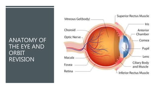
  • 1. ANATOMY OF THE EYE AND ORBIT REVISION
  • 2. WHAT BONES MAKE UP THE BOUNDARIES OF THE ORBIT? Wall Bones Superior • Orbital plate of frontal bone • LW sphenoid Inferior • Orbital surface of maxilla • Palatine bone • Orbital surface of zygomatic Medial • Ethmoid (orbital plate) • LW sphenoid • Frontal bone • Lacrimal bone • Frontal process of maxilla Lateral (thickest) • Zygomatic • GW sphenoid
  • 3. ORBIT SURROUNDED BY PARANASAL AIR- FILLED SINUSES  Air filled sinuses located within the bones of the skull and face  Functions:  Lightening weight of the head  Humidifying and heating inhaled air  Increasing resonance of speech  Protect vital structures in injury  Superior to the orbit- frontal sinus  Inferiorly- maxillary sinus  Medially- ethmoid sinus
  • 4. THE EYEBALL  Has 3 layers:  Fibrous outer layer:  Sclera: dense, white, avascular, continuous with the cornea  Vascular middle layer  Choroid: pigmented, vascular, continuous with ciliary body and iris  Ciliary body = muscular thickening that provides attachment for the lens. Secretes aq humor  Iris = on anterior surface of the lens. It is a thin contractile diaphragm  Inner retina layer  Light sensitive  Sensory neural layer
  • 5. THE PUPIL  Two involuntary muscles control the size of the pupil:  Dilator pupillae- sympathetic innervation (makes the pupil larger)  Sphincter pupillae- parasympathetic
  • 6. ACCOMMODATION  Ciliary m. control the shape of the lens- which enables the eye to focus (accommodation)  To focus on near objects:  Ciliary m. contract through parasympathetic innervation  Release tension on the zonal fibers  Lens becomes rounder/ convex- increased curvature shifts focal point closer to the eye
  • 7. MUSCLES OF THE EYELID  Levator palpebra superioris m.  Only in the UPPER eyelid  Raises the eyelid  Innervation via CN3  Inserts into the tarsus  In companion with the LPS m. is a collection of SM fibers = superior tarsal m.  Raises the eyelid  Innervation via the sympathetic fibers from superior cervical ganglion Thus, CN3 lesion/ sympathetic lesion leads to ptosis – drooping eyelid
  • 8. MUSCLES OF THE EYELID  Orbicularis Oris m.  A sphincter muscle located in the upper and lower eyelids  Palpebral part of the muscle close the eyelids during blinking and sleep  Orbital part: voluntary action- screws the eyes tightly shut for protection  Innervated by the zygomatic and temporal branches of CN7  Damage to CN7  paralysis  eyelids don’t close fully + eversion of the lower eyelid  drying + damage to cornea  Also assists in pumping tears into nasolacrimal duct system
  • 9. EXTRA-OCULAR MUSCLES Muscle Attachements Actions Innervation LPS From LW sphenoid to the superior tarsal plate of upper eyelid Elevates upper eyelid CN3 Superior rectus Superior part of the common tendinous ring to the superior and anterior sclera Elevation Adduction Medial rotation CN3 Inferior rectus Inferior common tendinous ring to the inferior and anterior sclera Depression Adduction Lateral rotation CN3 Medical rectus Medical part of common tendinous ring to the anteromedial sclera Adducts eyeball CN3 Lateral rectus Lateral part of the common tendinous ring to the anterolateral sclera Abducts CN4 Superior oblique Body of the sphenoid, attaches to the sclera posterior to superior rectus Depresses Abducts Medially rotates CN4 Inferior oblique Anterior aspect of the orbital floor to the sclera of the eye, posterior to lateral rectus Elevates Abducts Laterally rotates CN3 Located in the orbit, but extrinsic to the eyeball. They act to control movements of the eyeball and superior eyelid.
  • 10. EYELIDS, CONJUNCTIVA AND TEARS PROTECT THE EYE  Eyelids:  Dense connective tissue called Tarsus  Embedded in tarsus = tarsal glands  Sebaceous secretions which prevent tears from evaporating  Conjunctiva:  Provides protection and lubrication  Contains blood vessels to combat infection  Consists of an epithelial layer composed of stratified squamous and stratified columnar epithelium  Non-keratinized with interspersed goblet cells  Within the epithelial layer there are BVs, fibrous tissue, lymphatics, melanocytes, T/B cells, Langerhan's cells, accessory lacrimal glands Note: the suspensory ligament is hammock shaped thickening, enclosing the inferior rectus m. it supports the eyeball
  • 11. TEARS Tears/ lacrimal fluid from lacrimal gland produced by parasympathetic impulses from CN7 Secreted through lacrimal ducts to surface of conjunctiva When eyelids blink, it pushes tears medially over the cornea Fluid containing foreign material pushes to medial angle of the eye Drains through lacrimal canaliculi into the lacrimal sac Nasolacrimal duct conveys the fluid to the nasal cavity Function of tears:  Lubricate – basal tears  Removing irritants – reflex tears  Aiding immune system
  • 12. PARASYMPATHETIC AUTONOMIC INNERVATION OF THE EYE  Parasympathetic preganglionic fibers for the eye are axons of cells in the accessory oculomotor nucleus.  They run in the CN3 and exit in the motor root of the ciliary ganglion  The axons of the ciliary root ganglion are postganglionic parasympathetic fibers which reach the eye in the short ciliary nerves and are distributed to the sphincter pupillae, ciliary m. and BVs
  • 13. SYMPATHETIC AUTONOMIC INNERVATION OF THE EYE  Some of the filaments passing through the superior orbital fissure form the sympathetic root of the ciliary ganglion  Some pass through the ganglion as short ciliary nerves.  Other filaments join the ophthalmic nerve or its nasociliary branch and reach the eye in long ciliary nerves that supply the dilator pupillae  Both long and short ciliary nerves also contain afferents to the cornea, iris and choroid
  • 14. SYMPATHETIC AUTONOMIC INNERVATION OF THE EYE  Some of the fibers in the short ciliary nerves pass through a communicating ramus from the ciliary ganglion to the nasociliary nerve-  This ramus is called the sensory root of the ciliary ganglion  Involved in reflex actions  Other sympathetic fibers from the internal carotid plexus reach the eye through the ophthalmic periarterial plexus
  • 15. SUPERIOR ORBITAL FISSURE AND OPTIC CANAL  ”What structures run through the foramina of the orbit?”  Superior orbital fissure: CN3, 4, 6 and sensory innervation to the eye  Optic canal: CN2 and opothalmic a. and optic n.
  • 16. BLOOD SUPPLY TO THE ORBIT  Ophthalmic a:  First branch of the internal carotid, runs anteriorly through the optic canal along with optic n. (inferolaterally)  Branches supply all structures in the orbit in addition to the nose, face, meninges  Central retinal a. is the first branch of the ophthalmic a. which runs in the dura mater inferior to the optic n.  Supplies the internal retinal layers  Second branch is the lacrimal a. which runs along the superior edge of the lateral rectus m.  Supplies the lacrimal gland, eyelids and conjunctiva
  • 17. VENOUS DRAINAGE OF THE ORBIT  Ophthalmic v.  Superior and inferior subdivisions- pass through the superior orbital fissure and enter the cavernous sinus  Central retinal v.  Drains blood from the capillaries of the retina into the superior ophthalmic v. or the cavernous sinus directly