
Recommended
More Related Content
What's hot
What's hot (20)
Similar to Cleft Lip and Palate
Similar to Cleft Lip and Palate (20)
More from Hadi Munib
More from Hadi Munib (20)
Recently uploaded
Recently uploaded (20)
Cleft Lip and Palate
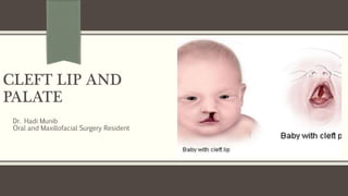
- 1. CLEFT LIP AND PALATE Dr. Hadi Munib Oral and Maxillofacial Surgery Resident
- 2. Outline Embryology Causative Factors Problems with individuals with oral clefts Treatment of patients with oral clefts Dental needs for individuals with oral clefts References
- 3. Introduction A cleft is a congenital abnormal space or gap in the upper lip, alveolus, or palate. Harelip – Demeaning Clefts of the lip and palate are the most common serious congenital anomalies to affect the orofacial region 1 in 700 births in the US. Less frequently in blacks but more so in Asians. Cleft Lip and Palate; Male: Female Ratio 3:2 Clefts of the palate; Female > Male Cleft Lip; Male > Female Three fourths of clefts are unilateral deformities; one fourth is bilateral - Left
- 4. Teams involved General or pediatric dentist Orthodontist Prosthodontist Oral-maxillofacial surgeon Plastic surgeon Audiologist Otorhinolaryngologist Pediatrician Speech pathologist Psychiatrist and a social worker.
- 5. Submucosal Clefts Submucosal clefts of the soft palate are called occult clefts. “Ah”; lifts the soft palate, a furrow in the midline is seen where the muscular discontinuity is present. Posterior aspect of the hard palate; characteristic absence of the posterior nasal spine. Hypernasal speech without an obvious soft palatal cleft, suspect a submucosal cleft
- 6. Types
- 8. Embryology 5th to 10th week of fetal life
- 9. Embryology Clefts of the primary palate result from a failure of the mesoderm to penetrate into the grooves between the medial nasal and maxillary processes. Clefts of the secondary palate are caused by a failure of the palatine shelves to fuse with one another. The causes for this are speculative and include failure of the tongue to descend into the oral cavity
- 10. Embryology
- 11. Causative Factors Unknown in most cases. Isolated clefts and clefts associated with other birth disorders or syndromes. A syndrome is a set of physical, developmental, and sometimes behavioral traits that occur together. Syndromes account for approximately 15% of the total number of cases of cleft lip and cleft palate but nearly 50% of cases of isolated cleft palate. Genetics in only 20% to 30% of patients with cleft lip or palate. Multigenetic. Nutritional deficiencies, radiation, several drugs, hypoxia, viruses, and vitamin excesses or deficiencies can produce clefting in certain situations.
- 12. Problems with individuals with clefts Dental Problems Congenital absence of teeth Hypomineralization Supernumerary teeth
- 13. Problems with patients with Clefts Malocclusion Class III malocclusion, seen in most cases, is caused by many factors. Mandibular prognathism - Relative. Generally, the operative trauma of the cleft closure and the resultant fibrosis (i.e., scar contracture) severely limit the amount of maxillary growth and development that can take place. Orthodontic treatment may be necessary throughout the individual’s childhood and adolescent years. Comprehensive orthodontic care is deferred until later, when most of the permanent teeth have erupted.
- 15. Problems with patients with clefts Nasal Deformity Deformity of normal nasal architecture is commonly seen in individuals with cleft lips. The alar cartilage on the affected side is flared The columella of the nose is pulled toward the side without the cleft. Surgical correction of nasal deformities should usually be deferred until associated problems have been corrected to prevent alteration of the osseous foundation of the nose.
- 16. Problems with patients with Clefts Ear Problems Middle ear infections. Suppurative Otitis Media The levator veli palatini and the tensor veli palatine normally inserted into the same muscles on the opposite side and are left unattached when the soft palate is cleft. These muscles have their origins directly on or near the auditory tube. The auditory tube in infants is at an angle that does not promote dependent drainage. Myringotomy; a hole through the inferior aspect of the tympanic membrane and inserts a small plastic tube, performs this procedure, which drains the ear to the outside instead of the nasopharynx Chronic serous otitis media is common among children with cleft palate
- 17. Problems with patients with Clefts Speech problems Four speech problems are usually created by cleft lip and palate deformity. Retardation of consonant sounds (i.e., “p,” “b,” “t,” “d,” “k,” and “g”) is the most common The velopharyngeal mechanism cannot function because of the discontinuity of the musculature from one side to the other. The soft palate cannot elevate to make contact with the pharyngeal wall. The result of this constant escape of air into the nasal cavity is hypernasal speech.
- 18. Problems with patients with Clefts Associated Anomalies 20 times more likely to have another congenital anomaly than a normal child. Of those children who have associated anomalies, 38% have isolated cleft palate, and 21% have cleft lip, with or without cleft palate. 30% have other anomalies in addition to the facial cleft, ranging from clubfoot to neurologic disturbances. O10% have congenital heart disease 10% have some degree of mental retardation.
- 19. Treatment The aim of treatment of cleft lip and palate is Correction of the cleft and associated problems surgically. Surgically producing a face that does not attract attention Vocal apparatus that permits intelligible speech Dentition that allows optimal function and esthetics. Operations begin early in life and may continue for several years.
- 20. Cheilorrhaphy Surgical correction of cleft lip “Rule of 10”. 10 weeks of age 10 lbs. body weight At least 10 g/dL hemoglobin in blood.
- 21. Cheilorrhaphy The cleft of the upper lip: Disrupts the important circumoral orbicularis oris musculature. Uncoordinated growth patterns At birth, the alveolar process on the unaffected side may appear to protrude from the mouth. Restoration of this muscular sphincter with lip repair has a favorable effect on the developing alveolar segments. Objectives Functional; Restore Orbicularis Oris Muscle function Esthetic; normally appearing upper lip
- 22. Cheilorrhaphy Surgical techniques; unilateral cases The unaffected side serves as a guide for lip length and symmetry. In lips closed in a linear fashion, scar contracture causes a characteristic notching of the upper lip.
- 23. Cheilorrhaphy
- 25. Palatorrhaphy One operation and occasionally in two. In two operations, the soft palate closure (i.e., staphylorrhaphy) is usually performed first and the hard palate closure (i.e., uranorrhaphy) is performed second. Objectives. Creation of a mechanism capable of speech and deglutition without significantly interfering with subsequent maxillary growth. Creation of a competent velopharyngeal mechanism and partitioning of the nasal and oral cavities.
- 26. Palatorrhaphy Surgical techniques; Each cleft of the palate is unique. Hard Palate Closure with soft tissues only. No effort is made to create an osseous partition between the nasal and oral cavities. Some tissues are atrophic and not particularly useful. Soft tissues are incised along the cleft margin and are dissected from the palatal shelves until approximation over the cleft defect is possible. Lateral relaxing incisions close to the dentition
- 29. Palatorrhaphy – Soft Palate Closure Access is the largest problem because the soft palate is toward the back of the oral cavity. The combination of difficulty with light, retraction, and the fact that the clinician can work only from the oral side. Extremely thin, atrophic tissues and yet produce a closure that will hold together under function. The soft palate is always closed in three layers: (1) nasal mucosa, (2) muscle, and (3) oral mucosa. Incised from the posterior end of the hard palate to at least the distal end of the uvula. The nasal mucosa is then dissected free from the underlying musculature and sutured to the nasal mucosa of the opposite side. The musculature of the cleft soft palate is inserted posteriorly and laterally along the margins of the hard palate. Re-approximation; Only then will the velopharyngeal mechanism have a chance to perform properly. If the quantity of muscular tissue is inadequate for approximation of the musculature in the midline, the pterygoid hamular processes can be infractured, thus releasing the tensor palatini muscles toward the midline.
- 30. Palatorrhaphy – Soft Palate Closure Incomplete palatal clefts; those of the soft palate only. The palate can be closed in a manner that not only brings the two lateral halves together in the midline but also gains palatal length. The so-called W-Y push-back procedure (Wardill) and U-shaped push-back procedure (Dorrance and Brown) are commonly used. The mucoperiosteum of the hard palate is incised and elevated in a manner that allows the entire soft tissue elements of the hard and soft palate to extend posteriorly, thus gaining palatal length.
- 33. Alveolar Cleft Grafts The alveolar cleft defect is usually not corrected in the original surgical correction of the cleft lip or the cleft palate. Residual oro-nasal fistulae in this area, and the maxillary alveolus will not be continuous because of the cleft. Because of this, five problems commonly occur: 1. Oral fluids escape into the nasal cavity 2. Nasal secretions drain into the oral cavity 3. Teeth erupt into the alveolar cleft 4. The alveolar segments collapse 5. If the cleft is large, speech is adversely affected.
- 34. Alveolar Cleft Grafts - Advantages 1. They unite the alveolar segments and help prevent collapse and constriction of the dental arch. 2. Alveolar cleft bone grafts provide bone support for teeth adjacent to the cleft and for those that will erupt into the area of the cleft. 3. Closure of the oronasal fistula. 4. Facilitates the use of dental prostheses by creating a more suitable supporting base. 5. The creation of a solid foundation for the lip and alar base of the nose.
- 35. Alveolar Cleft Grafts Timing of graft procedure; the patient is between ages 6 and 10 years, a major portion of maxillary growth has occurred, and the alveolar cleft surgery should not adversely affect the future growth of the maxilla. Surgical procedure. Intact mucoperiosteal flaps on each side must cover bone grafts placed into the alveolar cleft. The bone placed into the alveolar cleft is usually obtained from the patient’s ilium or cranium; however, some surgeons are using allogeneic bone (i.e., homologous bone from another individual) and recently bone morphogenetic proteins (BMP) The grafts are made into a particulate consistency and are packed into the defect once the nasal and palatal mucosa has been closed. The labial mucosa is then closed over the bone graft.
- 39. Dental Needs for individuals with Clefts Interdisciplinary approach. Teeth are so frequently missing in the patient. In patients who have failed to obtain velopharyngeal competence with surgical corrections, a speech aid appliance can be made by the dentist to decrease hypernasal speech. Acrylic Bulb attached to a toothborne appliance The bulb is fitted to project onto the undersurface of the soft palate and lifts the soft palate superiorly. If this bulb does not give adequate function, another projection of acrylic (i.e., bulb obturator) can be placed to extend to the posterior aspect of the palate. This narrows the pharyngeal isthmus, and the size can be adjusted for maximal effectiveness.
- 40. Dental Needs for patients with Clefts This type of appliance is used in two instances: 1. Before a pharyngeal flap procedure to develop muscle action 2. If the secondary surgical procedures are not successful in producing velopharyngeal competence.
- 41. References Chapter 28: Management of patients with Clefts
- 42. THANK YOU
Editor's Notes
- NOTE: To change the image on this slide, select the picture and delete it. Then click the Pictures icon in the placeholder to insert your own image.