FRENAL ATTACHMENT & ITS MANAGEMENT
•Download as PPTX, PDF•
8 likes•4,002 views

This document discusses frena, their development and classifications. It describes abnormal frenal attachments and their associated complications like loss of papilla and recession. Ankyloglossia (tongue-tie) is discussed in detail along with its classification and clinical features. Various techniques for treating abnormal frena are presented, including frenectomy, frenotomy, Z-plasty and laser frenectomy. Post-operative instructions are provided. The document emphasizes that proper technique selection based on frenal attachment type can achieve functional and aesthetic outcomes.

Recommended
More Related Content
What's hot
What's hot (20)
Similar to FRENAL ATTACHMENT & ITS MANAGEMENT
Similar to FRENAL ATTACHMENT & ITS MANAGEMENT (20)
More from DR.MD.SHADAB ANWAR
Recently uploaded
Recently uploaded (20)
FRENAL ATTACHMENT & ITS MANAGEMENT
- 2. 2 CONTENTS INTRODUCTION DEVELOPMENT CLASSIFICATION VARIATIONS DIAGNOSIS COMPLICATIONS OF ABNORMAL FRENUM SYNDROMES ASSOCIATED WITH ABNORMAL FRENUM ANKYLOGLOSSIA COMPLICATIONS OF ANKYLOGLOSSIA CLASSIFICATION TREATMENT CONCLUSION REFERENCES
- 3. 3 What is a frenum? Frenum is a thin fold of mucous membrane with enclosed muscle fibers that attach the lips to the alveolar mucosa and underlying periosteum. ( Carranza 10th edition) A frenulum is a small frenum. There are several frena that are usually present in a normal oral cavity, most notably the maxillary labial frenum, the mandibular labial frenum, and the lingual frenum. Their primary function is to provide stability of the upper and lower lip and the tongue. INTRODUCTION
- 4. 4 DEVELOPMENT The maxillary labial frenum develops as a post eruptive remnant of the ectolabial bands which connects the tubercle of the upper lip into the palatine papilla. It extends over the alveolar process in infants and forms a raphe that reaches the palatal papilla. Through the growth of alveolar process as the teeth erupt, this attachment generally changes to assume the adult configuration.
- 5. 5 Depending upon the extension of attachment of fibers, frena have been classified as i. Mucosal – when the frenal fibers are attached up to mucogingival junction ii. Gingival – when fibres are inserted within attached gingiva iii. Papillary – when fibres are extending into inter dental papilla; and iv. Papilla penetrating – when the frenal fibres cross the alveolar process and extend up to palatine papilla. Placek’s Classification Classification
- 6. 6
- 7. 7
- 8. 8 Simple frenum with anodule [Figure 5] Simple frenum with appendix [Figure 6] Simple frenum with nichum [Figure 7] Bifid labial frenum Persistent tectolabial frenum Double frenum Wider frenum [Figure 8] Frenum with two or more variations at the same time Sewerin’s Classification ( Based on morphotypes) Other variations of normal frenal attachment include:
- 9. 9
- 10. 10
- 11. 11 Tests for frenal attachment: 1. Tension Test OR 2. Blanch Test. Miller et al(1985) recommended that the frenum should be characterised as pathogenic when it is unusually wide or there is no apparent zone of attached gingiva along the midline or the interdental papilla shift when the frenum is extended. DIAGNOSIS
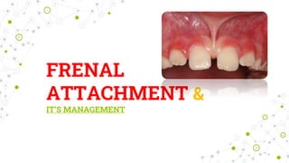
- 12. 12 Abnormal or aberrant frena are detected visually, by applying tension over it to see the movement of papillary tip or blanching produced due to ischemia of the region. Clinically, papillary and papilla penetrating frena are considered as pathological and have been found to be associated with loss of papilla, recession, diastema, difficulty in brushing, malalignment of teeth and it may also prejudice the denture fit or retention leading to psychological disturbances to the individual.
- 13. 13 A frenum can become a significant problem if tension from lip movement pulls the gingival margin away from the tooth, or if the tissue inhibits the closure of a diastema during orthodontic treatment. Frenal attachment that encroach on the marginal gingiva distend the gingival sulcus, fostering plaque accumulation, increasing the rate of progression of periodontal recession and and thereby leading to recurrence after treatment.
- 14. 14 Blanch Test OR Tension test Proposed by CRABER in 1961. To demonstrate- a continuity of the tissue fibers of the labial frenum through the diastema to the palatine papilla. Detected visually- by applying tension over it to see the movement of papillary tip or blanch produced due to ischemia of the region.
- 15. 15 Accomplished by lifting the upper lip upward and forward until the frenum is tightly streched. If the procedure produces a blanching or change of contour in this area, the frenum is consider to be an abnormal.
- 16. 16 COMPLICATIONS OF ABNORMAL FRENUM A frenum becomes a problem if the attachment is too close to the marginal gingiva. Tension on the frenum may pull the gingival margin away from the tooth. This condition may be conducive to plaque accumulation and inhibit proper tooth brushing. Abnormal frenum has been found to be associated with: • Loss of papilla. • Recession. • Persistence of midline diastema. • Difficulty in brushing. • Malalignment of teeth . • Compromised denture fit or retention
- 17. 17 Syndromes associated with different frenal attachments: a. Ehlers-Danlos syndrome b. Infantile hypertrophic pyloric stenosis, c. Holoprosencephaly, d. Ellis-van Creveld syndrome, and e. Oro-facial-digital syndrome. f. Pallister-hall syndrome g. Opitz C syndrome
- 18. 18 a. Ehlers-Danlos syndrome It is a genetic disorder characterized by hyper extensive skin and hyper mobile joints with no gender predilection. Absence of the inferior labial and lingual frena has been described in this disorder.
- 19. 19 b. Infantile hypertrophic pyloric stenosis Occurs commonly in males at a ratio of 4.5 to 1 with an unknown etiology. There is a disturbance in the frenum formation. The absence or hypoplasia of mandibular frenum represents an important diagnostic tool in detection of this disease.
- 20. 20 c. Holoprosencephaly It is characterized by defects including cyclopia, single nostril, single central incisor and premaxillary agenesis. Absence of labial maxillary frenum is one of the characteristic features of this condition. Holoprosencephaly is an abnormality of brain development in which the brain doesn't properly divide into the right and left hemispheres. The condition can also affect development of the head and face. Absence of frenum single central incisor
- 21. 21 d. Ellis-van Creveld syndrome It is an autosomal recessive disorder mainly affecting enamel, hair and nails. Patients with this syndrome characteristically present with congenitally missing teeth, abnormal frenal attachment, microdontia and hexadactyly. The most common finding is fusion of the anterior portion of the upper lip to the maxillary gingival margin, as a result of which no mucobuccal fold exists, causing the upper lip to present a slight V- shaped notch in the middle (partial hare lip or lip-tie).
- 22. 22 Nonsyndromic conditions Aberrant frenal attachments may be seen after orthognathic surgeries. Problems are probably caused by errors in the surgical technique. The design of the soft tissue incisions is critical, vertical incisions in the area of osteotomy will predictably create periodontal problems.
- 23. 23 END OF PART- 1
- 25. 25 Ankyloglossia or Tongue-tie is an uncommon congenital anomaly that occurs as a result of a short, tight, lingual frenulum causing difficulty in speech articulation due to limitation of tongue movement. WALLACE et al 1963 defined tongue-tie as “a condition in which the tip of the tongue cannot be protruded beyond the lower incisor teeth because of a short frenulum linguae, often containing scar tissue.” INTRODUCTION
- 26. 26 FREE-TONGUE: The term free-tongue is defined as the length of tongue from the insertion of lingual frenum from the base of the tongue to the tip of the tongue. Clinically acceptable, normal range of free-tongue is greater than 16 mm. (Kotlow et al 1999)
- 27. 27 CLASSIFICATION Ankyloglossia can be classified into 4 classes based on Kotlow’s assessment in 1999 (based on length of tongue from insertion of lingual frenum at base of the tongue to the tip of the tongue) as follows: Clinically accepted >16mm i. CLASS I: MILD ANKYLOGLOSSIA (12 to 16 mm) ii. CLASS II: MODERATE ANKYLOGLOSSIA (8 to 11mm) iii. CLASS III: SEVERE ANKYLOGLOSSIA (3 to 7 mm) iv. CLASS IV: COMPLETE ANKYLOGLOSSIA (< 3mm) Kotlow’s assessment
- 28. 28 During early development, the tongue is fused to the floor of the mouth. Cell death and resorption free the tongue, with the frenulum left as the only remnant of the initial attachment. Tongue-tie is the result of a short fibrous lingual frenulum or a highly attached genioglossus muscle. Etiology
- 29. 29 CLINICAL FEATURES & COMPLICATION OF ANKYLOGLOSSIA Ankyloglossia leads to : i. Limited mobility of tongue. ii. Difficulty in swallowing. iii. Difficulty in speech articulation which is evident for consonants like “s, z, t, d, l, j, zh, ch, th, dg” and it is especially difficult to roll an “r”. iv. Notched or “heart-shaped” tongue when it is protruded.
- 30. 30 v. Dentition- causes a pulling effect on the gingiva away from the teeth and even cause a mandibular diastema. Usually occurs after 8-10 years . vi. Cosmetics- looks abnormal and tongue has a forked or serpent look . vii. Feeding problems-approx 25% of newborns with ankyloglossia have feeding problems. As the child grows older, he may have difficulty moving a bolus in the oral cavity and clearing food from the sulci and molars. This leads to chronic halitosis and dental decay.
- 31. 31 TREATMENT Techniques for removal of aberrant frenum are : Frenotomy Frenectomy Frenectomy : Refers to the complete removal of frenum, including its attachment to the underlying bone. It is required in the correction of abnormal diastema between maxillary central incisors (Friedman 1957). Frenotomy: Is the incision of the frenum. It is usually done to relocate the frenal attachment so as to create a zone of attached gingiva between the gingival margin and the frenum.
- 32. 32 FRENECTOMY 1. Gingival or papillary frenal attachment: Where frenal fibres radiate into marginal gingiva producing gingival retraction and localized gingival recession. 2. High frenal attachment: Where oral hygiene is hindered by shallow vestibule caused by high frenal attachment. 3. Ankyloglossia: When lingual frenum interferes with speech. INDICATIONS
- 33. 33 TECHNIQUES OF FRENECTOMY Conventional (classical) frenectomy (SIMPLE EXCISION TECHNIQUE) Miller’s technique V-Y plasty Z plasty Frenectomy by using electrocautery Laser frenectomy A localized vestibuloplasty with secondary epithelialization
- 34. 34 CLASSICAL FRENECTOMY The classical technique was introduced by Archer et al 1961 and Kruger et al 1964. This approach was advocated in midline diastema cases with an aberrant frenum to ensure the removal of muscle fibres which were supposedly connecting the orbicularis oris with the palatine papilla.
- 35. 35 ARMAMENTARIUM
- 36. 36 STEPS:-
- 37. 37 Causes un-aesthetic labial tissue scarring. This may become a matter of concern in case of high smile line exposing the anterior gingiva. DISADVANTAGES
- 38. 38 MILLER’S TECHNIQUE This technique was advocated by Miller PD et al in 1985. This was proposed for post-orthodontic diastema cases. The ideal time for performing this surgery is after the orthodontic movement is complete and about 6 weeks before the appliances are removed. This allows healing and tissue maturation.
- 39. 39 STEPS:-
- 40. 40 Post-operatively, on healing, there is a continuous band of gingiva across the midline, that gives a bracing effect than the scar tissue, thus preventing orthodontic relapse. The transseptal fibres are not disrupted surgically and so, there is no loss of interdental papilla. ADVANTAGES OF MILLER’S TECHNIQUE
- 41. 41 Z- PLASTY TECHNIQUE This technique is indicated when: a) There is hypertrophy of the frenum with a low insertion, associated with distema. b) There is a short vestibule.
- 42. 42 STEPS:- Excision of the fibrous tissue by making small elliptical excision of mucosa(vertically) & underlying loose connective tissue.
- 43. 43 Two oblique incisions are made in a Z fashion, one at each end of the previous area of excision. Two pointed flaps are then gently undermined and rotated to close the initial vertical incision horizontally.
- 44. 44 Closure with interrupted sutures.
- 45. 45
- 46. 46 This technique can be used for lengthening the localized area, like a broad frena. This technique is mostly employed in a case of a papilla type of frenal attachment V-Y PLASTY TECHNIQUE
- 47. 47
- 48. 48 Localized vestibuloplasty with secondary epithelialization: Wide V-type of incision made at most inferior portion of frenal attachments through mucosal tissue and underlying submucosal tissue, without perforating the periosteum
- 49. 49 A supraperiosteal dissection is completed by undermining the mucosal and submucosal tissue with scissors .
- 50. 50 After a clean periosteal layer is identified, the edge of the mucosal flap is sutured to the periosteum at the maximal depth of the vestibule and the exposed periosteum is allowed to heal by secondary epithelialization.
- 51. 51 ELECTROSURGERY This technique is recommended for patients with bleeding disorders and non-compliant patients.
- 52. 52 This technique offers the advantages of: Minimal time consumption. Minimal procedural bleeding. No need of sutures. Healing is by secondary intention as the wound edges are not approximated with sutures. ADVANTAGES WITH ELECTROCAUTERY DEVICE
- 53. 53 LASER -FRENECTOMY The benefits of a laser frenectomy are greater as compared to traditional techniques . These include : Reduced bleeding during surgery. Reduced operating time and rapid postoperative hemostasis, thus eliminating the need for sutures. The lack of need for sutures, as well as improved postoperative comfort and healing, make this technique particularly useful for very young patients.
- 54. 54 BIOLASE- LASER UNIT (DIODE- LASER) TIPS FOOTSWITCH
- 55. 55 PROCEDURE DONE BY BIOLASE- LASER Figure: Papilla penetrating - frenal attachment(Preoperative) Holding the muscle with foecep
- 56. 56 Figure:- Releasing the muscle under local anaesthesia with LASER( During procedure) Post-operative view
- 58. 58 POST- OPERATIVE INSTRUCTIONS NOT to eat anything until the anesthesia wears off, as there are chances of biting the lips, cheek or tongue. Avoid extremely hot foods for the rest of the day and do NOT rinse out your mouth, as these will often prolong the bleeding. If bleeding continues, apply light pressure to the area with a moistened gauze for 20-30 minutes.
- 59. 59 Avoid alcohol and smoking until after your post-operative appointment. Follow a soft food diet, taking care to avoid the surgical area when chewing. Chew on the opposite side and do NOT bite into food. Be sure to maintain adequate nutrition and drink plenty of fluids. Do NOT use a drinking straw, as the suction may dislodge the blood clot.
- 60. 60 Maintain normal oral hygiene measures in the areas of mouth not affected by the surgery. In areas where there is dressing, lightly brush only the biting surfaces of the teeth. Vigorous rinsing should be avoided! Do NOT pull down the lip or cheek.
- 61. 61 CONCLUSION Frenum may not regularly draw close scrutiny on routine dental examination. While an aberrant frenum can be removed by any of the modification techniques that have been proposed, a functional and an aesthetic outcome can be achieved by a proper technique selection, based on the type of frenal attachment.
- 62. 62 References Carranza 10th and 12th edition. Priyanka M, Sruthi R, Ramakrishnan T, Emmadi P, Ambalavanan N. An overview of frenal attachments. J Indian Soc Periodontol 2013. Mirko P, Miroslav S, Lubor M. Significance of the labial frenum attachment in periodontal disease in man. Part I. Classification and epidemiology of the labial frenum attachment. J Periodontol 1974 Devishree et al. Journal of Clinical and Diagnostic Research. 2012 November. Kotlow LA. Oral diagnosis of abnormal frenum attachments in neonates and infants: Evaluation and treatment of maxillary frenum using the Erbium YAG Laser. J Pediatr Dent Care 2004. De Felice C, Toti P, Di Maggio G, Parinni S, Bagnoli F. Absence of the inferior labial and lingual frenula in Ehlers-Danlos syndrome. Lancet 2001.
- 63. 63