
Recommended
More Related Content
What's hot
What's hot (20)
Similar to Cleft palate final.pptx
Similar to Cleft palate final.pptx (20)
Recently uploaded
Recently uploaded (20)
Cleft palate final.pptx
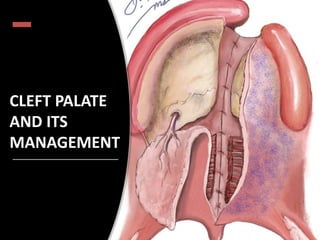
- 2. Introduction • A cleft is a birth defect that occurs wh en the tissues of the lip and palate of the fetus do not fuse in pregnancy • Cleft is derived from German word “Kluft” which means a fissure/ cleave • Cleft lip and palate are the second most common congenital deformity (after clubfoot). • Earlier cleft lip known as hare lip
- 3. Embryology
- 5. Anatomy of hard and soft palate
- 6. Anatomy of normal palate
- 9. Soft palate
- 10. Muscles of soft palate
- 12. In a cleft palate
- 14. In a cleft palate
- 16. Palatopharyngeus
- 17. Musculus uvulae
- 18. palatoglossus
- 20. THEORIES OF CLEFT LIP AND PALATE • Failure of fusion of process • Mesodermal migration theory. • The merging of prominences • Other theories
- 21. 1. FAILURE OF FUSION THEORY • Separate processes fuse to form central face- first advanced by Meckel in 1808 • Popularized by Dursy (1869) & Wilhelm His of Leipzig University (1901)
- 22. 2.MIGRATION OF MESODERM THEORY • Fleischmann of Erlangen, Germany (1910). • Veau (1934)- by penetration of mesoderm across groove, normal development was ensued while failure of mesodermal migration eventually led to breakdown & cleft formation. • Mesoderm gives rise to the fibromusculae layer of the lip.
- 23. • Bulging effect - “hills” & “valleys” • Failure of sufficient mesoderm to migrate - persistence of groove established cleft MIGRATION OF MESODERM THEORY
- 24. 3. THE MERGING OF PROMINENCES • Bradley M Patten - medial side of cleft there is small portion of prolabium • Patten also noted- when cleft of lip is relatively small - striking asymmetry in configuration of nose. • disturbance of potentialities of mesenchyme of nasal processes.
- 25. • Steininger(1939) proposed the theory of rupture of previously formed cysts in the soft tissue bridges. • Pfeifer (1966) proposed the possibility that clefts were formed not by failure of fusion but by the breakdown of lip once intact. OTHER THEORIES
- 26. CLASSIFICATION Classification of cleft lip Dewis and Richies classification Veau Classification Lashal classification WHO classification Spina classification Kernahan classification
- 27. DAVID AND RITCHIE (1922) on anatomical basis
- 28. VEAU (1931): In 1931, Victor Veau published his landmark Division Palatine, which described his approach to evaluation and management of cleft palate.
- 29. Karnahan’s Classification Millard’s Modification of Karnahan’s Classifcation CLASSIFICATION
- 31. Sub mucous cleft A palpable notch on the posterior border of the hard palate Bifd uvula- this may extend from a mere groove on the uvula to complete bifdity. Zona pellucida or a bluish tinge in the midline of the soft palate due the paucity of muscle bulk at this level.
- 32. Article: Prevalence of Dental Anomalies in a Population of Cleft Lip and Palate Patients Dental Nasal Feeding Skeletal Ear problems Speech
- 33. DENTAL Tooth agenesis, hypodontia (most common) Supernumerary teeth (2nd most common) Enamel hypoplasia (CI) Crossbites Ectopic eruption, transposition Taurodontism, dilacerations
- 34. Skeletal changes Maxillary retrognathism in a patient with unilateral cleft lip and palate exhibits increasing severity with age. A, At 10 years of age; B, 16 years; and C, 21 years. Facial Growth and Development in Individuals With Clefts John Daskalogiannakis, Gregory S. Antonarakis
- 35. Early profile convexity caused by protrusion of the premaxilla in a patient with bilateral cleft lip and palate gradually turns into a progressively worsening concave profile with age. A, At 5 years of age; B, 11 years; and C, 18 years.
- 37. Precautions • Do not flood the pharynx- provokes aspiration • Infant with a major cleft swallows air while feeding- feed more frequently than normal child. • Supplements required after two weeks of breast feeding
- 40. Presurgical orthopedics: Objectives : To reduce the severity of the original cleft deformity. To eliminate surgical columella reconstruction and reduce the resultant scar tissue in bilateral cleft lip and palate. To improve the surgical outcome of the primary repair in cleft lip and palate patients.
- 42. Timing of repair millard Rule of 10 • Wt - > 10 pounds • Hb - > 10 grams • Age - > 10 weeks • Initially – 3months of age • Later • Incomplete lip – 3 – 6 months • Lip adhesion – 3weeks • Definitive closure 6 – 8 months • Soft and hard palate – 1 1⁄2 - 2 1⁄2 year with soft palate repair. For the lip and nose repair, he waited for the incisors to erupt in order to avoid the inversion that frequently takes place if the operation is carried out before.
- 44. Objectives of the Cleft Palate Repair • To produce anatomical closure of the defect. • To create an apparatus for development and production of normal speech. • To minimize the maxillary growth disturbances.
- 45. Principles of Palatoplasty • Closure of the defect. • Repositioning of the abnormal position of the muscles of the soft palate. • Reconstruction of the muscle sling. • Favorable retro positioning of the soft palate and uvula so as to achieve velopharyngeal closure during speech. • Last and most important is tension-free suturing
- 46. Surgical techniques Von Langenbeck's bipedicle flap technique Veau-Wardill-Kilner Pushback technique Bardach's two-flap technique Furlow Double opposing Z-Plasty Two-stage palatal repair Hole in one repair Raw area free palatoplasty Alveolar extension palatoplasty (AEP) Intravelar veloplasty Vomer flap Buccal myomucosal flap
- 47. possibly the oldest palatoplasty still widely used today Commonly used for an incomplete cleft of the secondary palate without the presence of a cleft lip and alveolus. Von langenbeck
- 48. .
- 49. Ø Advantage:- Easy to perform. Requires less dissection. Results in decreased raw area of palate. Ø Disadvantages:- Cannot be used in wider and complete clefts. Failure to provide additional palatal length. Palatoplasty : Evolution and controversies Chang Gung medical journal 31(4):335-45·Nov 2007.
- 50. Veau- WARDILL- KILNER / V- y pushback palatoplasty
- 52. Cleft Palate Repair: Veau-Wardill-Kilner Technique – Pushback Palatoplasty Adversely affects midfacial growth in cleft palate patients because of scar tissue anteriorly. Higher rate of fistula in complete cleft palate than other techniques because it provides only a single nasal mucosa layer anteriorly Agrawal K. Cleft palate repair and variations. Indian Journal of Plastic Surgery : Official Publication of the Association of Plastic Surgeons of India. Advantages:- Lengthening the palate and repositioning the levator muscle in a more favourable position.
- 58. Cleft Palate Repair: Bardach Ø The design of this flap is entirely dependent on the greater palatine neurovascular pedicle and it provides greater versatility to cover the cleft. Advantages:- Complete closure of the entire palate in one stage. Creation of more physiologic soft palate muscle sling and a layered closure technique. Disadvantages:- Does not provide additional length to the repaired palate to allow normal speech production. Bardach J. Two flap palatoplasty Bardach Technique. Operative Techniques in Plastic and Reconstructive
- 60. Furlow Double Opposing Z-Plasty
- 61. Cleft Palate Repair: Furlow Ø Advantages:- No need to raise large mucoperiosteal flaps from the hard palate. The soft palate can be lengthened.[Good speech outcome] Ø Disadvantages:- Non anatomical palatal closure Ignores musculus uvulae Difficult to close wider clefts Large raw area - needs to be covered with buccal flap. Palatoplasty : Evolution and controversies Chang Gung medical journal 31(4):335- 45· Nov 2007.
- 63. vomer flap
- 64. Early complications: • Bleeding: Bleeding probably occurs most commonly from the lateral releasing incisions or from the margins of elevated flaps. Hemostasis needs to be meticulous. • Airway: Closure of the palate may compromise the airway,
- 65. • Dehiscence: Dehiscence of the repair is probably usually the result of either infection or hematoma, or closure under excess tension. Partial dehiscence results in fistula formation. • In the Wardill-Kilner pushback technique, these often occur at the junction of the flaps in the hard palate and are very difficult to close. In other repairs, they most commonly occur at the junction of the hard and soft palate. They can be minimized by the avoidance of hematoma and by avoiding undue tension. Intermediate Complications
- 66. Classification of post palatoplasty fistula
- 67. Hardwicke et al Fistula incidence after primarycleft palate repair: a systematic review of the literature. Plast Reconstr Surg 2014. In 2014, meta-analysis reported that oronasal fistulae occur in 8.6% of patients after cleft palate repair based on 9294 patients from 44 studies. 2014 In 2015, systematic review of 2505 patients over a 2-year period reported the incidence of fistulae as 4.9%. 2015
- 68. Bykowski MR et al: The rate of oronasal fistula following primary cleft palate surgery: a meta-analysis. Cleft Palate Craniofac J 2015.
- 69. management of palatal fistula Murthy J. Descriptive study of management of palatal fistula in one hundred and ninety-four cleft individuals. Indian J Plast Surg. 2011 Jan;44(1):41-6. doi: 10.4103/0970-0358.81447.
- 70. Tongue Flap
- 74. VPI Velopharyngeal insufficiency: inability to achieve complete closure of the velopharyngeal apparatus during speech. [Structural Defect] Velopharyngeal incompetence: Imperfect closure of velopharyngeal apparatus that is caused by neuromuscular dysfunction. i;e impaired motor programming of velopharynx. [Neurogenic Defect] Velopharyngeal inadequacy: Imperfect closure of the velopharyngeal apparatuscaused by the tissue defect. [Tissue insufficiency]
- 77. Pharyngeal Flap
- 79. Classification of IVV proposed by Andrades et al Type 0: No muscle dissection or suturing of muscle Type 1: No dissection, parallel suturing of muscle Type II a: Partial dissection (release from posterior palatal shelf but minimal dissection from nasal and oral mucosa) creating inverted-U muscle sling Type II b: Partial dissection (dissection from nasal mucosa but not oral mucosa) creating inverted-V muscle sling Type III: Complete dissection creating a transverse muscle sling (radical IVV) I b: Partial dissection (dissection from nasal mucosa but not oral mucosa) creating inverted-V muscle sling Type III: Complete dissection creating a transverse muscle sling (radical IVV)
- 80. VPI
- 81. Videofluoroscopy
- 82. Nasoendosopy
- 83. Conclusion • Cleft palate repair has undergone major changes in the past quarter century, the increased application of methods that incorporate reconstruction of the levator palatine muscles has produced much more predictable speech results.
- 84. References: • Maxillofacial surgery - Peter Ward Booth, 2nd Edition, Vol. II • Fonseca Oral and maxillofacial surgery - 2nd Edition, Vol. III • Textbook of oral and maxillofacial surgery for clinicians • Text book of Plastic surgery- Joseph Mc. Carthy – Vol. IV • Perterson’s principles of Oral and maxillofacial surgery - 3rdEdition, Vol. II • Janusz Bardach. Two flap palatoplasty- Bardach’s technique. Operative techniques in plastic and reconstructive surgery,1995;2(4):211-14.
- 85. Ask not for a spatula and torch to check your cleft palate repair, but listen to your patient speak.- Dr. Kilner Thank you!
Editor's Notes
- Isolated cleft palate occurs most commonly in females than in males 1/700 live births – with wide variability across geographic regions] Orientals , Caucasians, blacks
- The external human face develops between the 4th and 6th week of embryonic development. The development of the face is completed by the 6th week. Between the 6th and 8th week, the palate begins to develop. Consequently, this causes a distinction between the nasal and oral cavities. This development is completed by the 12th week. Frrontonasal , medial , lateral, max , mand processes
- primary , SECONDARY PALTE As the nose forms, the fusion of the medial nasal prominence with its contralateral counterpart creates the intermaxillary segment – which forms the primary palate (becomes the anterior 1/3 of the definitive palate). The intermaxillary segment also contributes to the labial component of the philtrum and the upper four incisors. The maxillary prominences expand medially to give rise to the palatal shelves. These continue to advance medially, fusing superior to the tongue. Simultaneously, the developing mandible expands to increase the size of the oral cavity; this allows the tongue to drop out of the way of the growing palatal shelves. The palatal shelves then fuse with each other in the horizontal plane, and the nasal septum in the vertical plane, forming the secondary palate.
- the palate is a bony/muscular partition that forms the roof of the oral cavity and the floor of the nasal cavities. It consists of two main parts; the hard palate and soft palate. The hard palate is the anterior bony portion, while the soft palate is the posterior muscular part.
- It comprises the anterior two-thirds of the palate. The hard palate is formed by the fusion of two pairs of facial bones in the midline, the maxillae (upper jaw bones) and palatine bones. The palatine processes of the maxillae form the anterior three-quarters of the hard palate, while the horizontal plates of the palatine bones form the remaining posterior oneIncisive fossa, greater and lesser palatine foramina. -quarter.
- BLOOD VESSEL LIES IN THE PLANE OF GLANDULAR TISSUE HARD PALATE –THIS PLANE SITUATED BETWEEN MUCOSA AND PERIOSTEUM SOFT PALATE-THIS PLANE SITUATED BETWEEN MUCOSA AND MUSCLE The oral aspect of the hard palate is covered by oral mucosa that is firmly bound to the underlying bone and overlies the mucus-secreting palatine glands and neurovascular structures. The nasal aspect on the other hand is covered by respiratory mucosa.
- GREATER PALATINE ARTERY SUPPLIES HARDPALATE LESSER PALATINE ARTERY -ANTERIOR SURFACE OF SOFT PALATE ASCENDINGPALATINE-BRANCH OF FACIAL ARTERY AND ASCENDING PHARYNGEAL ARTERY – SUPPLIES SOFT PALATE
- The soft palate (velum) is the posterior muscular portion of the palate that continues from the posterior border of the hard palate. It is a mobile soft tissue flap that curves posteriorly and inferiorly into the pharynx, demarcating the nasopharynx from the oropharynx. On its posterior free margin, the soft palate bears a conical projection in the midline known as the uvula. The epithelial lining of the oral surface of the soft palate contains a small number of taste buds.
- Extrinsic muscles Levator veli palatine Tensor veli palatine Palatoglossus Palatopharyngeus Intrinsic muscle Musculus uvulae
- Originates from the scaphoid fossa at the base of the medial pterygoid plate, spina angularis of the sphenoid and from the cartilaginous part of the auditory tube he muscle descends anteroinferiorly and winds around the pterygoid hamulus, to which some fbers are attached, and passes into a tendon that fans out to form the palatine aponeurosis few fbers are attached to the maxillary tuberosity, the palatine aponeurosis The main role of the tensor is to dilate the Eustachian tube and its role in speechIn a cleft palate child is defcient, especially medially is insignificant
- In a cleft palate child, the palatine aponeurosis is defcient, especially medially
- Arises from the petrous The muscle descends on either side, enters the intermediate 40% of the soft palate, and forms a muscular sling with its counterpart on the other sidepart of the temporal bone and from the cartilaginous part of the Eustachian tube When the paired muscle contracts, it elevates the soft palate superiorly and posteriorly to enable closure of the velopharyngeal sphincter. The latter is formed by the soft palate anteriorly, the posterior pillars of the fauces on either
- In the normal palate, the levator forms a muscular sling that suspends the soft palate from the cranial base. Running from its origins at the cranial base to its insertion into its partner in the velum, the levator takes a downward, forward, and medial course that facilitates a cranial, posterior, and lateral pull on the soft palate during velopharyngeal closure.3 a cleft child, as there is obviously no continuity across the midline due to the cleft, there are abnormal attachments of the levator muscle to the palatopharyngeus muscle posteriorly, to the edge of the cleft medially, to the tensor veli palatini, and to the posterior edge of the hard palate anteriorlyside, and the posterior pharyngeal wall posteriorl
- This clefted configuration prevents the levator, the principal motor of the velar component of velopharyngeal closure, from exerting its upward, backward, and lateral pull. Moreover, in the cleft palate, the levator has 3 abnormal associations that must be addressed in repairing the defect: an insertion onto the posterior medial edge of the hard palate, associations with the aponeurosis of the tensor veli palatini, and lateral adhesions to the superior pharyngeal constrictor
- Has a palatine portion, a pterygopalatine portion and the salpingopharyngeal part. It arises from the lateral and posterior part of pharynx and attaches into the velum. Its superior fbers arise from complex intermingling with the superior constrictor muscle It is found in the posterior pillar of the fauces. The fbers pass horizontally into the posterior three fourths of the soft palate inferior to the fbers of the levator palatini muscle The muscle helps to narrow the velopharyngeal opening by bringing the palatopharyngeal arches together he muscle is relatively well developed in the cleft palate child and ends partly along the cleft edge and partly along the posterior edge of the hard palate. Some fbers pass along the edge of the cleft along with the levator to form the muscle of veau
- Arises from the posterior nasal spine and the palatine aponeurosis, inserts into the junction of the proximal and middle thirds of the uvula. he presence and extent of the muscle in a cleft palate child is disputed. It is also supposed to be absent in occult submucous cleft patients
- arises from the tongue inserts into the soft palate The paired muscle forms the anterior part of the sphincter and narrows the isthmus. It is antagonistic to the levator in its action, drawing the palate inferiorly This muscle doesn’t play a major role in cleft palate Nerve supply
- The theory that separate processes fuse to form the central face Was first advanced by meckel in 1808 and later supported by Baer in 1868, german anatomist dursy in 1869 and german biologist Wilhelm his of leipzig university in 1901 working on chick Embryos popularized the theory of embryological development Of the midface by the fusion of five facial processes around the Rim of the primitive oral cavity or stomodeum. superiorly there Is the frontonasal process laterally there are the paired maxillary Processes and inferiorly the paired mandibular processes. according to the classical theory all of these processes grow forward As fingerlike projections to fuse with each other to form the Normal face between the fifth and eighth weeks. the frontonasal Process gives rise to three processes the frontal nasomedial Globular and nasolateral responsible for the development of Nose prolabium and premaxilla. the maxillary process by fusing With the nasomedial process forms the lateral upper lip and Cheek. the mandibular processes meet to form the lower jaw, Chin and lower lip. this hypothesis reigned some 30 years as the accepted basis of facial formation. the failure of fusion of These processes seemed to explain the formation of the various Degrees of unilateral and bilateral clefts and even the rare midline Upper and lower lip cleft. Thomas mullen of san francisco in 1931 following the Dursyhis hypothesis described it this way, Embryologically the growth toward the median line of the processes going Into the formation of the mouth and lips progresses until in the seventh Week , the manner in which the processes unite is similar To the healing of wounds the ectodermic coverings of the processes unite And the mesodermic elements spread across the line of epithelial union To give rise to the muscles and connective tissue of the adult structure. Epithelial ingrowths separate the lips from the alveolar portion of the jaw.Thirty years later that is in 1961 pruzanskys book congenital Anomalies of the face and associated structures presented this Schematic design of the fusion hypothesis. The fusion theory is no longer in vogue the term process Implies fingerlike projection of tissue and fusion implies That the projections meet. their epithelial walls disappear and They then grow together as shown as early as 1910 by pohlmann.
- Zoology professor Fleischmann of Erlangen, Germany in 1910 had hypothesis that cleft palate is the arrest of the disappearance of the epithelial membrane which remains intact or left unpenetrated by the adjacent mesoderm. this mesodermal penetration theory appealed to Victor Veau who admitted that until 1930, at the age of 60 he had never even looked at an embryo. in 1934 Veau disenchanted with the old facial process theory which he now considered ‘myth’, . Thus Veau endorsed the theory that with the penetration of Mesoderm across the groove, normal development ensued while failure of the mesodermal migration eventually led to Breakdown and cleft formation. His acceptance of the importance of mesodermal penetration might however explain his enthusiasm for wire approximation of the muscles across the cleft
- This theory proposes that with invagination of the oral cavity and nasal pits there is ‘heaping up’ of the adjacent tissue. As oral and nasal cavities deepen there is increase in sizes of the prominences due to the penetration of mesoderm. As more mesoderm enters, the bulging effect is increased thus transforming the wall of tissue with ectoderm on one side and endoderm on the other into “hills” and “valleys” Failure of sufficient mesoderm to migrate into specific area would be responsible for the persistence of groove which leads to established cleft
- Bradley M Patten - medial side of the cleft there is small portion of the prolabium that could have been derived from the nasomedial process. He believed that it is the prime mover in this important union and that when its growth is inadequate cleft will remain. Patten also noted that even when the cleft of the lip is relatively small there is striking asymmetry in the configuration of the nose. This means that there is disturbance of the potentialities of the mesenchyme of the nasal processes.
- GROUP I – PRE-ALVEOLAR CLEFTS UNILATERAL , BILATERAL OR MEDIAN GROUP II – POST-ALVEOLAR CLEFTS SOFT PALATE ONLY SOFT AND HARD PALATES SUBMUCOUS CLEFT GROUP III – ALVEOLAR CLEFTS UNILATERAL BILATERAL OR MEDIAN
- ) Cleft of soft palate only. 2 ) Cleft of hard and soft palate extending no further than incisive foramen, thus involving secondary palate alone. 3) Complete unilateral cleft, extending from the uvula to the incisive foramen in the midline, then deviating to one side and usually extending through alveolus at the position of the future lateral incisor tooth. 4) Complete bilateral cleft, resembling Of note, while Veau discusses the most intricate of anatomical findings in Division Palatine, he purposefully chose to exclude ‘‘confounding’’ details (such as severity) from the classification system itself, preferring simple groupings. In his subsequent work dedicated to cleft lip, Bec-de-Lievres ` (Veau and Recamier, 1938), Veau eschews ´ taxonomical groupings altogether and instead advocates a clear and concise description of the labial defect (including laterality [unilateral/bilateral/median] and extent [simple/ total]). The minimalism, morphologic basis, and clinical relevance of Veau’s approach to classification made it very attractive to contemporary surgeons.g Group III with two clefts extending forwards from the incisive foramen through the alveolus
- Kernahan and Stark proposed three groups: 1. Clefts of structures anterior to the incisive foramen 2. Clefts of structures posterior to the incisive foramen 3. Clefts affecting structures anterior and posterior to the incisive foramen
- Occult submucous cleft palate is diagnosed by multiview videofluoroscopy and nasal endoscopy during workup for VPI.
- Congenitally Missing teeth, Hypodontia, Hyperdontia, Oligodontia Presence of natal and neonatal teeth Anomalies of tooth morphology like microdontia, macrodontia Poor periodontal support, early loss of teeth
- An oral feeding should be completed within 20–30 minutes. Longer feedings may lead to a net caloric loss due to excessive energy output Isolated cleft lip – feed quite well Cleft palate – difficulty Feeding by keeping baby at 45-60 degree angulations reduces risk of aspiration Frequent burping –Aerophagia Specialized nipples Close attention to weight gain In 24 hours…2 – 3 ounces of milk for each pound of weight Intermittent episodes of breast feeding and specialized bottle feeding.
- Tendsor opens the eustachian tube whilemeating, releases fliod’’due to defect , no drainage of ear fluids Otitiis media
- 1689, Hoffmann demonstrated the use of facial binding to narrow the cleft and prevent postsurgical dehiscence. A similar technique helps was used by Desault in 1790 to retract the maxilla before surgical repair in patients with bilateral cleft repair The modern school of presurgical orthopaedic treatment in cleft lip and plate was started by McNeil in 1950- series of plates to mould the alveolar segments, populaprized by Burston, an orthodontist.
- This technique closes the incomplete cleft of the hard and soft palates without lengthening the palate by mobilizing bipedicle mucoperiosteal flaps medially.
- incision along the attached gingiva that runs posteriorly to a point lateral to the hamulus, approximately 1 cmposterior to the maxillary tuberosity. A mucosal incision along the border of the cleft, between the oral and nasal mucosa, marks the flap’s medial extent. Nasal mucosa flaps are sutured to one another in the midline to repair the nasal lining defect (often incorporating a vomer flap). The bipedicled hard palate flaps are advanced to close the oral side of the defect. Closure is done with a 4–0 Vicryl starting with the nasal layer in a simple interrupted fashion, incorporating vomer flaps anteriorly. The apex of the uvula is closed with a single horizontal mattress suture. Next, the intravelar veloplasty is completed. Interrupted or horizontal mattress sutures are used to reorient the levator veli palatini along the posterior velum to create the levator sling. Oral closure is then done with simple interrupted sutures using a 4–0 Vicryl working posteriorly to anteriorly. Finally, the relaxing incisions are stabilized with interrupted sutures through the medial gingival edge and microfibrillar collagen hemostatic agent is placed in the open defects laterally.
- Ø The Von Langenbeck technique is similar to Bardach palatoplasty but preserves an anterior pedicle for increased blood supply to the flaps. Ø Used in isolated cleft palates.
- Derived from a modification of the von Langenbeck technique Velopharyngeal incompetence is relatively common following palatoplasty either because there is insufficient mobility of the soft palate or because the length of the repaired palate is inadequate to reach the posterior pharyngeal wall. To increase the anteroposterior length of the palate at the time of primary palatoplasty, various mucoperiosteal flap maneuvers in the hard palate have been described in the literature.
- * It can be used to increase the palatal length. The Veau-Wardill-Kilner pushback palatoplasty can be suitably used for incomplete clefts of the hard palate. The flap design is similar to the von Langenbeck palatoplasty. The essence of this technique is the V to Y incision and closure on the hard palate (Fig. 2). The pushback technique has the advantage of lengthening the palate and repositioning the levator muscle in a more favorable position. However, this modification involves extensive dissection. At the free anterior end, the mucoperiosteal flaps can then be approximated directly or in a V-Y closure to lengthen the soft palate.
- Bardach- intiatily anteriorly onlyLate* The more extensive two-flap palatoplasty is a modification of the Langenbeck technique, extending the relaxing incisions along the alveolar margins to the edge of the cleft. This designs flaps entirely dependent on the circulation from the palatine vessels but also much more versatile in terms of their placement.. Intravelar veloplasty is an essential part of this closure
- In a complete unilateral cleft, the mucoperiosteal flap from the medial segment can be shifted across the cleft and closed directly behind the alveolar margin. The fistula in the anterior hard palate can be virtually eliminated by this technique.(22) Two-flap palatoplasty also has a minimal effect on subsequent maxillofacial growth due to the limited area of bone denudation on the hard palate when the mucoperiosteal flaps are elevated.(23,24) The limitation of this technique is that it does not provide additional length to the repaired palate to allow normal speech production. A variation from the standard technique of twoflap palatoplasty has been reported using supraperiosteal flaps instead of the mucoperiosteal technique for palatal closure.(25) Although this new approach improves speech outcome, it still requires further evaluation in a larger series to ascertain its applications.
- George Dorrance (1877–1949) of Philadelphia realized that a distinct number of patients with cleft would develop velopharyngeal dysfunction caused by inability of the soft palate to touch the posterior pharyngeal wall. In fact, he advocated muscle transposition but did so by fracturing the hamulus, which he believed would change the vector of muscle contraction and in combination with techniques of Langenbeck would lengthen the palate.
- The technique is based on the concept of lengthening the soft palate by use of “Double-opposing Z-plasties.”.” This involves mirror-image Z-plasties to the oral and nasal layers while also retropositioning and reconstructing the levator muscle mechanism by keeping the levator palatini muscle on each side attached to the limb of the Z-plasty that moves posteriorly (FIG 1).1 Randall has described a variation for wider CP that involves using Langenbeck-type lateral releasing incisions to relieve the tension on the oral Z-plasty BV
- Alternating the reversing Z-plasties of the nasal and oral flaps and repositioning the levator veli palatine muscle within the posteriorly mobilized flaps. Ø Effective for primary closure of a submucous cleft palate and secondary correction of marginal velopharyngeal insufficiency.
- In a bilateral complete cleft palate, a midline incision along the free margin of vomer is required to create two septal-mucosal flaps in opposite directions. These two flaps are used to bridge the gap between the free edges of the nasal mucosa. The two-flap palatoplasty combined with a vomer flap results in a four-flap palatoplasty that can be applied for simultaneous closure of the nasal and oral defects in the cleft palate. This technique results in a twolayer closure with a low fistula rate and less maxillary growth retardation. The long-term effect of this technique on facial growth is minimal This flap has a high incidence of maxillary retrusion, presumably from injury from vomer-premaxillary sutures, and a high fistula rate
- because of postoperative swelling New airway mechanism
- Cohen et al. [5] classified them according to their site as pre-alveolar, alveolar, post-alveolar, hard palate, hard-soft palate junction, soft palate and uvula. The commonest site for fistulae is the junction of the hard and soft palate. Tension , infection ,
- SOMERLAND PALATOPLASTY Age of Surgery • Hard Palate: 3 months • Soft palate: 1 Year Technique • Hard Palate: Single layer vomer flap at the time of lip closure • Soft Palate: Intravelar veloplasty
- Ø Guerrero-Santos and Altamirano, were the first to report on the use o tongue flaps for palatal defect closure.The tongue flap is easy and reproducible with excellent esthetical and functional results. Advantages: The advantages are th use of adjacent tissue, the excellen blood supply and the low morbidity idonor site.Ø Disadvantage: Inability in swallowing and speech until depedicling of the flap and in some cases the attachment of the flap can be lost due to traction.
- BMMF is a vascular and dependable flap. Vascular supply of the flap is consistent and profuse. Ø The buccinator myomucosal flap is effective in reducing/eliminating hypernasality in patients with cleft palate and velopharyngeal insufficiency. Ø Advantages: Flap congestion is occasional and necrosis is rare. It tolerates stretching, folding, and twisting. Ø Disadvantages: Fibrosis. Secondary healing. Parotid duct orifice injury.
- Late Complications • Maxillary retrusion: Cleft palate surgery in the developing child is known to be associated with Hypolpastic maxilla The decreased prominence of maxillary complex could be caused mainly by the shortened maxillary length; meanwhile, posterior position of the maxillary body may have some influence on the maxillary protrusion changes produced in the anatomy of the pharynx by closure. This is particularly relevant in babies with Pierre Robin sequence. Some surgeons use a temporary tunnel stitch, which does allow immediate control of the tongue position and potential improvement in the airway in the first postoperative hours. The use of an optimally positioned nasopharyngeal airway can be very helpful in the postoperative period.
- Inability to achieve complete closure of the velopharyngeal apparatus during speech. Velopharyngeal apparatus (Regulation of airflow from the lungs and larynx)
- Secondary Palatoplasty aims at correcting the velopharyngeal inadequacy (VPI) VPI is defined as inadequate closure of the soft palate during speech to the posterior pharynx during speech, resulting in air leak up into the nasopharynx (Lindsey and Davis, 1996). 5% to 36% of patients who have undergone primary palatoplasty have persisiting VPI (Dorf and Curtin, 1982; Bardach and Morris, 1990; Peat et al., 1994; Hudson et al., 1995)
- To improve the speech in children with cleft palate, primary pharyngeal flap pharyngoplasty is performed in a few center's This procedure is not popular presently, as it unnecessarily subjects the patients to the disadvantages of pharyngeal flap surgery like sleep apnea, hypo nasality etc. This creates an abnormal anatomy in all the cleft palate patients, which is not acceptable to most surgeons.[22] Since the majority of these patients will not develop velopharyngeal incompetence after classical palatoplasty, this procedure seems to be an overkill.
- Dissection of the Levator Palati from the posterior border of the hard palate, nasal and oral mucosa and posterior repositioning. Ø Suturing of the muscle with that of the opposite side for the reconstruction of the Levator sling. Sommerlad dissects the levator palatini belly separately and sutures independently as the Levator is the dominant muscle for elevation of the soft palate during speech. Also tensor tenotomy is performed. Ø Court Cutting transects the Tensor Palati and to keep its function intact, the cut end is transfixed with the hook of the hamulus.
- he lateral view shows the anatomy of the soft palate and provides information on the movement of the tongue, palate, and posterior pharyngeal wall as well as demonstrating any Passavant ridge (The lateral view cannot demonstrate movement of the lateralpharyngeal walls or the sphincteric movement of the velopharynx. Lateral pharyngeal walls can move incongruously with respect to the
- This child has demonstrates very limited velopharyngeal (VP) closure which is significantly impacting speech intelligibility.