Cleft palate Lecture notes ppt
•Download as PPT, PDF•
4 likes•724 views

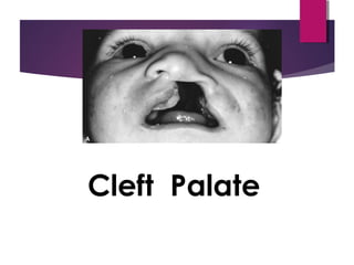
cleft palate is a common congenital disorder which has numerous associated problems, including anatomical defects

Recommended
More Related Content
What's hot
What's hot (20)
Similar to Cleft palate Lecture notes ppt
Similar to Cleft palate Lecture notes ppt (20)
Recently uploaded
Recently uploaded (20)
Cleft palate Lecture notes ppt
- 1. Cleft Palate
- 2. INTRODUCTION common congenital disorder have numerous associated problems, including basic anatomic deformity, deficient facial growth, dental problems speech problems otologic problems psychologic disorders, and additional congenital anomalies.
- 3. History 400BC Hippocrates and 150AD Galen mentioned cleft lip 500 AD 1st Operation on palate due to inflammation of the uvula 1564 Pare described the use of obturators for palatal perforations 1556Cleft palate recognised as a congenital disorder by Franco. 1764 LeMonnier, a French dentist performed 1st successful repair of a cleft velum. 1834Dieffennbach performed the 1st closure of the hard palate 1861Von Langenbeck described the use of mucoperiosteal flaps in cleft palate closure 1930Kilner and Wardill independently developed the ‘pushback’ procedure. 1944 Schweckendiek began closing cleft defects in young patients.
- 4. Epidemiology and Genetics The incidence of cleft palate only is half the incidence of CL/P (more than 1 : 2000 live births). There is usually little difference between racial groups. The incidence is much higher (about 4 : 1) in females than in males. About 20% to 30% of human cleft palate only are syndromic. Pierre-Robin Sequence, Treacher-Collins Malformation, Trisomies 13 and 18, Apert’s Syndrome, Stickler’s Syndrome and Waardenburg’s Syndrome.
- 5. Epidemiology and Genetics Familial risk One sibling with cleft, no parent with cleft 2% One sibling and one parent- 10-17% No sibling with cleft and are parent with cleft 7% Environmental factors exerting their teratogenic effects Those with proven clefting potential in humans include phenytoin, retinoic acid (vitamin A) derivatives, folic acid antagonists., cigarette smoking creating fetal hypoximia alcohol use in the first trimester of pregnancy
- 6. Anatomy Primary palate or premaxilla is a triangular area of the anterior hard palate extending from the anterior to the incisive foramen and to a point just lateral to each lateral incisor tooth. It includes the alveolar ridge containing the four incisor teeth. Secondary palate:- Consist of all remaining hard palate and are the soft palate. Hard palate- Formed by palatine processes of maxilla and horizontal lamina of palatine bones. Blood supply internal maxillary art- greater palative Ar. Soft Palate A fibromuscular shelf made up of several muscles attached like a sling to the posterior portion of the hard palate Function- close off the nasopharynx
- 7. EMBRYOLOGY The primary palate develops between 4 and 5 weeks gestation secondary palate between 8 and 9 weeks gestation. The primary palate, lip and alveolus develop as a mesodermal and ectodermal proliferation of the frontonasal and maxillary processes. A primary cleft is a failure of proliferation, not a failure to meet in the midline. The secondary palate develops as a medial ingrowth of the lateral maxillae with fusion in the midline.
- 8. EMBRYOLOGY
- 9. CLASSIFICATION no standard classification is universally accepted. Cleft palate A. Location in reference to incisive foramen 1. Primary—anterior 2. Secondary—posterior B. Unilateral or bilateral C. Complete or incomplete Submucous cleft palate
- 10. Classification (Iowa system) Group I - cleft of lip only Group II - Cleft of palate only Group III - Clefts of lip alveolus Group IV - Cleft of the lip and alveolus+ Palate Group V - Miscellaneous
- 11. GENERAL MANAGEMENT The multidisciplinary cleft team approach The comprehensive multidisciplinary team surgeon (plastic, pediatric surgery, oromaxillofacial surgery, or otolaryngology) otolaryngologist/otologist, dentist (pedodontist, orthodontist, prosthodontist, oral surgeon), speech language pathologist, audiologist, geneticist, Social worker, nurse, nutritionist, and psychiatrist/psychologist.
- 12. Management: Neonatal Period. The pediatrician is ideally suited for counseling in the immediate postpartum period. Children with clefts of the secondary palate usually have severe difficulties feeding. A soft bottle with a large opening is usually required. palatal prosthesis. A nasogastric tube is rarely required. palatal prosthetic plates. plates mold the palate into a more ideal position, leading to less difficulty feeding and an easier surgical repair. improve facial growth and children require less intervention throughout time
- 13. Management: The timing of the repair is controversial Palatal repair is performed anywhere from 6 months onward. delay in palatal repair results in improved midfacial growth earlier repair results in better speech
- 14. Management: Toddler Years. The most important process in the toddler years is the development of adequate speech requires speech therapy The child is trained to speak slowly and forcefully in order to maximize palatal elevation and minimize hypernasality up to 20% of patients still require pharyngoplasty to reduce VPI recurrent or chronic ear infections growth hormone deficiency is 40 times more common in this population
- 15. Management: School Years orthodontic management psychological growth psychotherapist is essential to ensure adequate social development high rate of depression and poor self esteem Child abuse is also more common
- 16. Management: Teenage Years. The teenage growth spurt is marked by midface retrusion, altering appearance. Palatal repair is likely responsible ideal timing for palatal repair so that midface retrusion is minimized is currently an area of intense research, with many conflicting studies in the literature The treatment requires maxillary advancement through a combination of LeForte I, II and III osteotomies. The treatment of midfacial retrusion requires maxillary advancement through a combination of LeForte I, II and III osteotomies. usually performed in young adulthood, after growth is complete Rhinoplasty, when required, is usually the last surgery performed.
- 17. Surgical Techniques The child with CLAP usually undergoes numerous surgical procedures. The first procedure is repair of the lip, which occurs around 10 weeks. The palate is usually repaired between 6 and 18 months, with newer studies advocating earlier repair Pharyngeal flaps, if required, are performed around age 4. Alveolar bone grafting occurs between ages 9 and 11, definitive orthodontics at age 12-13. Maxillary advancement and rhinoplasty are delayed until the later teenage years.
- 18. Surgical Techniques Palatoplasty The basic surgical techniques of palate repair include Schweckendiek’s procedure or primary veloplasty, Von Langenbeck’s palatoplasty, V-Ypush-back double reversing Z-plasty (Furlow’s palatoplasty), and two-flap palatoplasty. A combination of palatoplasty and pharyngeal flap may be used for the repair of extremely large clefts15 and occasionally for primary palate repair.
- 19. • Pre-Surgical Orthopedics allowing gingivoperiosteoplasty. A: Wide unilateral cleft lip with separation of the arch and collapse of the lesser segment. B: Improved arch alignment and nasal shape after presurgical orthopedic treatment. C: Results after rotation-advancement lip repair, nasal repair, and gingivoperiosteoplasty, all performed at the first operation, when the child is approximately 3 months of age.
- 20. Palatoplasty Technique. A: Cleft palate closure after healing of gingivoperiosteoplasty at 11–12 months of age. Bilateral, unipedicle mucoperiosteal flaps based on the greater palatine arteries are elevated. B: Anteriorly, the nasal floor is repaired by suturing the vomerine mucosa to the nasal mucosa on the cleft side.
- 21. Palatoplasty Technique. The levator muscles are dissected free from the oral and nasal mucosa and released from the posterior edge of the hard palate. The levator muscles are approximated to each other in the midline. D: The oral mucosa is reapproximated in the midline with interrupted horizontal mattress sutures.
- 22. Double opposing Z-plasty closure of an isolated cleft palate. A: Design of the incisions
- 23. Double opposing Z-plasty closure of an isolated cleft palate. B: Muscle included in the posteriorly based flap.
- 24. Double opposing Z-plasty closure of an isolated cleft palate. C: Final result with recreation of the levator sling.
- 25. SUBMUCOUS CLEFT PALATE The submucous cleft palate is traditionally defined by a triad of deformities: a bifid uvula, a notched posterior hard palate, and muscular diastasis of the velum. A submucous cleft palate can vary considerably, however, and muscular diastasis can occur in the absence of a bifid uvula. The majority of patients with are asymptomatic, although approximately 15% will develop velopharyngeal insufficiency. Velopharyngeal incompetence (VPI) correlates with short palatal length, limited mobility, and easy fatigability of the palate. Since the majority of patients remain asymptomatic, a nonoperative approach is recommended until speech can be
- 26. Otologic Disease Children with cleft palate suffer from a greater incidence of middle ear disease and hearing loss, the primary pathology being eustachian tube dysfunction due to abnormal insertion of the palatal musculature Robinson et al performed a prospective study of 150 cleft palate children whose palates were repaired between 2 and 18 months. They found 92% of these children developed chronic otitis media with effusion which required tympanostomy tubes Muntz et al, who found 96% of cleft palate children developed chronic middle ear effusions decreasing incidence of middle ear disease with age.
- 27. Otologic Disease An aggressive approach is required in the management of middle ear disease including frequent myringotomy and tympanostomy tube placement the goal of the management of ear disease in cleft palate patients is to provide adequate hearing, maintain ossicular continuity, Maintain adequate middle ear space, and prevent deterioration of the tympanic membrane until the patient develops adequate eustachian tube function.
- 28. Otologic Disease Indications for myringotomy and insertion of ventilating tubes include persistent middle ear effusion with a conductive hearing loss despite adequate medical management, recurrent acute otitis media failing prophylaxis, a combination of both, or presence of a retraction pocket
- 29. Otologic Disease Abnormal insertion of the levator and tensor veli palatini muscles into the posterior margin of the hard palate and the muscular hypoplasia appears to be the primary cause of eustachain tube dysfunction in cleft patients. Recent evidence that the nature of middle ear effusions in cleft patients is different from that in noncleft patients. Hutton and others recently published a study on the characterization of mucin from the effusions of cleft patients. When compared with mucoid and serous effusions from noncleft patients, the mucin in the middle ears of cleft patients, contained larger glycopeptides with a larger hydrodynamic size. This suggests that infections in cleft patients may be different from those in noncleft patients.