SlideShare a Scribd company logo
1 of 57
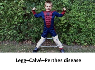
Legg–Calvé–Perthes disease
Learning objective
• Anatomy of femoral head
• Pathogenesis
• Pathological process
• Clinical features
• Classification
• Prognostic
• Managment
Endochondral ossification
Anatomy
Introduction
Osteonecrosis of epiphysis femoral head
Common in 4-8 years
Epidemiology
• Incidence : 1:1000
• Usual age : 4-8 years
• Boys:girls – 5:1
• Higher incidence in Caucasian, Chinese,
Japanese, Inuits, Northern Europe
• Etiology remains unknown .
• But some of the contributing factors are ;
– Antithrombotic factors deficiencies
– hypofibrinolysis
• Ischemia of femoral head
Pathogenesis
Up to 4 months 1. Metaphyseal vessels
2. Lateral epiphyseal
3. Scanty vessels in ligamentum
teres
4-7 years 1. Lateral epiphyseal vessels
2. Metaphyseal supply DISSAPEAR
7 years 1. Vessels in ligamentum teres have
developed
Susceptible to ischemia,
as it depend entirely on
lateral epiphyseal vessel.
Pathology
Stage 1 – ischemia and bone death
• All/part of bony nucleus of femoral head is dead
• Cartilaginous part – remains viable and thicker
• Thickening and edema of synovium and capsule
Pathological process  3-4 years
Stage 2 – revascularization and repair
Stage 3 – Distortion and remodeling
• Repair process
- Rapid and complete :
shape is restored
- Tardy : bony collapse
and growth distortion
Symptoms
• Typically boy – 4-8 years
• Painless limping – continues for week or recur
intermittently
• Pain in groin, thigh and knee – activity related,
relieved by rest
• Urogenital anomaly (4% cases)
Clinical feature
Signs
• Hip pain with passive range of movement
• Reduced range of movement (abduction &
internal rotation)
• Hip flexion contracture
• Leg length discrepancy
• Mild muscle wasting – thigh, calf,
buttock
• Tredenlenburg test ; positive
• X-ray of both hip (AP & Frog lateral view)
• Bone scan
• Ultrasound – joint effusion
• CT scan – follow up
• Arthrography : to see congruity, head deformity
and determine method of treatment
• Blood inflammatory marker
- FBC
- ESR
- CRP
Investigation
• Widening of joint space
• Sclerosis
• Necrotic phase : increase density of ossific nucleus
• Fragmentation : alternating patches of density and
lucency
• Lateral uncovering of femoral head
• Sagging rope sign
• Acetabular remodelling
X-ray
Waldenström classification based on
radiographic changes
Stage 1 ( increased density)
- ossific nucleus smaller and denser
- Gage’s sign
- subchondral fracture
- radiolucencies in the metaphysis
Stage 2 (fragmentation and revascularization)
- lucency in epiphysis
- pillars are demarcated
- metaphyseal changes resolve
-acetabular contour change
Stage 3 (healing or reossification stage)
- new bone formation
- homogenous epiphysis
Stage 4 (remodelling)
- femoral head is reossified and remodels
- acetabular remodelling
Sclerosis of epiphysis and widening of joint space
in the early stages
V shape defect
laterally
Slight lateral
displacement of
femoral head
Fragmentation of the femoral capital epiphysis
Eight-year-old boy with right hip pain for 5 months.
(a) AP pelvis radiograph showing a smaller epiphysis due to growth arrest of the
epiphysis. Note the increased radiodensity of the epiphysis. The femoral head is in
the initial stage of LCPD.
(b) AP pelvis radiograph obtained 5 months later showing fragmented and resorptive
changes in the affected epiphysis with further flattening of the femoral head. Most
of the deformity occurs during the stage of fragmentation
Caffey’s sign
Subchondral fracture
in the anterolateral
aspect of the femoral
capital epiphysis
Produces crescentic
radiolucency
Sagging rope sign
Curvilinera sclerotic line
running horizontally across
femoral neck
In a mature hip with
Perthe’s
According to stage of disease –
Waldenström classification
According to prognostic outcome –
– Caterall classification
– Salter and Thompson
– Herring lateral pillar
According to defining outcome –
Stulberg classification
Classification
The Catterall (1982)
Catterall group 1
Catterall group 2
Note the
intact lateral
pillar
Catterall group 3
• involvement of
the lateral pillar as
well as the
subchondral
radiolucent zone
taken 8 months
after onset of
symptoms
Catterall group 4
• 14 months—
fragmentation
18 months—early
reossification
25 months—late
reossification
52 months—healed.
• Note also the
growth arrest line
and evidence of
reactivation of the
growth plate along
the femoral neck.
5225181412
The Herring – based on severity of structural
lateral pillar not
affected
>50% of height of
lateral pillar preserved
<50% of height of
lateral pillar preserved
Salter Thompson – extent of subchondral fracture
<50 % of femoral head
= Caterall I & II
>50% of fermoral head
= Caterall III & IV
• Child under 6 years – excellent
• Older – less good
• Female
• Femoral head involvement (head at risk signs)
Prognostic features
Caterall ‘head at risk’ –
Clinical
– Progressive loss of hip motion more so abduction
– Fixed flexion deformity and adduction deformities
of hip
– Obese child
– Age on higher side
Radiographical
– Progressive uncovering of the epiphysis
– Calcification in the cartilage lateral to ossific
nucleus
– Radiolucent area at the lateral edge of bony
epiphysis (gage ‘s sign)
– Severe metaphyseal resorption
• Transient synovitis
• Juvenile rheumatoid arthritis
• Sickle-cell disease
• Septic arthritis
• Morquio disease, cretinism, Gauchers’ disease
• Old perthes deformities in adult :
– Multiple epiphyseal dysplasia
Differential diagnosis
Perthes Transient synovitis
Average duration of symptom is 6-8 weeks days
Synovitis thickening Synovitis with capsular distension
Bony changes No bony changes
Perthes Epiphyseal dysplasia
Unilateral Bilateral
No involvement of other joints Involvement of othe joints or spine
Acetabulum not involved Involved
Principle
1. Prevent deformity to femoral head before
remodelling phase
2. Restore and maintain ROM
3. Concept of containment
4. Relief of symptoms
Management
Guidelines to treatment
• Decision are based on :
– Stage of disease
– Prognostic x-ray classification
– Age and clinical feature particularly range of
abduction and extension
Guidelines by Herring (1994)
– Child <6 years : symptomatic treatment
– Bone age at/or below 6 years
• Group A and B – symptomatic
• Group C – abduction brace
– Bone age over 6 years
• Group A and B – abduction brace or osteotomy
• Group C – outcome probably unnaffected by treatment
– Child 9 years and older : operative contaiment
Symptomatic
• Pain control
• Hospitalization for bed rest and short period
traction
• Gentle exercise to maintain movement and
regular reassessment
• Preservation of abduction, with formal
stretching
Containment
Harrison and Menon stated ;
‘if the head is contained within the acetabular
cup, then like jelly poured into a mold the head
should be the same as the cup when it is
allowed to come out after reconsitution ‘
Containment – non operative
• >60% does not require surgery
• NSAIDS
• Nightime abduction splinting
• Physiotherapy
• Monitoring Indication
- <6 years age
-Herring A and B
– Orthosis
– Biphosphonate
(delay resorption,
prevent collapse)
– Bone
morphogenetic
protein - 2
Petrie cast and A frame orthosis
Containment – surgical
• Done before irreversible deformation of femoral
head occurs (early in fragmentation stage )
Ideal patient  6-9 years, Caterall II-III,
good ROM,
Surgical
containment
Femoral VDRO
osteotomies
Varus 20
Derotation 20-30
Pelvic
osteotomies
Salters
osteotomies
Femoral varus derotational osteotomy
Original technique
of Pauwel
Modified Mullers
Technique
Original Mullers
technique
• Advantage
– Seats the head deeply in acetabulum
– Removes vulnerable portion from acetabular edge
– Decrease joint reaction forces
• Disadvantage
– Excessive varus angulation
– Limb shortening
– Non union
– Tredelenburg gait
Pelvic osteotomy
• Advantage
– Anterolateral coverage of the femoral head
– Lengthening of extremity
– Avoidance of second operation
• Disadvantage
– Increase in acetabular and hip joint pressure
– Increase in leg length on operated side
Summary
Questions
• What are the blood supply of head in child?
• What are the head at risk sign ?
• How do you classify Perthe’s ?
REFERENCES
• Apley ‘s System of Orthopedics and Fractures 9th Edition
• Turek’s Orthopedic, Principle and their Application, 6th
Edition.
• Lovell and Winter's Pediatric Orthopaedics, 5th Edition
• https://www.researchgate.net/publication/224929158_
Bone_Structure_Development_and_Bone_Biology_Bone
_Pathology
• https://radiologykey.com/legg-calve-perthes-disease-
pathology-pathophysiology-and-pathogenesis-of-
deformity/

More Related Content

What's hot

Perthes ’ disease
Perthes ’ diseasePerthes ’ disease
Perthes ’ diseaseMannan Ahmed
 
Tuberculosis of hip
Tuberculosis of hipTuberculosis of hip
Tuberculosis of hipHardik Pawar
 
Perthes disease ADOLESCENT COXA VARA
Perthes disease ADOLESCENT COXA VARAPerthes disease ADOLESCENT COXA VARA
Perthes disease ADOLESCENT COXA VARADr ashwani panchal
 
Congenital vertical talus BY DR.NAVEEN RATHOR
Congenital vertical talus BY DR.NAVEEN RATHORCongenital vertical talus BY DR.NAVEEN RATHOR
Congenital vertical talus BY DR.NAVEEN RATHORDR.Naveen Rathor
 
Tendoachilles rupture and its management
Tendoachilles rupture and its managementTendoachilles rupture and its management
Tendoachilles rupture and its managementRohan Vakta
 
Perthes disease by DR.NAVEEN RATHOR
Perthes disease by DR.NAVEEN RATHORPerthes disease by DR.NAVEEN RATHOR
Perthes disease by DR.NAVEEN RATHORDR.Naveen Rathor
 
Classification & management of legg calve perthes disease
Classification & management of legg calve perthes diseaseClassification & management of legg calve perthes disease
Classification & management of legg calve perthes diseaseSitanshu Barik
 
Slipped capital femoral epiphysis (scfe)
Slipped capital femoral epiphysis (scfe)Slipped capital femoral epiphysis (scfe)
Slipped capital femoral epiphysis (scfe)farranajwa
 
Flat foot By Dr.Mahbub
Flat foot By Dr.MahbubFlat foot By Dr.Mahbub
Flat foot By Dr.Mahbubdr_mhb21
 
Congenital talipes equinovarus (club foot/ctev) ppt by Dr Pratik
Congenital talipes equinovarus (club foot/ctev) ppt by Dr PratikCongenital talipes equinovarus (club foot/ctev) ppt by Dr Pratik
Congenital talipes equinovarus (club foot/ctev) ppt by Dr PratikDr. Pratik Agarwal
 
Clinical Examination of the Hip
Clinical Examination of the HipClinical Examination of the Hip
Clinical Examination of the Hiporthoprince
 
Leg Calve Perthes disease
Leg Calve Perthes disease Leg Calve Perthes disease
Leg Calve Perthes disease darshanck89
 
Cubitus varus by Dhrumil Patel
Cubitus varus by Dhrumil PatelCubitus varus by Dhrumil Patel
Cubitus varus by Dhrumil Pateldhrumil88
 

What's hot (20)

Perthes ’ disease
Perthes ’ diseasePerthes ’ disease
Perthes ’ disease
 
Tuberculosis of hip
Tuberculosis of hipTuberculosis of hip
Tuberculosis of hip
 
Cubitus varus deformity
Cubitus varus deformityCubitus varus deformity
Cubitus varus deformity
 
Perthes disease ADOLESCENT COXA VARA
Perthes disease ADOLESCENT COXA VARAPerthes disease ADOLESCENT COXA VARA
Perthes disease ADOLESCENT COXA VARA
 
Tb hip
Tb hipTb hip
Tb hip
 
Congenital vertical talus BY DR.NAVEEN RATHOR
Congenital vertical talus BY DR.NAVEEN RATHORCongenital vertical talus BY DR.NAVEEN RATHOR
Congenital vertical talus BY DR.NAVEEN RATHOR
 
Tendoachilles rupture and its management
Tendoachilles rupture and its managementTendoachilles rupture and its management
Tendoachilles rupture and its management
 
Perthes disease by DR.NAVEEN RATHOR
Perthes disease by DR.NAVEEN RATHORPerthes disease by DR.NAVEEN RATHOR
Perthes disease by DR.NAVEEN RATHOR
 
Classification & management of legg calve perthes disease
Classification & management of legg calve perthes diseaseClassification & management of legg calve perthes disease
Classification & management of legg calve perthes disease
 
Slipped capital femoral epiphysis (scfe)
Slipped capital femoral epiphysis (scfe)Slipped capital femoral epiphysis (scfe)
Slipped capital femoral epiphysis (scfe)
 
Flat foot By Dr.Mahbub
Flat foot By Dr.MahbubFlat foot By Dr.Mahbub
Flat foot By Dr.Mahbub
 
Congenital talipes equinovarus (club foot/ctev) ppt by Dr Pratik
Congenital talipes equinovarus (club foot/ctev) ppt by Dr PratikCongenital talipes equinovarus (club foot/ctev) ppt by Dr Pratik
Congenital talipes equinovarus (club foot/ctev) ppt by Dr Pratik
 
Clinical Examination of the Hip
Clinical Examination of the HipClinical Examination of the Hip
Clinical Examination of the Hip
 
Leg Calve Perthes disease
Leg Calve Perthes disease Leg Calve Perthes disease
Leg Calve Perthes disease
 
Pes planus
Pes planusPes planus
Pes planus
 
Hallux valgus.pptx
Hallux valgus.pptxHallux valgus.pptx
Hallux valgus.pptx
 
Ctev
CtevCtev
Ctev
 
Cubitus varus by Dhrumil Patel
Cubitus varus by Dhrumil PatelCubitus varus by Dhrumil Patel
Cubitus varus by Dhrumil Patel
 
Coxa vara
Coxa varaCoxa vara
Coxa vara
 
Blounts disease
Blounts diseaseBlounts disease
Blounts disease
 

Similar to Perthes disease

Legg-Calve-Perthes Disease.pptx
Legg-Calve-Perthes Disease.pptxLegg-Calve-Perthes Disease.pptx
Legg-Calve-Perthes Disease.pptxNellyPhiri5
 
Osteochondritis of different bones
Osteochondritis of different bonesOsteochondritis of different bones
Osteochondritis of different bonesPramod Govindraj
 
Perthes disease
Perthes diseasePerthes disease
Perthes diseaseAhmed Azim
 
Slipped capital femoral epiphysis
Slipped capital femoral epiphysisSlipped capital femoral epiphysis
Slipped capital femoral epiphysisDr. Ditesh Jain
 
Case discussion of perthes disease-Dr. Siddharth Deshwal PG Orthopaedics
Case discussion of perthes disease-Dr. Siddharth Deshwal PG OrthopaedicsCase discussion of perthes disease-Dr. Siddharth Deshwal PG Orthopaedics
Case discussion of perthes disease-Dr. Siddharth Deshwal PG OrthopaedicsSIDDHARTHDESHWAL3
 
Legg calve-perthes disease
Legg calve-perthes diseaseLegg calve-perthes disease
Legg calve-perthes diseaseDr Rajeev
 
SCFE / slipped capital femoral epiphysis
SCFE / slipped capital femoral epiphysis SCFE / slipped capital femoral epiphysis
SCFE / slipped capital femoral epiphysis Surya Vijay Singh
 
Legg Calve Perthes disease
Legg Calve Perthes diseaseLegg Calve Perthes disease
Legg Calve Perthes diseaseshahinhamza2
 
fdocuments.in_legg-calve-perthes-disease-2.ppt
fdocuments.in_legg-calve-perthes-disease-2.pptfdocuments.in_legg-calve-perthes-disease-2.ppt
fdocuments.in_legg-calve-perthes-disease-2.pptHusain91
 
Legg calve perthes disease
Legg calve perthes diseaseLegg calve perthes disease
Legg calve perthes diseaseBijay Mehta
 

Similar to Perthes disease (20)

Legg-Calve-Perthes Disease.pptx
Legg-Calve-Perthes Disease.pptxLegg-Calve-Perthes Disease.pptx
Legg-Calve-Perthes Disease.pptx
 
Perthes disease
Perthes diseasePerthes disease
Perthes disease
 
Osteochondritis of different bones
Osteochondritis of different bonesOsteochondritis of different bones
Osteochondritis of different bones
 
Perthes
PerthesPerthes
Perthes
 
Perthes disease
Perthes diseasePerthes disease
Perthes disease
 
Slipped capital femoral epiphysis
Slipped capital femoral epiphysisSlipped capital femoral epiphysis
Slipped capital femoral epiphysis
 
Congenital scoliosis
Congenital scoliosis Congenital scoliosis
Congenital scoliosis
 
Case discussion of perthes disease-Dr. Siddharth Deshwal PG Orthopaedics
Case discussion of perthes disease-Dr. Siddharth Deshwal PG OrthopaedicsCase discussion of perthes disease-Dr. Siddharth Deshwal PG Orthopaedics
Case discussion of perthes disease-Dr. Siddharth Deshwal PG Orthopaedics
 
Perthes disease
Perthes diseasePerthes disease
Perthes disease
 
Legg calve-perthes disease
Legg calve-perthes diseaseLegg calve-perthes disease
Legg calve-perthes disease
 
Perthes disease
Perthes diseasePerthes disease
Perthes disease
 
Perthes disease
Perthes diseasePerthes disease
Perthes disease
 
PERHTES DISEASE
PERHTES DISEASEPERHTES DISEASE
PERHTES DISEASE
 
SCFE / slipped capital femoral epiphysis
SCFE / slipped capital femoral epiphysis SCFE / slipped capital femoral epiphysis
SCFE / slipped capital femoral epiphysis
 
Legg Calve Perthes disease
Legg Calve Perthes diseaseLegg Calve Perthes disease
Legg Calve Perthes disease
 
Nitin perthes
Nitin perthesNitin perthes
Nitin perthes
 
Scfe seminar
Scfe seminarScfe seminar
Scfe seminar
 
fdocuments.in_legg-calve-perthes-disease-2.ppt
fdocuments.in_legg-calve-perthes-disease-2.pptfdocuments.in_legg-calve-perthes-disease-2.ppt
fdocuments.in_legg-calve-perthes-disease-2.ppt
 
Legg calve perthes disease
Legg calve perthes diseaseLegg calve perthes disease
Legg calve perthes disease
 
Spondylolisthesis
SpondylolisthesisSpondylolisthesis
Spondylolisthesis
 

More from Fadzlina Zabri

Operative procedure in obstetric
Operative procedure in obstetricOperative procedure in obstetric
Operative procedure in obstetricFadzlina Zabri
 
Examination of wrist and hand
Examination of wrist and handExamination of wrist and hand
Examination of wrist and handFadzlina Zabri
 
Examination of shoulder joint
Examination of shoulder jointExamination of shoulder joint
Examination of shoulder jointFadzlina Zabri
 
Examination of peripheral nerve injuries
Examination of peripheral nerve injuriesExamination of peripheral nerve injuries
Examination of peripheral nerve injuriesFadzlina Zabri
 
Examination of knee joint
Examination of knee jointExamination of knee joint
Examination of knee jointFadzlina Zabri
 
Examination of hip joint
Examination of hip jointExamination of hip joint
Examination of hip jointFadzlina Zabri
 
Examination of case of long bone nonunion lld
Examination of case of long bone nonunion lldExamination of case of long bone nonunion lld
Examination of case of long bone nonunion lldFadzlina Zabri
 
Examination of elbow joint
Examination of  elbow jointExamination of  elbow joint
Examination of elbow jointFadzlina Zabri
 
Occupational hazard report
Occupational hazard reportOccupational hazard report
Occupational hazard reportFadzlina Zabri
 
Health education report
Health education reportHealth education report
Health education reportFadzlina Zabri
 
Chronic white phosphorus poisoning
Chronic white phosphorus poisoningChronic white phosphorus poisoning
Chronic white phosphorus poisoningFadzlina Zabri
 
Drugs for iron def anemia
Drugs for iron def anemiaDrugs for iron def anemia
Drugs for iron def anemiaFadzlina Zabri
 
Calcium,vit d,osteoporosis
Calcium,vit d,osteoporosisCalcium,vit d,osteoporosis
Calcium,vit d,osteoporosisFadzlina Zabri
 
Ischemic Heart Disease
Ischemic Heart DiseaseIschemic Heart Disease
Ischemic Heart DiseaseFadzlina Zabri
 

More from Fadzlina Zabri (20)

Operative procedure in obstetric
Operative procedure in obstetricOperative procedure in obstetric
Operative procedure in obstetric
 
Child with convulsion
Child with convulsionChild with convulsion
Child with convulsion
 
Examination of wrist and hand
Examination of wrist and handExamination of wrist and hand
Examination of wrist and hand
 
Examination of shoulder joint
Examination of shoulder jointExamination of shoulder joint
Examination of shoulder joint
 
Examination of peripheral nerve injuries
Examination of peripheral nerve injuriesExamination of peripheral nerve injuries
Examination of peripheral nerve injuries
 
Examination of knee joint
Examination of knee jointExamination of knee joint
Examination of knee joint
 
Examination of hip joint
Examination of hip jointExamination of hip joint
Examination of hip joint
 
Examination of case of long bone nonunion lld
Examination of case of long bone nonunion lldExamination of case of long bone nonunion lld
Examination of case of long bone nonunion lld
 
Examination of elbow joint
Examination of  elbow jointExamination of  elbow joint
Examination of elbow joint
 
Occupational hazard report
Occupational hazard reportOccupational hazard report
Occupational hazard report
 
Health education report
Health education reportHealth education report
Health education report
 
Chronic white phosphorus poisoning
Chronic white phosphorus poisoningChronic white phosphorus poisoning
Chronic white phosphorus poisoning
 
Cannabis
CannabisCannabis
Cannabis
 
Cerbera odollam
Cerbera odollamCerbera odollam
Cerbera odollam
 
Arrythmias
ArrythmiasArrythmias
Arrythmias
 
Drugs for iron def anemia
Drugs for iron def anemiaDrugs for iron def anemia
Drugs for iron def anemia
 
Calcium,vit d,osteoporosis
Calcium,vit d,osteoporosisCalcium,vit d,osteoporosis
Calcium,vit d,osteoporosis
 
Antipsychotic drugs
Antipsychotic drugsAntipsychotic drugs
Antipsychotic drugs
 
Antidiarrheals drug
Antidiarrheals drugAntidiarrheals drug
Antidiarrheals drug
 
Ischemic Heart Disease
Ischemic Heart DiseaseIschemic Heart Disease
Ischemic Heart Disease
 

Recently uploaded

Hierarchy of management that covers different levels of management
Hierarchy of management that covers different levels of managementHierarchy of management that covers different levels of management
Hierarchy of management that covers different levels of managementmkooblal
 
Procuring digital preservation CAN be quick and painless with our new dynamic...
Procuring digital preservation CAN be quick and painless with our new dynamic...Procuring digital preservation CAN be quick and painless with our new dynamic...
Procuring digital preservation CAN be quick and painless with our new dynamic...Jisc
 
Framing an Appropriate Research Question 6b9b26d93da94caf993c038d9efcdedb.pdf
Framing an Appropriate Research Question 6b9b26d93da94caf993c038d9efcdedb.pdfFraming an Appropriate Research Question 6b9b26d93da94caf993c038d9efcdedb.pdf
Framing an Appropriate Research Question 6b9b26d93da94caf993c038d9efcdedb.pdfUjwalaBharambe
 
“Oh GOSH! Reflecting on Hackteria's Collaborative Practices in a Global Do-It...
“Oh GOSH! Reflecting on Hackteria's Collaborative Practices in a Global Do-It...“Oh GOSH! Reflecting on Hackteria's Collaborative Practices in a Global Do-It...
“Oh GOSH! Reflecting on Hackteria's Collaborative Practices in a Global Do-It...Marc Dusseiller Dusjagr
 
Types of Journalistic Writing Grade 8.pptx
Types of Journalistic Writing Grade 8.pptxTypes of Journalistic Writing Grade 8.pptx
Types of Journalistic Writing Grade 8.pptxEyham Joco
 
Introduction to ArtificiaI Intelligence in Higher Education
Introduction to ArtificiaI Intelligence in Higher EducationIntroduction to ArtificiaI Intelligence in Higher Education
Introduction to ArtificiaI Intelligence in Higher Educationpboyjonauth
 
Introduction to AI in Higher Education_draft.pptx
Introduction to AI in Higher Education_draft.pptxIntroduction to AI in Higher Education_draft.pptx
Introduction to AI in Higher Education_draft.pptxpboyjonauth
 
18-04-UA_REPORT_MEDIALITERAСY_INDEX-DM_23-1-final-eng.pdf
18-04-UA_REPORT_MEDIALITERAСY_INDEX-DM_23-1-final-eng.pdf18-04-UA_REPORT_MEDIALITERAСY_INDEX-DM_23-1-final-eng.pdf
18-04-UA_REPORT_MEDIALITERAСY_INDEX-DM_23-1-final-eng.pdfssuser54595a
 
Blooming Together_ Growing a Community Garden Worksheet.docx
Blooming Together_ Growing a Community Garden Worksheet.docxBlooming Together_ Growing a Community Garden Worksheet.docx
Blooming Together_ Growing a Community Garden Worksheet.docxUnboundStockton
 
Employee wellbeing at the workplace.pptx
Employee wellbeing at the workplace.pptxEmployee wellbeing at the workplace.pptx
Employee wellbeing at the workplace.pptxNirmalaLoungPoorunde1
 
Roles & Responsibilities in Pharmacovigilance
Roles & Responsibilities in PharmacovigilanceRoles & Responsibilities in Pharmacovigilance
Roles & Responsibilities in PharmacovigilanceSamikshaHamane
 
ECONOMIC CONTEXT - PAPER 1 Q3: NEWSPAPERS.pptx
ECONOMIC CONTEXT - PAPER 1 Q3: NEWSPAPERS.pptxECONOMIC CONTEXT - PAPER 1 Q3: NEWSPAPERS.pptx
ECONOMIC CONTEXT - PAPER 1 Q3: NEWSPAPERS.pptxiammrhaywood
 
Crayon Activity Handout For the Crayon A
Crayon Activity Handout For the Crayon ACrayon Activity Handout For the Crayon A
Crayon Activity Handout For the Crayon AUnboundStockton
 
Alper Gobel In Media Res Media Component
Alper Gobel In Media Res Media ComponentAlper Gobel In Media Res Media Component
Alper Gobel In Media Res Media ComponentInMediaRes1
 
Difference Between Search & Browse Methods in Odoo 17
Difference Between Search & Browse Methods in Odoo 17Difference Between Search & Browse Methods in Odoo 17
Difference Between Search & Browse Methods in Odoo 17Celine George
 
AmericanHighSchoolsprezentacijaoskolama.
AmericanHighSchoolsprezentacijaoskolama.AmericanHighSchoolsprezentacijaoskolama.
AmericanHighSchoolsprezentacijaoskolama.arsicmarija21
 
Painted Grey Ware.pptx, PGW Culture of India
Painted Grey Ware.pptx, PGW Culture of IndiaPainted Grey Ware.pptx, PGW Culture of India
Painted Grey Ware.pptx, PGW Culture of IndiaVirag Sontakke
 

Recently uploaded (20)

Hierarchy of management that covers different levels of management
Hierarchy of management that covers different levels of managementHierarchy of management that covers different levels of management
Hierarchy of management that covers different levels of management
 
Procuring digital preservation CAN be quick and painless with our new dynamic...
Procuring digital preservation CAN be quick and painless with our new dynamic...Procuring digital preservation CAN be quick and painless with our new dynamic...
Procuring digital preservation CAN be quick and painless with our new dynamic...
 
Framing an Appropriate Research Question 6b9b26d93da94caf993c038d9efcdedb.pdf
Framing an Appropriate Research Question 6b9b26d93da94caf993c038d9efcdedb.pdfFraming an Appropriate Research Question 6b9b26d93da94caf993c038d9efcdedb.pdf
Framing an Appropriate Research Question 6b9b26d93da94caf993c038d9efcdedb.pdf
 
“Oh GOSH! Reflecting on Hackteria's Collaborative Practices in a Global Do-It...
“Oh GOSH! Reflecting on Hackteria's Collaborative Practices in a Global Do-It...“Oh GOSH! Reflecting on Hackteria's Collaborative Practices in a Global Do-It...
“Oh GOSH! Reflecting on Hackteria's Collaborative Practices in a Global Do-It...
 
Model Call Girl in Bikash Puri Delhi reach out to us at 🔝9953056974🔝
Model Call Girl in Bikash Puri  Delhi reach out to us at 🔝9953056974🔝Model Call Girl in Bikash Puri  Delhi reach out to us at 🔝9953056974🔝
Model Call Girl in Bikash Puri Delhi reach out to us at 🔝9953056974🔝
 
Types of Journalistic Writing Grade 8.pptx
Types of Journalistic Writing Grade 8.pptxTypes of Journalistic Writing Grade 8.pptx
Types of Journalistic Writing Grade 8.pptx
 
Introduction to ArtificiaI Intelligence in Higher Education
Introduction to ArtificiaI Intelligence in Higher EducationIntroduction to ArtificiaI Intelligence in Higher Education
Introduction to ArtificiaI Intelligence in Higher Education
 
Introduction to AI in Higher Education_draft.pptx
Introduction to AI in Higher Education_draft.pptxIntroduction to AI in Higher Education_draft.pptx
Introduction to AI in Higher Education_draft.pptx
 
18-04-UA_REPORT_MEDIALITERAСY_INDEX-DM_23-1-final-eng.pdf
18-04-UA_REPORT_MEDIALITERAСY_INDEX-DM_23-1-final-eng.pdf18-04-UA_REPORT_MEDIALITERAСY_INDEX-DM_23-1-final-eng.pdf
18-04-UA_REPORT_MEDIALITERAСY_INDEX-DM_23-1-final-eng.pdf
 
Blooming Together_ Growing a Community Garden Worksheet.docx
Blooming Together_ Growing a Community Garden Worksheet.docxBlooming Together_ Growing a Community Garden Worksheet.docx
Blooming Together_ Growing a Community Garden Worksheet.docx
 
9953330565 Low Rate Call Girls In Rohini Delhi NCR
9953330565 Low Rate Call Girls In Rohini  Delhi NCR9953330565 Low Rate Call Girls In Rohini  Delhi NCR
9953330565 Low Rate Call Girls In Rohini Delhi NCR
 
Employee wellbeing at the workplace.pptx
Employee wellbeing at the workplace.pptxEmployee wellbeing at the workplace.pptx
Employee wellbeing at the workplace.pptx
 
Roles & Responsibilities in Pharmacovigilance
Roles & Responsibilities in PharmacovigilanceRoles & Responsibilities in Pharmacovigilance
Roles & Responsibilities in Pharmacovigilance
 
ECONOMIC CONTEXT - PAPER 1 Q3: NEWSPAPERS.pptx
ECONOMIC CONTEXT - PAPER 1 Q3: NEWSPAPERS.pptxECONOMIC CONTEXT - PAPER 1 Q3: NEWSPAPERS.pptx
ECONOMIC CONTEXT - PAPER 1 Q3: NEWSPAPERS.pptx
 
Crayon Activity Handout For the Crayon A
Crayon Activity Handout For the Crayon ACrayon Activity Handout For the Crayon A
Crayon Activity Handout For the Crayon A
 
TataKelola dan KamSiber Kecerdasan Buatan v022.pdf
TataKelola dan KamSiber Kecerdasan Buatan v022.pdfTataKelola dan KamSiber Kecerdasan Buatan v022.pdf
TataKelola dan KamSiber Kecerdasan Buatan v022.pdf
 
Alper Gobel In Media Res Media Component
Alper Gobel In Media Res Media ComponentAlper Gobel In Media Res Media Component
Alper Gobel In Media Res Media Component
 
Difference Between Search & Browse Methods in Odoo 17
Difference Between Search & Browse Methods in Odoo 17Difference Between Search & Browse Methods in Odoo 17
Difference Between Search & Browse Methods in Odoo 17
 
AmericanHighSchoolsprezentacijaoskolama.
AmericanHighSchoolsprezentacijaoskolama.AmericanHighSchoolsprezentacijaoskolama.
AmericanHighSchoolsprezentacijaoskolama.
 
Painted Grey Ware.pptx, PGW Culture of India
Painted Grey Ware.pptx, PGW Culture of IndiaPainted Grey Ware.pptx, PGW Culture of India
Painted Grey Ware.pptx, PGW Culture of India
 

Perthes disease

  • 2. Learning objective • Anatomy of femoral head • Pathogenesis • Pathological process • Clinical features • Classification • Prognostic • Managment
  • 4.
  • 5.
  • 6. Introduction Osteonecrosis of epiphysis femoral head Common in 4-8 years
  • 7. Epidemiology • Incidence : 1:1000 • Usual age : 4-8 years • Boys:girls – 5:1 • Higher incidence in Caucasian, Chinese, Japanese, Inuits, Northern Europe
  • 8. • Etiology remains unknown . • But some of the contributing factors are ; – Antithrombotic factors deficiencies – hypofibrinolysis
  • 9. • Ischemia of femoral head Pathogenesis
  • 10. Up to 4 months 1. Metaphyseal vessels 2. Lateral epiphyseal 3. Scanty vessels in ligamentum teres 4-7 years 1. Lateral epiphyseal vessels 2. Metaphyseal supply DISSAPEAR 7 years 1. Vessels in ligamentum teres have developed Susceptible to ischemia, as it depend entirely on lateral epiphyseal vessel.
  • 11. Pathology Stage 1 – ischemia and bone death • All/part of bony nucleus of femoral head is dead • Cartilaginous part – remains viable and thicker • Thickening and edema of synovium and capsule Pathological process  3-4 years
  • 12.
  • 13. Stage 2 – revascularization and repair
  • 14. Stage 3 – Distortion and remodeling • Repair process - Rapid and complete : shape is restored - Tardy : bony collapse and growth distortion
  • 15. Symptoms • Typically boy – 4-8 years • Painless limping – continues for week or recur intermittently • Pain in groin, thigh and knee – activity related, relieved by rest • Urogenital anomaly (4% cases) Clinical feature
  • 16. Signs • Hip pain with passive range of movement • Reduced range of movement (abduction & internal rotation) • Hip flexion contracture • Leg length discrepancy • Mild muscle wasting – thigh, calf, buttock • Tredenlenburg test ; positive
  • 17. • X-ray of both hip (AP & Frog lateral view) • Bone scan • Ultrasound – joint effusion • CT scan – follow up • Arthrography : to see congruity, head deformity and determine method of treatment • Blood inflammatory marker - FBC - ESR - CRP Investigation
  • 18. • Widening of joint space • Sclerosis • Necrotic phase : increase density of ossific nucleus • Fragmentation : alternating patches of density and lucency • Lateral uncovering of femoral head • Sagging rope sign • Acetabular remodelling X-ray
  • 19. Waldenström classification based on radiographic changes Stage 1 ( increased density) - ossific nucleus smaller and denser - Gage’s sign - subchondral fracture - radiolucencies in the metaphysis Stage 2 (fragmentation and revascularization) - lucency in epiphysis - pillars are demarcated - metaphyseal changes resolve -acetabular contour change
  • 20. Stage 3 (healing or reossification stage) - new bone formation - homogenous epiphysis Stage 4 (remodelling) - femoral head is reossified and remodels - acetabular remodelling
  • 21. Sclerosis of epiphysis and widening of joint space in the early stages
  • 22. V shape defect laterally Slight lateral displacement of femoral head
  • 23. Fragmentation of the femoral capital epiphysis
  • 24. Eight-year-old boy with right hip pain for 5 months. (a) AP pelvis radiograph showing a smaller epiphysis due to growth arrest of the epiphysis. Note the increased radiodensity of the epiphysis. The femoral head is in the initial stage of LCPD. (b) AP pelvis radiograph obtained 5 months later showing fragmented and resorptive changes in the affected epiphysis with further flattening of the femoral head. Most of the deformity occurs during the stage of fragmentation
  • 25. Caffey’s sign Subchondral fracture in the anterolateral aspect of the femoral capital epiphysis Produces crescentic radiolucency
  • 26. Sagging rope sign Curvilinera sclerotic line running horizontally across femoral neck In a mature hip with Perthe’s
  • 27. According to stage of disease – Waldenström classification According to prognostic outcome – – Caterall classification – Salter and Thompson – Herring lateral pillar According to defining outcome – Stulberg classification Classification
  • 30. Catterall group 2 Note the intact lateral pillar
  • 31. Catterall group 3 • involvement of the lateral pillar as well as the subchondral radiolucent zone taken 8 months after onset of symptoms
  • 32. Catterall group 4 • 14 months— fragmentation 18 months—early reossification 25 months—late reossification 52 months—healed. • Note also the growth arrest line and evidence of reactivation of the growth plate along the femoral neck. 5225181412
  • 33. The Herring – based on severity of structural lateral pillar not affected >50% of height of lateral pillar preserved <50% of height of lateral pillar preserved
  • 34. Salter Thompson – extent of subchondral fracture <50 % of femoral head = Caterall I & II >50% of fermoral head = Caterall III & IV
  • 35. • Child under 6 years – excellent • Older – less good • Female • Femoral head involvement (head at risk signs) Prognostic features
  • 36. Caterall ‘head at risk’ – Clinical – Progressive loss of hip motion more so abduction – Fixed flexion deformity and adduction deformities of hip – Obese child – Age on higher side
  • 37. Radiographical – Progressive uncovering of the epiphysis – Calcification in the cartilage lateral to ossific nucleus – Radiolucent area at the lateral edge of bony epiphysis (gage ‘s sign) – Severe metaphyseal resorption
  • 38. • Transient synovitis • Juvenile rheumatoid arthritis • Sickle-cell disease • Septic arthritis • Morquio disease, cretinism, Gauchers’ disease • Old perthes deformities in adult : – Multiple epiphyseal dysplasia Differential diagnosis
  • 39. Perthes Transient synovitis Average duration of symptom is 6-8 weeks days Synovitis thickening Synovitis with capsular distension Bony changes No bony changes Perthes Epiphyseal dysplasia Unilateral Bilateral No involvement of other joints Involvement of othe joints or spine Acetabulum not involved Involved
  • 40. Principle 1. Prevent deformity to femoral head before remodelling phase 2. Restore and maintain ROM 3. Concept of containment 4. Relief of symptoms Management
  • 41. Guidelines to treatment • Decision are based on : – Stage of disease – Prognostic x-ray classification – Age and clinical feature particularly range of abduction and extension
  • 42. Guidelines by Herring (1994) – Child <6 years : symptomatic treatment – Bone age at/or below 6 years • Group A and B – symptomatic • Group C – abduction brace – Bone age over 6 years • Group A and B – abduction brace or osteotomy • Group C – outcome probably unnaffected by treatment – Child 9 years and older : operative contaiment
  • 43. Symptomatic • Pain control • Hospitalization for bed rest and short period traction • Gentle exercise to maintain movement and regular reassessment • Preservation of abduction, with formal stretching
  • 44. Containment Harrison and Menon stated ; ‘if the head is contained within the acetabular cup, then like jelly poured into a mold the head should be the same as the cup when it is allowed to come out after reconsitution ‘
  • 45. Containment – non operative • >60% does not require surgery • NSAIDS • Nightime abduction splinting • Physiotherapy • Monitoring Indication - <6 years age -Herring A and B
  • 46. – Orthosis – Biphosphonate (delay resorption, prevent collapse) – Bone morphogenetic protein - 2
  • 47. Petrie cast and A frame orthosis
  • 48. Containment – surgical • Done before irreversible deformation of femoral head occurs (early in fragmentation stage ) Ideal patient  6-9 years, Caterall II-III, good ROM, Surgical containment Femoral VDRO osteotomies Varus 20 Derotation 20-30 Pelvic osteotomies Salters osteotomies
  • 50.
  • 51. Original technique of Pauwel Modified Mullers Technique Original Mullers technique
  • 52. • Advantage – Seats the head deeply in acetabulum – Removes vulnerable portion from acetabular edge – Decrease joint reaction forces • Disadvantage – Excessive varus angulation – Limb shortening – Non union – Tredelenburg gait
  • 54. • Advantage – Anterolateral coverage of the femoral head – Lengthening of extremity – Avoidance of second operation • Disadvantage – Increase in acetabular and hip joint pressure – Increase in leg length on operated side
  • 56. Questions • What are the blood supply of head in child? • What are the head at risk sign ? • How do you classify Perthe’s ?
  • 57. REFERENCES • Apley ‘s System of Orthopedics and Fractures 9th Edition • Turek’s Orthopedic, Principle and their Application, 6th Edition. • Lovell and Winter's Pediatric Orthopaedics, 5th Edition • https://www.researchgate.net/publication/224929158_ Bone_Structure_Development_and_Bone_Biology_Bone _Pathology • https://radiologykey.com/legg-calve-perthes-disease- pathology-pathophysiology-and-pathogenesis-of- deformity/

Editor's Notes

  1. A,B – long bones dev thru endochondorl ossification where mesenchyme stem cells condense and differentiate into chondrocytes to form the cartilaginous model of the bone C - Chondrocytes ft undergo hypertrophy and apoptosis and their death allows blood vessels to enter that region (primary ossification center) D,E – blood vessel brings in osteoblast, which lay down new bon ematrix F,H – secondary ossification centre forms as blood vessel enter the near end bone. Bone Structure, Development and Bone... (PDF Download Available). Available from: https://www.researchgate.net/publication/224929158_Bone_Structure_Development_and_Bone_Biology_Bone_Pathology [accessed Apr 15 2018].
  2. Growth cartilage Responsible for circumferential growth of secondary center until full size of femoral head reach
  3. Osteonecrosis of epiphysis femoral head , Childhood avascular necrosis (AVN) of the femoral head 
  4. Elderly will not be at risk developing AVN, why? Fat cells become smaller in elderly person, sapce between the fat cells with a loose reticulum and mucoid fluid, are resistant to AVN. This is know as gelatinous marrow
  5. Trueta’s hypothesis – postulates that solitary blood supply during 4-8 years makes vulnerable for AVN of head
  6. Pathological process  3-4 years
  7. Weeks of infarction - few changes seen Dead marrow replaced by granulation tissue Stage where there is collapse and loss of strucrural integrity of femoral head as it softened dt bone resoption, collapse of bone
  8. Antalgic gait , O/E :- hips may look normal, little wasting - early on : all movements are diminished - later : movements ull,abduction and IR is limited
  9. Arthrography is not routinely used
  10. waldenström sign (hip)  Widening of joint space
  11. Necrotic trabecular debris is resorbed and replaced by vascular fibrous tissue  alternating areas of sclerosis and fibrosis on xray as fragmentation of epiphysis
  12. Is an early radiographic feature best seen on frog lateral projectiom
  13. 1- only AL quadrant affected 2- ant 2/3 or half of femoal 3- up to ¾ of femoral head affected 4 – whole head affected
  14. The greater the degree of femoral head involvement, the worse the outcome
  15. Gage sign – rarefaction in lateral part of the epipysis and subjacent metaphysis
  16. Child 6-8 years : bone age is more important than chronological age
  17.  Containment means keeping the softened part of the femoral head within the acetabulum (socket) so the acetabulum can act as a round mold and help keep the femoral head round.  This can be accomplished many different ways. Holding the hips widely abducted, in plaster or in a removable brace (ambulation, though awkward, is just possible, but the position must be maintained for at least a year)
  18. Orthosis - Abduction bracing / casting Discontinued when disease enteres healing phse
  19. Orthosis Non ambulatory weight relieving : abduction plaster cast, hip spica cas Ambulatory both limbs : petrie cast, toronto orthosis. Birmingham brace Ambulatory unilateral : tachdjian
  20. Pelvic osteotomies : salters, pemberton, steele <8 years  proximal femur varus osteotomy >8 years  pelvic osteotomy
  21. Varus derotational osteotomy (see text) A, Level of osteotomy. B and C, Insertion of guide pin. D, Reaming of femur. E, First depth marking flush with lateral cortex. F, Removal of wedge to customize fit. G-I, Plate and compression screw application. J-L, Insertion of bone screws.