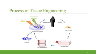
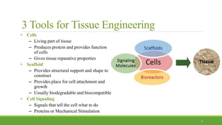
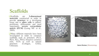
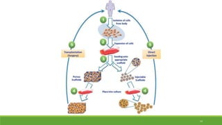
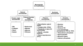
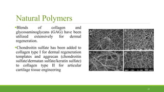
Tissue Engineering:
Scaffold Materials
BY: ELAHEH ENTEZAR-ALMAHDI
1
References:
Hollander AP, Hatton PV. Biopolymer methods in tissue engineering: Springer; 2004.
Shoichet MS, Hubbell JA. Polymers for tissue engineering. Journal of Biomaterials Science,
Polymer Edition. 1998;9(5):405-6.
Yang S, Leong K-F, Du Z, Chua C-K. The design of scaffolds for use in tissue engineering. Part I.
Traditional factors. Tissue engineering. 2001;7(6):679-89.
Shoichet MS. Polymer scaffolds for biomaterials applications. Macromolecules. 2009;43(2):581-
91.
Araci IE, Brisk P. Recent developments in microfluidic large scale integration. Current opinion in
biotechnology. 2014;25:60-8.
Coluccino L, Stagnaro P, Vassalli M, Scaglione S. Bioactive TGF-β1/HA alginate-based scaffolds
for osteochondral tissue repair: design, realization and multilevel characterization. Journal of
applied biomaterials & functional materials. 2016;14(1).
2
Characterizing Regenerative Medicine
1.Regenerative medicine is a broad definition for innovative medical therapies that will enable the
body to repair, replace, restore and regenerate damaged or diseased cells, tissues and organs. (Mayo
Clinic)
2.Tools and Procedures (Biofabrication or Additive Manufacturing) of Regenerative Medicine
Tissue Engineering: Tissue Repair/Replacement and Lab Grown Organs
Technologies
Stem cells
Natural and Synthetic Scaffolds
3-D Printing and Chip Technologies
3
Regenerative Medicine
1. Artificial organs
4
Regenerative Medicine
2. Tissue Engineering and Biomaterials
5
The Beginning…
Joseph Vacanti (Harvard Stem Cell Institute) And Robert Langer (MIT) 1993
6
Regenerative medicine pioneer
Dr. Ali Khademhosseini (Wyss Institute at Harvard)
Organs in the lab
Lab on a chip
7
Process of Tissue Engineering
8
3 Tools for Tissue Engineering
• Cells
– Living part of tissue
– Produces protein and provides function
of cells
– Gives tissue reparative properties
• Scaffold
– Provides structural support and shape to
construct
– Provides place for cell attachment and
growth
– Usually biodegradable and biocompatible
• Cell Signaling
– Signals that tell the cell what to do
– Proteins or Mechanical Stimulation
9
Stem Cells
Stem cells are cells that:
(1) can self-renew
(2) have the potential to differentiate along one or two lineages.
1. Totipotent  Can produce all cell types
2. Pluripotent  Can produce most cell types
3. Multipotent  Can produce more than one cell type
4. Unipotent  Can produce one cell type
10
Cell Sources
Autologous: Come from the person that needs the new cells.
Allogeneic: Come from a body from the same species.
Xenogenic: Come from a different species then the organism they’re going into.
Isogenic (Syngenic): Come from identical twins.
11
Scaffolds
Scaffolds are 3-dimensional
materials constructed in order to
provide structure to a developing
tissue and to allow cells to adhere,
proliferate, differentiate and most
importantly, secrete extracellular
matrix (ECM) (Leong MF. et al.,
2009).
Many different materials have been
investigated in order to construct
scaffolds such as polymers (PLA,
PGA, PCL, PEG), bioactive ceramics
(HA, TCP) as well as natural
polymers (Collagen, GAGs,
Chitosan).
12
Scaffold Construction Requirements
Scaffolds should provide void volume for vascularization, new tissue formation
and remodeling so as to facilitate host tissue integration upon implantation.
Should have high porosity and have suitable pore sizes, and the pores should be
interconnected.
the biomaterials should also be degradable upon implantation at a rate matching
that of the new matrix production by the developing tissue.
The biomaterials used to fabricate the scaffolds need to be compatible with the
cellular components of the engineered tissues and endogenous cells in host tissue.
Scaffolds provide mechanical and shape stability to the tissue defect.
13
14
15
Scaffolds Synthesis
Nanofiber self-assembly:
Molecular self-assembly is one of the few methods for creating biomaterials with
properties similar in scale and chemistry to that of the natural in vivo extracellular
matrix (ECM), a crucial step toward tissue engineering of complex tissues.
Moreover, these hydrogel scaffolds have shown superiority in in vivo toxicology
and biocompatibility compared to traditional macro scaffolds and animal-derived
materials (Cassidy JW, 2014).
16
Scaffolds Synthesis
Textile technologies:
These techniques include all the approaches that have been successfully
employed for the preparation of non-woven meshes of different polymers. In
particular, non-woven polyglycolide structures have been tested for tissue
engineering applications: such fibrous structures have been found useful to grow
different types of cells. The principal drawbacks are related to the difficulties in
obtaining high porosity and regular pore size(Ekevall E, 2004).
17
Scaffolds Synthesis
Freeze- drying:
First, a synthetic polymer is dissolved into a suitable solvent (e.g. polylactic acid
in dichloromethane) then water is added to the polymeric solution and the two
liquids are mixed in order to obtain an emulsion. Before the two phases can
separate, the emulsion is cast into a mold and quickly frozen by means of
immersion into liquid nitrogen. The frozen emulsion is subsequently freeze-dried
to remove the dispersed water and the solvent, thus leaving a solidified, porous
polymeric structure (Haugh MG, 2010).
18
Scaffolds Synthesis
CAD/CAM (3D-Printing):
19
Scaffolds Synthesis
Electrospinning:
A highly versatile technique that can be used to produce continuous fibers from
submicrometer to nanometer diameters. In a typical electrospinning set-up, a solution is
fed through a spinneret and a high voltage is applied to the tip. The buildup of
electrostatic repulsion within the charged solution, causes it to eject a thin fibrous
stream. A mounted collector plate or rod with an opposite or grounded charge draws in
the continuous fibers, which arrive to form a highly porous network. The primary
advantages of this technique are its simplicity and ease of variation. At a laboratory
level, a typical electrospinning set-up only requires a high voltage power supply (up to
30 kV), a syringe, a flat tip needle and a conducting collector. For these reasons,
electrospinning has become a common method of scaffold manufacture in many labs. By
modifying variables such as the distance to collector, magnitude of applied voltage, or
solution flow rate researchers can dramatically change the overall scaffold architecture
(Lannutti J, et al., 2007).
20
Electrospinning
21
Poly-α-hydroxy acid
22
Methods:
 Drying
 Extrusion
 Knitting
 Gamma sterilization
Poly-α-hydroxy acid
Extensive research has been performed in developing a full range of PLGA
polymers.
Both L- and DL-lactides have been used for co-polymerization.
The ratio of glycolide to lactide at different compositions allows control of the
degree of crystallinity of the polymers.
When the crystalline PGA is co-polymerized with PLA, the degree of
crystallinity is reduced and as a result this leads to increases in rates of hydration
and hydrolysis.
In general, the higher the content of glycolide, the quicker the rate of
degradation. However, an exception to this rule is the 50:50 ratio of PGA: PLA,
which exhibits the fastest degradation.
23
24
Fibrin microbeads (FMB)
Fibrinogen exerts adhesive effects on cultured fibroblasts and other cells.
Specifically, fibrin(ogen) and its various lytic fragments (e.g., FPA, FPB,
fragments D and E) were shown to be chemotactic to macrophages, human
fibroblasts, and endothelial cells
Micro-carrier beads made of some plastic polymers or glass provide cells with a
surface area on the order of 104 cm2/L for cell attachment, which is one order of
magnitude larger than the area available with stack plates or multi-tray cell-
culture facilities
microparticles from plasma proteins, such as albumin or fibrinogen, generally
using glutaraldehyde to cross-link the proteins.
25
Setup for producing FMB by oil emulsion
method
26
Natural Polymers
Blends of collagen and
glycosaminoglycans (GAG) have been
utilized extensively for dermal
regeneration.
Chondroitin sulfate has been added to
collagen type I for dermal regeneration
templates and aggrecan (chondroitin
sulfate/dermatan sulfate/keratin sulfate)
to collagen type II for articular
cartilage tissue engineering
27
Hyaluronan
 Composed of repeated disaccharide units
of D-glucuronic acid and N-
acetylglucosamine
 The unique properties of HA are
manifested in its mechanical function in
the synovial fluid, the vitreous humor of
the eye, and the ability of connective
tissue to resist compressive forces, as in
articular cartilage.
 Plays a fundamental role during
embryonic development and in wound
healing
28
Hyaluronan scaffold for central neural
tissue engineering
29
Collagen
In the form of collagen sponge
Porosity, biodegradability, and biocompatibility
Can be modified using growth factors or other manipulations to promote chondrocyte
growth and cartilage matrix formation
Scaffolds made from a single collagen type or composites of two or more types
Disadvantages
Poor dimensional stability. Variability in drug release kinetics.
Poor mechanical strength.
30
Chitosan
It consists of β-1-4 linked 2 amino-2-deoxy
gluco –pyranose moieties.
Commercially manufactured by N-deacetylation
of Chitin which is obtained from Mollusc shells.
It is soluble only in acidic pH i.e. when amino
group is protonated.
Thereby it readily adheres to bio membranes.
It is degraded mainly by Glycosidases &
lysozymes.
31
Chitosan Scaffold
Fabrication of bulk porous chitosan scaffolds
Freezing of a chitosan-acetic acid solution
Subsequent lyophilization
Scaffold microstructure will depend on the shape of the mold used for freezing
and on the freezer temperature.
32
Chitosan and Collagen-chitosan scaffold
33
Hydrogels as scaffold
Cells are suspended within or adhered to the 3D hydrogel framework during or
after formulation as scaffolds
RGD (arginine–glycine–aspartic acid) adhesion peptide sequence. Inclusion of
these RGD domains in hydrogels has shown improved cellular migration,
proliferation, growth, and organization in tissue regeneration applications.
Cells have been shown to favorably bind to the RGD-modified hydrogel
scaffolds. These cells include endothelial cells (ECs), fibroblasts, smooth muscle
cells (SMCs), chondrocytes and osteoblasts.
34
Alginate as Scaffold
35
Pure Alginate as Scaffold
36
Bioactive TGF-β1/HA alginate-based
scaffolds for osteochondral tissue repair
37
38

Tissue Engineering: Scaffold Materials

  • 1.
  • 2.
    References: Hollander AP, HattonPV. Biopolymer methods in tissue engineering: Springer; 2004. Shoichet MS, Hubbell JA. Polymers for tissue engineering. Journal of Biomaterials Science, Polymer Edition. 1998;9(5):405-6. Yang S, Leong K-F, Du Z, Chua C-K. The design of scaffolds for use in tissue engineering. Part I. Traditional factors. Tissue engineering. 2001;7(6):679-89. Shoichet MS. Polymer scaffolds for biomaterials applications. Macromolecules. 2009;43(2):581- 91. Araci IE, Brisk P. Recent developments in microfluidic large scale integration. Current opinion in biotechnology. 2014;25:60-8. Coluccino L, Stagnaro P, Vassalli M, Scaglione S. Bioactive TGF-β1/HA alginate-based scaffolds for osteochondral tissue repair: design, realization and multilevel characterization. Journal of applied biomaterials & functional materials. 2016;14(1). 2
  • 3.
    Characterizing Regenerative Medicine 1.Regenerativemedicine is a broad definition for innovative medical therapies that will enable the body to repair, replace, restore and regenerate damaged or diseased cells, tissues and organs. (Mayo Clinic) 2.Tools and Procedures (Biofabrication or Additive Manufacturing) of Regenerative Medicine Tissue Engineering: Tissue Repair/Replacement and Lab Grown Organs Technologies Stem cells Natural and Synthetic Scaffolds 3-D Printing and Chip Technologies 3
  • 4.
  • 5.
    Regenerative Medicine 2. TissueEngineering and Biomaterials 5
  • 6.
    The Beginning… Joseph Vacanti(Harvard Stem Cell Institute) And Robert Langer (MIT) 1993 6
  • 7.
    Regenerative medicine pioneer Dr.Ali Khademhosseini (Wyss Institute at Harvard) Organs in the lab Lab on a chip 7
  • 8.
    Process of TissueEngineering 8
  • 9.
    3 Tools forTissue Engineering • Cells – Living part of tissue – Produces protein and provides function of cells – Gives tissue reparative properties • Scaffold – Provides structural support and shape to construct – Provides place for cell attachment and growth – Usually biodegradable and biocompatible • Cell Signaling – Signals that tell the cell what to do – Proteins or Mechanical Stimulation 9
  • 10.
    Stem Cells Stem cellsare cells that: (1) can self-renew (2) have the potential to differentiate along one or two lineages. 1. Totipotent  Can produce all cell types 2. Pluripotent  Can produce most cell types 3. Multipotent  Can produce more than one cell type 4. Unipotent  Can produce one cell type 10
  • 11.
    Cell Sources Autologous: Comefrom the person that needs the new cells. Allogeneic: Come from a body from the same species. Xenogenic: Come from a different species then the organism they’re going into. Isogenic (Syngenic): Come from identical twins. 11
  • 12.
    Scaffolds Scaffolds are 3-dimensional materialsconstructed in order to provide structure to a developing tissue and to allow cells to adhere, proliferate, differentiate and most importantly, secrete extracellular matrix (ECM) (Leong MF. et al., 2009). Many different materials have been investigated in order to construct scaffolds such as polymers (PLA, PGA, PCL, PEG), bioactive ceramics (HA, TCP) as well as natural polymers (Collagen, GAGs, Chitosan). 12
  • 13.
    Scaffold Construction Requirements Scaffoldsshould provide void volume for vascularization, new tissue formation and remodeling so as to facilitate host tissue integration upon implantation. Should have high porosity and have suitable pore sizes, and the pores should be interconnected. the biomaterials should also be degradable upon implantation at a rate matching that of the new matrix production by the developing tissue. The biomaterials used to fabricate the scaffolds need to be compatible with the cellular components of the engineered tissues and endogenous cells in host tissue. Scaffolds provide mechanical and shape stability to the tissue defect. 13
  • 14.
  • 15.
  • 16.
    Scaffolds Synthesis Nanofiber self-assembly: Molecularself-assembly is one of the few methods for creating biomaterials with properties similar in scale and chemistry to that of the natural in vivo extracellular matrix (ECM), a crucial step toward tissue engineering of complex tissues. Moreover, these hydrogel scaffolds have shown superiority in in vivo toxicology and biocompatibility compared to traditional macro scaffolds and animal-derived materials (Cassidy JW, 2014). 16
  • 17.
    Scaffolds Synthesis Textile technologies: Thesetechniques include all the approaches that have been successfully employed for the preparation of non-woven meshes of different polymers. In particular, non-woven polyglycolide structures have been tested for tissue engineering applications: such fibrous structures have been found useful to grow different types of cells. The principal drawbacks are related to the difficulties in obtaining high porosity and regular pore size(Ekevall E, 2004). 17
  • 18.
    Scaffolds Synthesis Freeze- drying: First,a synthetic polymer is dissolved into a suitable solvent (e.g. polylactic acid in dichloromethane) then water is added to the polymeric solution and the two liquids are mixed in order to obtain an emulsion. Before the two phases can separate, the emulsion is cast into a mold and quickly frozen by means of immersion into liquid nitrogen. The frozen emulsion is subsequently freeze-dried to remove the dispersed water and the solvent, thus leaving a solidified, porous polymeric structure (Haugh MG, 2010). 18
  • 19.
  • 20.
    Scaffolds Synthesis Electrospinning: A highlyversatile technique that can be used to produce continuous fibers from submicrometer to nanometer diameters. In a typical electrospinning set-up, a solution is fed through a spinneret and a high voltage is applied to the tip. The buildup of electrostatic repulsion within the charged solution, causes it to eject a thin fibrous stream. A mounted collector plate or rod with an opposite or grounded charge draws in the continuous fibers, which arrive to form a highly porous network. The primary advantages of this technique are its simplicity and ease of variation. At a laboratory level, a typical electrospinning set-up only requires a high voltage power supply (up to 30 kV), a syringe, a flat tip needle and a conducting collector. For these reasons, electrospinning has become a common method of scaffold manufacture in many labs. By modifying variables such as the distance to collector, magnitude of applied voltage, or solution flow rate researchers can dramatically change the overall scaffold architecture (Lannutti J, et al., 2007). 20
  • 21.
  • 22.
    Poly-α-hydroxy acid 22 Methods:  Drying Extrusion  Knitting  Gamma sterilization
  • 23.
    Poly-α-hydroxy acid Extensive researchhas been performed in developing a full range of PLGA polymers. Both L- and DL-lactides have been used for co-polymerization. The ratio of glycolide to lactide at different compositions allows control of the degree of crystallinity of the polymers. When the crystalline PGA is co-polymerized with PLA, the degree of crystallinity is reduced and as a result this leads to increases in rates of hydration and hydrolysis. In general, the higher the content of glycolide, the quicker the rate of degradation. However, an exception to this rule is the 50:50 ratio of PGA: PLA, which exhibits the fastest degradation. 23
  • 24.
  • 25.
    Fibrin microbeads (FMB) Fibrinogenexerts adhesive effects on cultured fibroblasts and other cells. Specifically, fibrin(ogen) and its various lytic fragments (e.g., FPA, FPB, fragments D and E) were shown to be chemotactic to macrophages, human fibroblasts, and endothelial cells Micro-carrier beads made of some plastic polymers or glass provide cells with a surface area on the order of 104 cm2/L for cell attachment, which is one order of magnitude larger than the area available with stack plates or multi-tray cell- culture facilities microparticles from plasma proteins, such as albumin or fibrinogen, generally using glutaraldehyde to cross-link the proteins. 25
  • 26.
    Setup for producingFMB by oil emulsion method 26
  • 27.
    Natural Polymers Blends ofcollagen and glycosaminoglycans (GAG) have been utilized extensively for dermal regeneration. Chondroitin sulfate has been added to collagen type I for dermal regeneration templates and aggrecan (chondroitin sulfate/dermatan sulfate/keratin sulfate) to collagen type II for articular cartilage tissue engineering 27
  • 28.
    Hyaluronan  Composed ofrepeated disaccharide units of D-glucuronic acid and N- acetylglucosamine  The unique properties of HA are manifested in its mechanical function in the synovial fluid, the vitreous humor of the eye, and the ability of connective tissue to resist compressive forces, as in articular cartilage.  Plays a fundamental role during embryonic development and in wound healing 28
  • 29.
    Hyaluronan scaffold forcentral neural tissue engineering 29
  • 30.
    Collagen In the formof collagen sponge Porosity, biodegradability, and biocompatibility Can be modified using growth factors or other manipulations to promote chondrocyte growth and cartilage matrix formation Scaffolds made from a single collagen type or composites of two or more types Disadvantages Poor dimensional stability. Variability in drug release kinetics. Poor mechanical strength. 30
  • 31.
    Chitosan It consists ofβ-1-4 linked 2 amino-2-deoxy gluco –pyranose moieties. Commercially manufactured by N-deacetylation of Chitin which is obtained from Mollusc shells. It is soluble only in acidic pH i.e. when amino group is protonated. Thereby it readily adheres to bio membranes. It is degraded mainly by Glycosidases & lysozymes. 31
  • 32.
    Chitosan Scaffold Fabrication ofbulk porous chitosan scaffolds Freezing of a chitosan-acetic acid solution Subsequent lyophilization Scaffold microstructure will depend on the shape of the mold used for freezing and on the freezer temperature. 32
  • 33.
  • 34.
    Hydrogels as scaffold Cellsare suspended within or adhered to the 3D hydrogel framework during or after formulation as scaffolds RGD (arginine–glycine–aspartic acid) adhesion peptide sequence. Inclusion of these RGD domains in hydrogels has shown improved cellular migration, proliferation, growth, and organization in tissue regeneration applications. Cells have been shown to favorably bind to the RGD-modified hydrogel scaffolds. These cells include endothelial cells (ECs), fibroblasts, smooth muscle cells (SMCs), chondrocytes and osteoblasts. 34
  • 35.
  • 36.
    Pure Alginate asScaffold 36
  • 37.
    Bioactive TGF-β1/HA alginate-based scaffoldsfor osteochondral tissue repair 37
  • 38.