Rectal prolapse surgical approaches
•Download as PPTX, PDF•
7 likes•1,696 views

Presented in Bir Hospital, NAMS during the pandemic of the covid

Recommended
Recommended
More Related Content
What's hot
What's hot (20)
Similar to Rectal prolapse surgical approaches
Similar to Rectal prolapse surgical approaches (20)
More from Dr. Kiran Pandey
Recently uploaded
Recently uploaded (20)
Rectal prolapse surgical approaches
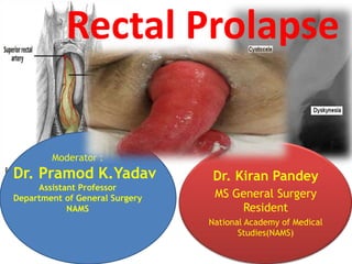
- 1. Dr. Kiran Pandey MS General Surgery Resident National Academy of Medical Studies(NAMS) Rectal Prolapse Moderator : Dr. Pramod K.Yadav Assistant Professor Department of General Surgery NAMS
- 2. Objective • Rectal anatomy • Introduction • Presentation • Surgical Approach • References
- 3. Approximately 12–18 cm in length Upper third: Mobile Peritoneal covering anteriorly and laterally; Middle third: Peritoneum covers anterior and part of the lateral surfaces Lower third: Lies deep in the pelvis surrounded by fatty mesorectum Rectal Anatomy
- 4. Rectal Anatomy Begins at sacral promentry Taenia coli joins to form a continuous outer longitudinal muscle layer Follows the curve of the sacrum Ends at the anorectal junction. The puborectalis muscle encircles the posterior and lateral aspects of the junction, creating th anorectal angle (normally 120°).
- 5. Rectal Anatomy The rectum has three lateral curvatures: Upper and Lower: convex to the right Middle : Convex to the left. On the luminal Three curves are marked by semicircular folds(Houston’s valves) Separated by adjacent structures by fascial layers. Denonvilliers’ fascia seperates from the prostate/vagina Waldeyer’s fascia seperates from the coccyx and lower two sacral vertebrae They are a barrier to malignant invasion.
- 6. Rectal Anatomy Blood supply Superior rectal artery Middle rectal artery Inferior rectal artery Venous drainage Superior rectal vein Middle rectal vein
- 7. Rectal Anatomy Lymphatic drainage • The lymphatics of the mucosal lining communicate with muscular layers. • The usual flow is upwards • Surgical clearance of malignant disease Concentrates mainly wide resection of proximal lymph nodes.
- 8. Rectal Prolapse : Introduction Defined as drooping of the rectum or Falling down of hind gut. • First described by Papyrus in 1500 BC • Types: Occult (internal) rectal prolapse : Rectal wall has prolapsed but does not protrude through the anus. Mucosal prolapse only of the rectal and anal mucosa Complete rectal prolapse or rectal procidentia: The prolapse is delivered through the anal canal leading to a protrusion of all layers of rectum
- 9. Rectal Prolapse : Introduction Factors associated Sliding hernia through a defect in the pelvic fascia Intussusception Complex, redundant rectosigmoid colon Deep pouch of doglus Loose rectal fixation Pelvic fascia defect, resulting in a sliding hernia. Constipation and difficulty in defecation, Pelvic floor atony Patulous anus, Excessive inhibition reflex of pelvic floor. Pudendal neuropathy Abnormal hindgut motility associated with Reduced high- amplitude propagated contractions, Increased resting colonic pressure, Slow colonic transit
- 10. Rectal Prolapse : Introduction
- 11. Rectal Prolapse : Introduction Factors preventing prolapse: Curvature of sacrum Tilt of pelvis Serpentine course of rectum Levator ani muscles- fixes rectum Puborectalis sling-Tilt and elevate lower end of rectum
- 12. Rectal Prolapse : Introduction • Bimodal distribution – one peak occurs in children within the first 3 years of life – second peak occurs after the seventh decade • In the elderly, rectal prolapse is more common in women (80% to 90%), and the prevalence of this abnormality increases with age • Institutionalized patients who have neurologic or psychiatric comorbidities (15%)
- 13. Clinical features • Mass coming out of anal canal during straining/coughing • Mucous leakage • Rectal bleeding • Dull perineal pain or pressure • Tenesmus • Difficult defecation • Fecal incontinence
- 14. Assessment • Detailed history and examination • Flexible Sigmoidoscope/ total colonoscopy • Defecating Proctography • Electromyography and measurement of the pudendal nerve terminal motor latency
- 15. Treatment of Mucosal Prolapse In infants and young children Digital repositioning. The parents are taught to replace the protrusion, and any underlying causes are addressed. Submucosal injections. If digital repositioning fails after 6 weeks’ trial, injections of 5 per cent phenol in almond oil are carried out under general anaesthetic. Aseptic inflammation following these injections, the mucous membrane becomes tethered to the muscle coat. Surgery. Occasionally, surgery is required and, in such cases, The child is placed in the prone jack-knife position, the retrorectal space is entered, and the rectum is sutured to the sacrum.
- 16. Treatment of Mucosal Prolapse In Adults: Local treatments. Submucosal Injections of phenol In almond oil or the application of rubber bands Excision of the prolapsed mucosa.
- 17. Surgical Approach • Surgery definitive treatment • Therapeutic options – narrowing of anal orifice – obliteration of pouch of douglas – restoration of pelvic floor – resection of redundant bowel – Suspension / fixation of rectum to sacrum Garely et.al., Rectal prolapse. In: Cameron JL, Cameron AM (editors): Current surgical therapy. Elsevier Saunders: Philadelphia; 2014 Rickert et al. Laparoscopic surgery for rectal prolapse and pelvic floor disorders. World J Gastrointest Endosc 2015
- 18. Surgical treatment • Perineal approach – Thiersch – Delorme’s – Altemeier’s • Abdominal approach – Conventional laprotomy • Well’s Repair • Frykman and Goldberg procedure – Laproscopic surgery
- 19. Perineal Approach Perineal procedures include • anal encirclement (Thiersch procedure), • mucosal sleeve resection (Delorme procedure), and • perineal rectosigmoidectomy (Altemeier procedure). The choice of operation depends on the • patient’s age, • comorbidities, • operative risk, • associated • anatomic or physiologic conditions, • history of previous surgery for prolapse.
- 20. Perineal Approach Since affeccted patients are ,numerous comorbidities and high operative risk, Perineal approach is excellent treatment strategy. Perineal procedures allow the use of spinal or locoregional anesthesia associated with decreased perioperative morbidity rates, Shorter hospital stay and recovery to normal activity, less pain, avoidance of peritoneal adhesions, lower risk of injury to the pelvic nerves, when compared with abdominal approaches
- 21. Perineal Approach • In patients with large prolapses, perineal rectosigmoidectomy may result in decreased recurrence rates as compared with the Delorme procedure. • In small rectal prolapse, the Altemeier procedure may be technically difficult and inadequate mobilization of the descending colon may lead to ischemia with high risk of anastomotic dehiscence
- 22. Incarcerated and strangulated rectal prolapse Reduction can be attempted under sedation. If successful, the patient is referred to elective defi nitive treatment. Prolapsed bowel is viable Emergency perineal rectosigmoidectomy is required with or without fecal diversion. Ruptured prolapse and perineal evisceration of small bowel is a critical condition that requires immediate transabdominal repair including closure of peritoneal defect and suture rectopexy bowel is not viable
- 23. Anal Encirclement (Thiersch Procedure) Avoids exteriorization of the rectum by encircling and narrowing the anal canal. It was initially proposed by Thiersch using a silver wire, But prosthetic materials such as silastic rods and polypropylene meshes have been used. Two small 2-cm incisions are made anteriorly and posteriorly or laterally on both sides of the anus. submucosal tunnel created around the anus using a Kelly clamp, prosthetic mesh is placed in the subcutaneous layer, Leading to partial closure and narrowing of the anal canal Placement of the mesh in the ischiorectal space may improve results. High recurrence rates and fecal impaction (80% of the patients.) Considered only for extremely high-risk patients.
- 24. Anal Encirclement (Thiersch Procedure)
- 25. Mucosal Sleeve Resection (Delorme Procedure) fi rst described by Edmond Delorme in 1900 and Involves rectal muscle layer plication and mucosal layer resection relaps rates ranging from 7% to 23% Sphincteroplasty can be added to the Delorme procedure in patients with fecal incontinence to improve results. Complications may include bleeding, leakage, stricture, fecal incontinence, and recurrence Incision Th e rectum is pulled down to the full extent of the prolapse using Babcock clamps. Injection of an epinephrine solution (1:200,000) in the submucosal layer facilitates dissection and decreases bleeding. A circumferential incision is made with electrocautery, 2 cm proximal to the dentate line to preserve anorectal sensorial area, exposing the circular muscular layer.
- 26. Delorme (Mucosal sleeve resection)
- 27. Mucosal Sleeve Resection (Delorme Procedure) Mucosal Sleeve Resection Muscular Plication • After the mucosal sleeve is stripped from the muscularis, nonabsorbable longitudinal sutures are placed in the muscular layer to createm an accordion eff ect. • Th is plication begins at the proximal end of the mucosectomy and ends at the distal edge of the internal sphincter close to the dentate line Mucosal Anastomosis Once muscular plication is finished, the stripped mucosa is resected and a mucosal anastomosis is performed using 4-0 absorbable sutures, beginning with four sutures placed in the four quadrants Once the anastomosis is complete it usually is pulled back spontaneously into the anal canal
- 28. Delorme (Mucosal sleeve resection)
- 29. Delorme (Mucosal sleeve resection)
- 30. Perineal Rectosigmoidectomy with Coloanal Anastomosis (Altemeier Procedure) • In 1889, Mickulicz was the first to describe perineal rectosigmoidectomy for treating rectal procidentia, • In a subsequent study, they also included anterior levatorplasty and high ligation of the pouch of Douglas to the procedure. • Prolapse Delivery delivered through the anus using Babcock clamps Epinephrine solution (1:200,000) is injected circumferentially the submucosal layer, 1.5 cm proximal to the dentate line to minimize bleeding.
- 31. Perineal Rectosigmoidectomy with Coloanal Anastomosis (Altemeier Procedure) • Incision • A circumferential incision is initiated at the • posterior outer rectal wall, 1.5 to 2 cm above • the dentate line, preserving the entire transitional zone of the anal canal. • The incision is made through all layers of the outer rectal wall and deepened until the perirectal fat is encountered and the mesorectum identifi ed. • Care should be taken not to damage the mesorectal vessels and to avoid simultaneous transection of the inner rectal wall Rectal Mobilization • Mobilization of the rectum proceeds cranially with careful isolation and ligation of mesorectal vessels close to the intestinal wall. Levatorplasty • Posterior plication of the levator muscles is • one modifi cation to the Altemeier procedure • proposed to include the treatment of fecal incontinence by restoring the angles of the pelvic floor. Resection and Anastomosis • Before sectioning the proximal colon, arterial • patency is tested by dividing the terminal branches before routinely perform in all intestinal anastomoses. • Anastomosis can also be performed using circular staples • Th e anastomosis should be located above the pectinate line, thus preservin the columns of Morgagni that play an importan role in preserving fi ne anal continence and decreasing the risk of postoperative stenosis. clamping them, as we cilitate sigmoid mo-
- 32. Perineal Rectosigmoidectomy with Coloanal Anastomosis (Altemeier Procedure)
- 33. Perineal Rectosigmoidectomy with Coloanal Anastomosis (Altemeier Procedure)
- 34. Perineal Rectosigmoidectomy with Coloanal Anastomosis (Altemeier Procedure)
- 35. COMPLICATIONS Deaths related to perineal rectosigmoidectomy rare. In the literature vary from 0% to 32%. They may occur in the early or immediate Postoperative period and include pelvic hematomas, Anastomotic dehiscence, abscesses, and bleeding. The most frequently observed late postoperative complications are anal strictures, which may be managed by anal dilation.
- 36. Open abdominal approach • Anterior Mesh Sling Or Ripstein Repair • Posterior Mesh Sling Or Wells Repair • Sigmoid Resection And Suture Proctopexy
- 37. BOWEL PREPARATION A bowel devoid of stool is easier to handle especially if a resection is anticipated. At present, two sachets of oral Pico-Salax are used the day prior to operation. Antibiotics are also administered preoperatively and our current choice is Timentin 3.1-g i.v. within 1 hour of incision and two postoperative doses q8h.
- 38. WELL’S REPAIR Anterior mesh sling or Ripstein repair : most popular repair for rectal prolapse Ripstein himself abandoned it, for posterior sling d/t complication Complication reduced by half, if the sling were placed posterior to the rectum leaving the anterior one-fourth to one third of the circumference of the rectum free to expand, a principle expounded by Wells. valon sponge (polyvinyl alcohol), (polypropylene) and Teflon are the most common materials used. Th e abdomen entered by infraumbilical transverse incision. Care is taken to protect the ureters
- 39. The retropresacral space is entered at the level of the sacral promontory, and the rectum is gently mobilized from the sacral hollow Ensure that the mesorectum is anterior to the plane of dissection or the intestine may be devascularized and there may be annoying bleeding. At the same time, one should avoid the presacral nerves. Dissection in the presacral plane should be performed with cautery taking care not to injure the rectal wall. The rectum is fully mobilized to the level of the coccyx. Anterior mobilization is begun by continuing the lateral peritoneal incisions distally until they meet in the deepest portion of the cul-de- sac.
- 40. Well’s Repair
- 41. Well’s Repair
- 42. Well’s Repair
- 43. Postoperative Care • Intravenous fl uids are given until the postoperative ileus has resolved, usually in 2 or 3 days. • Nasogastric suction is ordinarily not necessary. • Th e Foley catheter is removed on the second day. • ensure that the patient does not strain at defecation. • oral intake, a bulk-forming agent in the form of a psyllium seed preparation is given two or three times a day, • supplemented by milk of magnesia as necessary.
- 44. SIGMOID RESECTION AND SUTURE PROCTOPEXY Described by Frykman , performed through a transverse or midline abdominal incision. Foley catheter is inserted into the urinary bladder. The head of the operating room table is tilted down 30 to 40 degrees, depending on the shape of the pelvis. The entire small bowel is packed to the upper abdomen with moist lap pads. Mobilization of Sigmoid Colon and Rectum Th e lateral peritoneal reflection of the sigmoid colon (white line of Toldt) is incised by The gonadal vessels and left ureter are identified and swept posteriorly. The peritoneal incision is continued to the left side of the rectum, about 1 cm lateral to the rectal wall, and curves anteriorly to the rectovesical sulcus. Similarly, the peritoneum at the base of the sigmoid mesentery is incised by cautery and continued to the right side of the rectum to unite with the incision on the left side
- 45. SIGMOID RESECTION AND SUTURE PROCTOPEXY Entering the Retrorectal Space (Posterior Mobilization). Sweeping the Ureters and Pelvic Nerves Anterior Mobilization . Resection of the Redundant Sigmoid and Anastomosis Proctopexy Sutures
- 46. COMPLICATIONS: THEIR PREVENTION AND MANAGEMENT Th is review revealed a recurrence rate of 2.3%, and complications directly related to placement of the sling occurred in 16.5% of the patients. The overall reoperation rate was 4.1% Indications for reoperation included fecal impaction, small-bowel obstruction, stricture, pelvic abscess, rectal erosion, and hemorrhage. Presacral bleeding may range from nuisance bleeding to hemorrhages of terrifying proportions. Strictures were common, but only those that required further operation, either division or removal of the sling or resection of the segment of bowel, were included.
- 50. Laparoscopic approach Goals of surgery correct anatomic abnormalities associated with prolapse provide functional improvement of his or her symptoms. Laparoscopic surgical options include rectopexy with or without mesh, sigmoid resection alone, or combination of the two.
- 51. Laparoscopic approach • None operation is universal for prolapse patients • Must be tailored to the patient and his or her individual symptoms. A resection with rectopexy is indicated in sigmoid diverticular disease, a sigmoidocele, a redundant sigmoid colon, slow transit time, and/or constipation.
- 52. Laparoscopic approach • A rectopexy relatively contraindicated in constipation (high rates of persistence or worsening of constipation following this procedure. ) Absolute contraindication to a laparoscopic repair patient’s inability to tolerate general anesthesia Relative contraindications prior abdominal surgery irreducible prolapse
- 53. Preoperative Preparation • Clear liquid diet 2 days before surgery. • On the day prior to surgery, Two doses of an phosphosoda preparation. 1 g metronidazole and 1 g neomycin at 2 p.m., 3 p.m., and 11 p.m. • NPO the night before surgery. • Preoperative antibiotics prior induction of general anesthesia. • Positioned supine on split-leg table and sequential venous compression stockings are placed on both lower extremities.
- 54. Patient Positioning and Port Placement • Surgeon s on the patient’s right side and the assistant on the left. • Two monitors are positioned at the foot of the bed • The patient is then placed in steep Trendelenburg position secured with bean bag. Port Placement • Either a Veress needle or the Hassan technique to access the peritoneal cavity at or superior to the umbilicus. • A carbon dioxide pneumoperitoneum of 15 mm Hg is created, a 30-degree laparoscope is inserted, and the abdominal cavity is inspected.
- 55. Patient Positioning and Port Placement 5-mm port just lateral to the rectus sheath cephalad to thecamera port 12-mm port in the right iliac fossa at the McBurney point. (used in resection cases for the introduction of stapling devices and in rectopexy cases for intracorporeal suturing.) 5-mm port is placed on the left that mirrors the right upper port location.
- 57. Rectopexy without Mesh A complete mobilization of the rectum from the sacral promontory to the coccyx In the course of developing this plane, the right and left ureters are identified, and the hypogastric nerves are kept with the retroperitoneal tissue posterior to the rectum. Preservation of the lateral rectal ligaments to prevent disruption of the parasympathetic supply to the rectum, (high rate of constipation after rectopexy alone may be caused by disruption of these nerves.
- 58. Rectopexy without Mesh Once the rectum has been fully mobilized, the surgeon performs the rectopexy. Rectum is placed on cephalad tension, two or three nonabsorbable stitches are used to secure it to the presacral fascia at the level of the sacral promontory. These lateral rectal stitches are placed on both sides and should include the submucosa but not the mucosa. The presacral stitches should engage the fascia without piercing the presacral venous plexus. After the pelvis is examined for hemostasis, the ports are removed, the pneumoperitoneum is evacuated, and the incisions are closed
- 59. Rectopexy with Mesh After rectal mobilization, sacral fixation can be achieved with Mesh placed anteriorly (per Ripstein) or Mesh placed anteriorly posteriorly (per Wells). Ripstein procedure Anterior, circumferential fixation of the rectum by a piece of polypropylene mesh. Most surgeons have abandoned postoperative fibrotic reaction is often dense and leads to varying degrees of obstruction. Wells procedure posterior, partial mesh wrap. posterior rectopexy with the creation of a T-shaped or rectangulat piece of mesh. The long part of the “T” is sutured with nonabsorbable suture to the presacral with junction of “T” is at the level of the sacral promontory. The “arms” of the “T” are sewn to the lateral rectal walls with two or three nonabsorbable stitches. Th e arms do not completely encircle the rectum, and the final product resembles a hotdog (rectum) in a bun (mesh).
- 60. Sigmoid Resection with Rectopexy Sigmoid resections without rectopexy have been abandoned in favor of sigmoid resection with fixation. After complete rectal mobilization, sigmoid colon dissected free from its lateral attachments. Left ureter is identifid and a proximal margin of resection is chosen based on the ability of the colon to easily reach the rectum (which is placed on tension to eliminate redundancy). An articulating, linear, endoscopic stapler is used to divide the proximal rectum at the distal line of resection. The distal end of the specimen is eviscerated via a left lower quadrant muscle–splitting incision and the proximal line of resection is divided extracorporeally.
- 61. Sigmoid Resection with Rectopexy A mesh rectopexy is not recommended After removing the stapler from the rectum and inspecting the anastomotic rings for completeness, the pelvis is fi lled with irrigation fluid, and a flexible sigmoidoscope is used to inspect the anastomosis for hemostasis and integrity. If there are no air bubbles present upon insuffl ation of the rectum, the air is evacuated and the scope is withdrawn. A single absorbable suture is used to close the right lower quadrant 12- mm port site using a suture passer device. After a final inspection of the abdominal cavity, the 5-mm ports are removed as the pneumoperitoneum is evacuated. The skin is closed.
- 62. Postoperative Management • Early postoperative pain is controlled with parenteral narcotics and parenteral NSAIDs • On the day of surgery, patients are encouraged to ambulate and are allowed clear liquids. • The average length of stay for rectopexy patients is 1 to 2 days, but with resection remain hospitalized for longer. • Most patients begin to mobilize third-spaced fluids between postoperative day 1 and 2 and their diet is advanced with the passage of flatus. • The parenteral narcotics are replaced on postoperative day 2 with an oral narcotic/acetaminophen combination. • Most patients are discharged on postoperative day 3. • Resolution of incontinence may take as long as 6 months, so patients may need reassurance
- 63. Complications Lap. Rectopexy with or without mesh has been safe as open rectopexy. The complication rates range from 0% to 30% in most series. leak rates remain in the 1% to 2% range. Anal incontinence rates decrease by 40% to 60%, and constipation improves in 60% to 90% of patients after resection rectopexy. Reoperation • bleeding (1% to 2%) • fecal impaction (1%). Rectopexy alone is associated with a slightly lower reported rate in improvement in constipation than resection rectopexy. Surgical treatment for Internal rectal prolapse, or rectal intussusceptio has been ineffective (posterior rectopexy) or thought to be too risky (sigmoid resection with anastomosis) for a condition without a clear natural history toward full, external rectal prolapse.
- 64. Lap. Rectopexy Vs Rectopexy with resection Resection with rectopexy has become the preferred approach for rectAal prolapse repair, In relatively young and healthy and suff er from constipation. Resection with rectopexy is as effi cacious as rectopexy alone in resolving incontinence, but has decreased postoperative constipation rates. Eliminate the creation of an acute angle at the rectosigmoid junction by removing a redundant colon, Preserves the rectal reservoir and avoids damage to sphincters as opposed to the perineal approach. Reported prolapse recurrence rates of rectopexy both with and without resection are low and range from 0% to 7% over 2 to 10 years of follow-up.
- 65. Open Vs Laparoscopic Approach Laparoscopic procedures have been shown to be equivalent to the open variants in terms of safety, effectiveness, and recurrence rates. In centers with the necessary expertise in laparoscopic pelvic surgery, recurrences can be treated laparoscopically as well otherwise open approach. Laproscopic surgical procedures have following benefits over open • Minimal surgical stress • Decreased lengths of stay, • Minimal Postoperative pain • Decreased use of narcotics • High patient satisfaction.
- 66. Postoperative rates of morbidity, mortality and recurrence . Hori et. al. Surgical options for full-thickness rectal prolapse: current status and institutional choice. Annals of gastroenterology. 2018
- 68. Recent Advances Robotic Modified Orr Loygue • Involves standard ventral mesh with narrow mesh posteriorly. • Robotic dissection allows a long narrow tube to be dissected posterior to rectum in between hypogastric nerve down to rectal tube and waldeyers fascia allowing the mesh to be tied to the rectal muscle. Not known if it improves the outcome. Disadvantage :cost, prolong surgery and off sight injury
- 69. References Fischer’s mastery of surgery, 6th edition Bailey and love’s short practice of surgery, 27th edition Sabiston textbook of surgery, 20th edition Surgical options for full-thickness rectal prolapse: current status and institutional choice, Hori et. al.
Editor's Notes
- The mucous membrane and submucosa of the rectum protrude outside the anus for approximately 1–4 cm. When the prolapsed mucosa is palpated between the finger and the thumb, it is evident that it is composed of no more than a double layer of mucous membrane. In infants The direct downward course of the rectum, due to the as-yet undeveloped sacral curve, predisposes infants to this condition .In children, Mucosal prolapse often commences after an attack of diarrhoea, or from loss of weight and consequent loss of fat in the ischiorectal fossa. It may also be associated with cystic fibrosis and should be treated conservatively .In the adult often associated with third-degree haemorrhoids, when it is referred to as mucohaemorrhoidal prolapse . In the female, a torn perineum, and in the male straining from urethral obstruction, predispose to mucosal prolapse. In old age, both mucosal and full-thickness prolapse are associated with weakness of the sphincter mechanism.
- It is possible that chronic constipation and straining to defecate may be a contributing factor
- Benign disease : into their underwear and around the anus that causes variable degrees of perineal skin irritation. By the time the patients seek medical consultation, about 50% of them already have In a review of 32 young adults (<30 years), Sun et al. found 41% had chronic psychiatric disease requiring treatment and these patients experienced significantly more constipation than nonpsychiatric patients (83% vs. 50%) All patients in whom rectalprolapse is suspected should be examined while straining on the toilet. The diagnosis of rectal prolapse is easy if the protrusion comes through the anus
- to rule out associated abnormalities, especially polyps, cancer, and inflammatory bowel disease. Occasionally, a solitary rectal ulcer is found are useful to confirm the nerve damage to the external anal sphincter and the pelvic floor muscles. Patients with damage to the pudendal nerves usually continue to have fecal incontinence after successful repair of the rectal prolapse
- Abdominal procedures are preferred by most surgeons, because they are more effective and are associated with lower recurrence rates, even in older patients . Perineal approaches are less invasive and potentially advantageous.
- Cochrane Review