This document discusses parturition related disorders in sheep and goats. It describes the normal stages of labor and common disorders that can occur during pregnancy (pre-partum), at parturition, and after parturition (post-partum). Some of the most common pre-partum disorders discussed include abortion, pregnancy toxemia, vaginal prolapse, and hydrometra. Common parturient disorders include dystocia caused by fetal or maternal factors, prolonged gestation, and fetal mummies. Post-partum disorders discussed are retained placenta, metritis, uterine prolapse, and uterine rupture. The document provides details on the causes, diagnosis, and treatment of these various disorders.
Vet obst lecture 14 Postpartum complications in sheep and goats
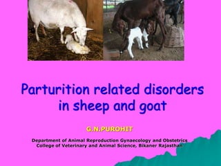
1. Parturition related disorders
in sheep and goat
G.N.PUROHIT
Department of Animal Reproduction Gynaecology and Obstetrics
College of Veterinary and Animal Science, Bikaner Rajasthan
2. Parturition – Successful culmination
of pregnancy
Ist stage of labor
lasts for 6 - 12 hr both in sheep and goat
The animal is restless and paws the ground.
2nd stage of labor lasts for 0.5 – 1 hr. The fetus is
delivered in this stage in anterior longitudnal
presentation sometimes in posterior presentation
3rd stage of labor lasts for 3-4 hr after the
delivery of last lamb. The placenta is expelled
and the uterus starts involution.
vocalization shown by goat
birth canal of goat more fragile
6. During pregnancy the fetus grows in utero deriving nutrition
from its mother and constantly changing its position and
excreting end products into maternal circulation. The feto-
maternal interactions when obstructed or altered result into
disorders.
Pre-Partum Disorders
8. Abortion- Delivery of fetus before full term which is
incapable of independent life
Incidence: 3 – 11%
9. Causes of Abortion are multiferous
Genetic disorders: Abortion is an inherited
disorder in older Angora goats.
Nutritional factors: Deficiency of energy, protein,
iodine, copper, selenium etc..
Toxic plants and pharmaceuticals: Ex. copper,
dexamethasone injections etc.
Stress or trauma: predator attacks, heat stress.
Infectious: Ex. Brucella ovis, akabane virus
10. Specimens to be submitted to
diagnostic laboratory
Fetus and placenta – chilled and in a clean container
Fresh chilled fetal heart blood,serum or thoracic fluid -
5mL
Frozen fetal abomasal contents
Blood samples from minimum 10% of aborting animals
Management of aborting flock
Remove pregnant ewes/goats from aborting
animals to a clean area.
Initiate specific control measures on the basis of
agents suspected
Send animals that have aborted and proposed for
culling directly to slaughter only after discharge
from reproductive tract have ceased.
11. Hydrometra(Pseudopregnancy)or
cloud out-burst
Seen in goats
Incidence: 3-14%
Etiology :High prolactin levels
Persistence of CL subsequent to
fetal death and reabsorption
commonly accepted cause
Diagnosis :Clinical cases discharge of large
quantity of fluid without fetal delivery
Cases referred for PD diagnosis by
ultrasonography
13. In a study hydrometra was diagnosed in 21 does and
out of these it was diagnosed in 5 does by
ultrasonography while in 16 does it was diagnosed in
goats presented for examination with history of
discharge without fetal delivery (Purohit, 2006)
Therapy:
Prostaglandin injections
Inj.Lutalyse 1.5 – 2.0 mL IM
Inj. Prostodin 125 µg IM
Anti-prolactins
Bromocryptine 1 mg SC twice daily for 6-10 days
14. Hydroallantois:
Accumulation of excessive fluid in allantois,
seen both in sheep and goat
Etiology: Legumes with high
estrogens
Hypothyroidism
Placental or uterine
disease
Clinical signs: Sudden
excessive abdominal enlargement
Difficulty in respiration
Diagnosis: Clinical signs,
ultrasonography
Therapy: Pregnancy
termination with prostaglandins,
caesarean section
Prognosis: Poor extreme care
necessary in therapy.
16. Clinical signs
Depression, recumbency,
tremors,circling, grinding of teeth, etc.
Diagnosis:
Ketone bodies in urine
Low blood glucose/ calcium
Therapy: Difficult pregnancy termination must be
considered
20-80 mL of 25 % glucose
0.5-2 mL of Insulin inj.
5-20 mL of calcium borogluconate
17. Rupture of the prepubic tendon/
ventral Hernia Occurs in animals
with multiple
fetuses
Pregnant females
with abdominal
trauma
THERAPY
Application of
canvas girdle
Reduction in salt
and trace minerals
in feed
19. Dystocia: Difficult birth Incidence 3-32%
Factor Incidence Reference
Age Not related with age.
Common in 2 years old
ewes
George (1975), Kloss et al. (2002).
Majeed and Taha (1989b)
Majeed et al. (1993)
Sharma et al. (1999)
Sex of fetus More in ewes with
male fetuses
Majeed and Taha (1989b) Majeed et al.
(1993)
Parity More in first and
second parity
Echternkamp and Gregory (1999)
Number of
fetuses
Common in ewes with
single fetus
George (1976)
Silva and Noakes (1984)
Krueger and Wassmuth (1974)
Echternkamp and Gregory (1999)
Season of
lambing
Common in spring and
winter lambings
George (1975), George (1976)
Cecilia et al. (1996)
Breed Higher in Texel ewes
Higher in Cheviot ewes
Krueger and Wassmuth (1974)
Whitelaw and Watchorn (1975)
Length of
Gestation
Higher in prolonged
gestation
Dennis (1974)
Health of ewes Higher in weak ewes George (1975)
25. Examination of animals for dystocia must not begin
before 30 min after beginning of contractions
Cleanliness, gentleness and lubrication in examination
and handling of dystocia are of utmost importance
Handling of dystocia Manual correction Due
care necessary in vaginal manipulation
especially in the goat to avoid rupture
26. Fetotomy Only partial fetotomy of one limb or head possible.
Caesarean section
Sites Midline
Flank
Paramedian
Oblique venterolateral
Anesthesia Local infiltration with
sedation
27. Subsequent to delivery the uterus must be examined to
explore any remaining fetus
Abdominal ballotment may be confusing because of
presence of ruminal foreign bodies/ bezoars
Prolonged
Gestation
Difficult to detect in animals
without breeding records
Cause
defects in hypothalamopituitarry
axis
Consumption of plant toxins
Viral diseases
Therapy
pregnancy termination using PG
+ dexa methasone
Caesarean section
Result Extra large fetuses
28. Fetal mummies
delivered with normal fetus or recognised on vaginal or
sonographic examination.
Therapy PG + dexamethasone or caesarean section.
30. Retained Placenta
Known to occur both in sheep
and goats
More prevalent in young goats
ETIOLOGY
Vitamin A deficiency
Obesity, hypocalcaemia
Infectious disease
THERAPY
Manual removal
Prostaglandin Injections
Oxytocin
Uterine echbolics
31. Post Parturient metritis
Uncommon in sheep common in goats
Cause: Poor hygiene at kidding/ lambing
Therapy: Intrauterine/ Parentral antibiotics
Prostaglandin injections
Post parturient disorders(Purohit et al., 2006)
Retained placenta 51.5%
Post-parturient metritis 38.3%
Vaginal prolapse 4.4%
Uterine prolapse 2.9%
Uterine rupture 2.9%
33. Cause Lack of exercise
High estrogenic feeds
Herreditary
Prepartum vaginal
prolapse common in sheep
and a commercial
accessory is available for
therapy
Post partum- replacement
calcium therapy
34. Uterine ruptures
Common subsequent to dystocia handling
by breeders using undue force on a
maldisposed fetus. Rarely spontaneous
rupture is possible.
Referred to vet when Intestinal loops
prolapse out
Emergency laparotomy suggested
Sometimes repair not possible
Potent antibiotic therapy is suggested
Prognosis is poor to fair
35. Pre and Post parturient care
Ultrasonography is a good preparturient
diagnostic tool. Regular scanning is useful.
Scanning at 3-4 months appears important, it
can give clues to conditions like twin fetuses,
fetal mummification, pseudopregnancy.
Evaluation of fetal viability can lead to decision
on the manner of dystocia handling.
Deworming and vaccinations are suggested 1
month before lambing
Supplementary feeding with mineral vitamins can
avoid subsequent retained placenta etc.
Close monitoring and timely help can prevent
dystocia
Post partum hygiene is important in prevention of
many problems