Neuro-otological aspects of Cerebellopontine angle SOL
•Download as PPTX, PDF•
6 likes•1,859 views

Brief description of Neuro-otological aspects of CP angle SOL

Recommended
More Related Content
What's hot
What's hot (20)
Similar to Neuro-otological aspects of Cerebellopontine angle SOL
Similar to Neuro-otological aspects of Cerebellopontine angle SOL (20)
More from Dr Fakir Mohan Sahu
More from Dr Fakir Mohan Sahu (19)
Recently uploaded
Recently uploaded (20)
Neuro-otological aspects of Cerebellopontine angle SOL
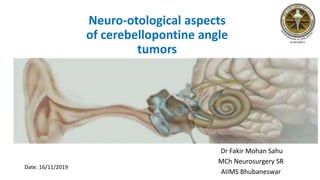
- 1. Neuro-otological aspects of cerebellopontine angle tumors Dr Fakir Mohan Sahu MCh Neurosurgery SR AIIMS Bhubaneswar Date: 16/11/2019
- 2. Cerebellopontine angle Anatomy Boundaries -Medial : lat. Surface of brain stem -Lateral : petrous bone -Superior : MCP and cerebellum -Inferior : arachnoid tissue of lower CN -Posterior : cerebellar peduncle Contents: CSF, arachnoid tissue, CN and their associated vessel (a)internal auditory canal, (b) anterior is VII nerve (c)posterior is VIII nerve P = Pons, mcp = Middle cerebellar peduncle CH = Cerebellar hemisphere
- 3. Internal acoustic meatus anatomy • Lateral wall: divided into superior and inferior halvesfalciform crest. • Upper compartment: Ant. area facial nerve Post. area superior vestibular nerve (by a sharp vertical ridge of bone known as ‘Bill’s bar’ after William House.) • Lower compartment: • Ant.Tractus spiralis foraminosus (cochlear nerve ) • Post.Inferior vestibular nerve (supplying the saccule)
- 4. Anatomy • Inner ear (labyrinth) - Bony : Perilymph - Membranous : Endolymph • Cochlea - 2 ¾ turns - Scalae • Organ of Corti • Semi-circular canals
- 5. Neurophysiology Neuron order Auditory pathway Ascending auditory pathway paathway 1st Bipolar nucleus of spiral ganglion and cochlear nerve 2nd Dorsal and ventral cochlear nuclei 3rd Superior olivary complex in pons. From here travels in lateral lemniscus in pons 4th Inferior colliculus in mid brain 5th Medial geniculate body in thalamus Auditory cortex (B. A 41 ,42) in temporal lobe through the auditory radiation in sublentiform part of internal capsule
- 6. Vestibular apparatus and function • Semicircular canals : rotational movements • Otoliths(utricle and saccule) : linear accelerations • Projection pathways • To cerebellum: muscle movements of the head, eyes, and posture. • To nuclei of CN III, IV, and VI. :Vestibulo-ocular reflex (VOR):stabilizing vision in motion • To the reticular formation: new posture of the body has taken on, and adjust circulation and breathing due to body position. • To the spinal cord :allow quick reflex reactions to both the limbs and trunk to regain balance. • To the thalamus: for head and body motor control as well as being conscious of body position
- 7. Cerebellopontine Angle Masses -Vestibular schwannoma (Acoustic Neuroma)--->80-90% -CPA Meningioma-->5-10% -Epidermoid Cyst--->5-7% -Trigeminal Schwannoma -Arachnoid Cyst -Aneurysm -Metastases -Skull base / Temporal bone tumors -Skull base infection -CPA Lipoma
- 8. Effects of CPA tumor on the Inner Ear • Cochlear changes (may result from interference with the arterial blood supply of the inner ear from pressure of tumour on branches of the internal auditory artery ). -Degeneration is more common in cochlea than otolith organs or SCC -Atrophy of the organ of Corti, (most frequently seen in the basal turn, but occasionally widespread or complete). -Vacuolization of the stria vascularis
- 9. Clinical presentation Auditory: U/L hearing impairment: sporadic (SNHL/retrocochlear) B/L hearing impairment (NF-2) Tinnitus Vestibular: Instability on movement of head Nystagmus(positional/spontaneous) Early stage- vestibular dysfunction: fine horizontal, directed away from side lesion Later stage- Brain stem compression: fine on C/L side and coarse on I/L Others: Raised ICP Headache, Mastoid pain/otalgia Facial numbness
- 10. Otological Stage -->Deafness And Tinnitus • Unilateral HL and tinnitus : over 90% of patients • Deafness : gradual in onset and slowly progressive over a period (few months to 20 years or more, but averaging about 2 years) • Variations in speech discrimination (conversing on the telephone) • 10% HL : sudden and may be profound (vascular accident to the cochlea) • Tinnitus : Non-pulsatile, high pitched Ipsilateral to the side of lesion Same time as, or precedes, the deafness.
- 11. Otological Stage -->Imbalance • Severe disturbances of equilibrium not seen (Destroys the vestibular nerve gradually--->central nervous system compensate-- >unilateral loss of peripheral input) • Total loss of caloric response without dysequilibrium seen in affected side • Slight imbalance/lightheadedness on change of head/body position, especially in the dark. • True rotatory vertigo. • Nystagmus: either vestibular or cerebellar dysfunction.
- 12. Otological Stage-->Facial Nerve Involvement • Facial nerve compressed/ attenuated by the expanding tumour but obvious facial weakness is uncommon. ( Motor neurons more resistant to pressure than sensory fibers) • Facial tic • Pain, pressure or numbness around the ear (sensory branch of the facial nerve) • Nervus intermedius :altered lacrimation/ sensation of taste
- 13. Investigation Auditory • Tuning Fork Tests: Rinne’s, Weber’s, ABC test, Schwabach test • Audiometry test :PTA, SDA, Loudness recruitment, Stapedius reflex assay • Evoked response: Brainstem electric response audiometry(BERA), Electrocochleography(ECOG) • Vestibular Rotational chair test Caloric test Electronystagmography
- 14. Tuning Fork Tests: • Tuning fork: frequency of 512 Hz. Higher frequency tends to decay quickly Lower frequency tends to enhance perception • Rinne Test • Positive Rinne’s test – AC > BC • Negative Rinne’s test – BC > AC (Conductive deafness > 25 dB )
- 15. False Negative Rinne • Absent hearing in the test ear but perceived from the BC stimulus of the contralateral (non-test) ear. • As there is no hearing by AC, labelled Rinne negative suggesting that the deafness is conductive in nature. • This mistaken impression of function in a non-functioning ear is called a false negative Rinne. • In such cases the diagnosis : Rinne and Weber test. • Non-test ear can be masked by a Barany noise box (a clockwork-driven sound generator of about 90dB)..
- 16. Weber Test • The tuning fork is struck and the base placed on either the forehead, vertex or upper incisor teeth. • The patient is asked where the sound is heard loudest. Unilateral SN deafness: good ear Conductive deafness :affected ear.
- 17. Modified Schwabach Test • Compares the BC of the patient a normal hearing person. • The tuning fork is placed on the mastoid with the meatus blocked. • When the patient no longer hears it, the fork is placed on the normal hearing person’s mastoid (usually the examiner’s), again with the meatus blocked. • If the examiner hears the note, the patient’s BC -->reduced.
- 18. Gelle Test • The air pressure in the EAM is altered using a Siegle’s speculurn. • Normal individual/SNHL-->increasing the meatal pressure results in a decreased sensation of loudness from a bone- conducted stimulus. • No alteration of bone- conduction thresholds indicates fixation of the stapes.
- 19. Bing Test • Increased loudness for bone-conducted stimuli < 2kHz, occurs in the normal /SNHL when the external meatus is occluded without altering meatal pressure. • There is no change when a conductive deafness is present
- 20. Test for vestibular components Caloric Testing • The classical Fitzgerald— Halipike bithermal caloric test • Supine with the head elevated to an angle of 30° to the horizontal. Lateral semicircular canal into the vertical plane. • C/I wax or perforation • Water at 44 °C and 30 °C (7 °C above and below normal body temperature) for 40 seconds. • Volume of water 300 ml. • The eyes are observed for Nystagmus with the patient focusing on a near object. • The end point of the nystagmus is noted, and its duration recorded.
- 21. Caloric Test • A normal caloric reaction results in nystagmus 90 - 140 seconds of irrigation, and prolongation by a further 60 seconds following the reduction of visual fixation. • The affected ear is stimulated with warm water, then the contralateral ear is tested first with warm water, then with cold. • The test concluded by cold water irrigation of the affected ear. • Between each irrigation a rest period of 7 minutes is allowed. • Endolymph flows toward the ampulla for warm irrigation and away from the ampulla for cold irrigation.(COWS) it indicates the fast phase of nystagmus.
- 22. Caloric Test • When the nystagmus in one direction is significantly greater after bithermal testing, it is termed ‘directional preponderance’. • Direction preponderance: In central lesion occurs towards the lesion peripheral lesion it occurs in away from lesion. • Canal paresis: less or no response is seen from particular side. • Significant canal paresis in well over 90% of patients with an acoustic neuroma
- 23. • Normal hearing • < 20 db HL (adults) • < 15 db HL (children) • Mild hearing loss = 20-40 db HL • Moderate hearing loss = 41-70 dB HL • Severe hearing loss = 71-90 db HL • Profound hearing loss = 90+db HL
- 24. Pure Tone Audiometry • Behavioral test measure, used to determine hearing sensitivity. • Involves the peripheral and central auditory systems. • Indicate the softest sound audible to an individual at least 50% of the time. • Hearing sensitivity is plotted on an audiogram, which is a graph displaying intensity as a function of frequency
- 25. Pure Tone Audiometry • Usually frequencies of 250-8000 Hz are used in testing because this range represents most of the speech spectrum. • It differentiates between conductive and sensorineural deafness. • In conductive deafness, the pure tone BC has a normal threshold, while pure tone AC has elevated thresholds. • In SNHL, both AC & BC thresholds are elevated. • Disadvantage: -purely subjective/cannot be performed in a child/noncooperative and in unconscious patients.
- 26. Speech discrimination audiometry: • It is another phenomenon which is associated with cochlear damage. • It is tested by using standardized monosyllables using a live voice or taped material. • Patients with both cochlear and retrocochlear lesions have low speech discrimination scores • Retrocochlear lesions having lower scores than those with cochlear lesions.
- 27. Speech Audiometry • By comparing speech comprehension with anticipated speech comprehension, inferences can be made about central processing and central hearing deficits. • Failure to comprehend < 90% of the presented words is considered an abnormal result. • Speech reception threshold(SRT) the lowest intensity level at which the patient can correctly identify 50% of common two-syllable words such as: baseball, airplane, mushroom.., • The pure tone average or PTA should match the SRT, within 5 dB, and the speech detection threshold (SDT), within 6-8 dB.
- 28. Modified Gardner-Robertson system of hearing grade
- 29. Stapedius Reflex Measurement • Retro cochlear pathology: the stapedius reflex threshold is elevated above normal levels • Cochlear deafness : the threshold is usually normal. • Significant elevation is 95 dB HL at 250, 500, 1000, 2000 and 3000 Hz, and 100dB HL at 1500 Hz and for the threshold to be abnormal it must be significantly raised at four out of the six test frequencies (250 Hz—3 kHz). • Reflex asymmetry: difference in the reflex threshold between the two ears of more than 15 dB should be regarded as abnormal. • In acoustic neuroma, the elevation of the reflex threshold was greater in the higher than the lower frequencies..
- 30. Stapedius Reflex Measurement • Stapedius reflex decay is the decline in amplitude of the reflex on prolonged stimulation. • In neural pathology the rate at which the decay occurs is increased. • Pathological decay is judged if the response amplitude declines by > 50% in 5 seconds at 500 Hz and at 1 kHz. • Abnormal stapedius reflex decay is a more specifically retro cochlear finding than elevation of the threshold.
- 31. Brainstem Electric Response Audiometry(BERA) • Its a recording of the synchronized response of a large number of neurons in the lower portions of the auditory pathways. • First described by Sohmer and Feinmesser in 1967 • The ABR is recorded using differential amplification with the active electrode positioned at the vertex or high forehead with reference electrodes positioned at the mastoids or the ear lobes.
- 32. Brainstem Electric Response Audiometry (BERA) • Within the first 7 milliseconds following acoustic stimulation, a series of five negative deflections appear. • Their sites of origin are thought to be as follows: N1 cochlear nerve N2 cochlear nucleus N3 superior olivary complex N4 lateral lemniscus N5 inferior colliculus
- 33. Brainstem Electric Response Audiometry • The normal latency for wave V is between 5 and 5.7 ms. • The inter wave period between the wave I and wave V may be used to detect a retrocochlear lesion. • The maximum interaural latency difference between waves I and V in the normal population is no more than 2 ms. • Advantage • Non-invasive techniques and is • Easy to record, • Not strongly affected by attention/sleep/sedation/anaesthesia / age.
- 34. Electrocochleography(ECoG) • ECoG is a short latency evoked potential that reflects the summed activity of a large number of peripheral auditory nerve fibres as well as the response of generators located within the cochlea itself. • The ECoG consists of three distinct evoked potentials: 1. The cochlear microphonic (CM) 2. The summating potential (SP) 3. Compound action potential (AP) • Broadening of the eighth nerve action potential • Good preservation of the cochlear microphonic • Preservation of the action potential at stimulus intensities that are inaudible to the patient.
- 35. Clinical Applications of ECoG To assess hearing in the paediatric age group. Tool to assess auditory status in patients suspected of having Ménière’s disease. As a part of retrocochlear assessment. During otoneurologic surgery.
- 36. Loudness Recruitment • Differentiation of neural from cochlear lesions. • The phenomenon of ‘decruitment’ (tone decay): seen in acoustic neuroma, that is the sensation of loudness grows more slowly in the affected ear than the normal ear. • Recruitment, a supposed end-organ phenomenon, is “connected with hair cell changes resulting from occlusion of the cochlear blood supply’’. • The procedure is, if possible, carried out at more than one frequency
- 37. Auditory adaptation • Sound presented to the ear at a level just greater than threshold, will become inaudible after a short period of time, the length of which has a predictable value in normal ears. • In ears with cochlear deafness, values are similar that of normal subjects. • In neural pathology, speed of this adaptation is classically greatly increased. • This phenomenon forms the basis of Car hart's tone decay test (1957).
- 38. Electronystagmography(ENG) • Examination of eye movements during several manoeuvres that elicits inappropriate eye movement or nystagmus • The most useful test in patients having suspected of having an acoustic neuroma is Barany’s calorics stimulation test • Small tumors: ipsilateral reduced response • Large tumors: failure of fixation suppression, slowing of opticokinetic nystagmus, saccadic pursuit
- 39. Impedance Audiometry • Major advancements in the field of otology and neuro-otology in recent The uses of impedance can briefly be summarized as: Objective differentiation between conductive and sensorineural hearing loss. Measurement of middle ear pressure (tympanometry) and evaluation of Eustachian tube function. Differential diagnosis of whether the lesion is cochlear or retrocochlear. Identification of site of lesion in facial palsy and certain brainstem pathologies (stapedial reflex test).
- 41. Hearing Preservation in CP Angle tumours • An attempt at hearing preservation: compromise of gross total removal of the tumour increased risk of recurrence and post-operative morbidity and mortality. • However Most commonly used criteria for preservation: speech reception threshold <50 db speech discrimination score >50%. • Idea to preserve hearing in unilateral CP Angle tumour to provide binaural hearing. to perceive stereophonic sound to localise the sound and to suppress background noise.
- 42. Contd… • Hearing preservation will be useful only to aidable ear. Pure tone audiometry average of at least 70 db Speech discriminationscore of 70% with a normal dynamic range • Suboccipital and the middle cranial fossa approach are used in hearing preservation. • Intra-operative monitoring is extremely useful when hearing preservation is attempted. (BERA and ECoG together, monitor the entire auditory system.)
- 43. Syndrome of delayed post-operative HL • Patients who have initial hearing preservation gradually lose hearing in the post-operative period. • Exact cause of deterioration-not known. • Postulated that combination of the effects: Cerebellar retraction Disturbances in the microcirculation in the vasa nervorum during mechanical manipulation of the cochlear nerve An increased permeability of the endo-neural vessels after mechanical compression trauma
- 44. Conclusion Neuro-otological examination in CPA tumours: To detect the level of HL (Conductive/SNHL) To detect the involvement of vestibular component To decide the approach for CP angle tumour To preserve hearing in aidable ear To monitor during oto-neurological surgery Pre-op workup for later comparison with post-op/follow up For rehabilitation during and after Sx by cochlear and brainstem implant
- 45. THANK YOU