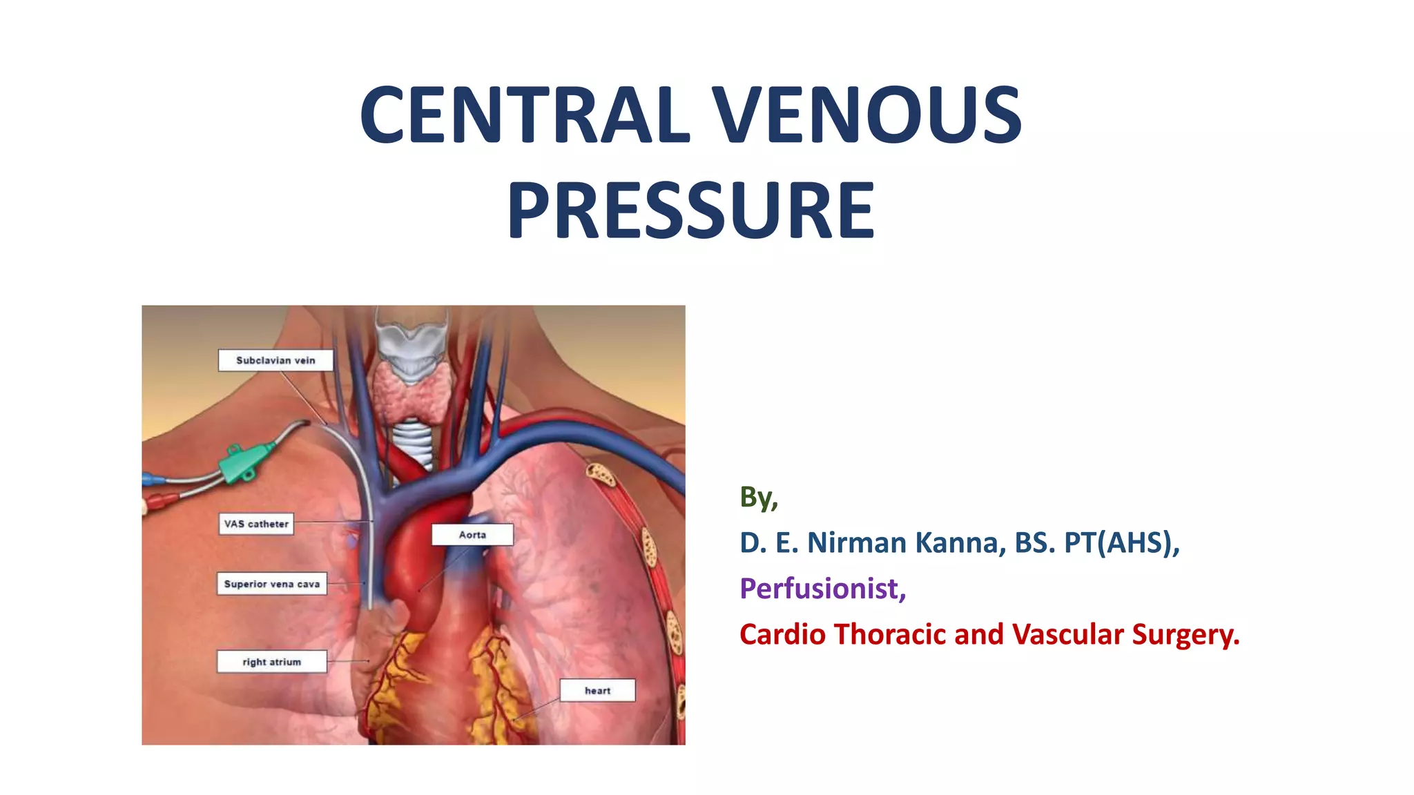
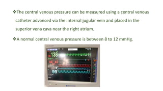
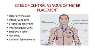
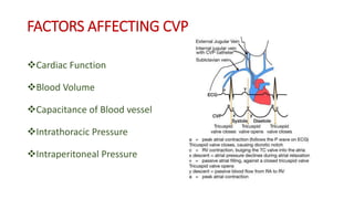
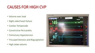
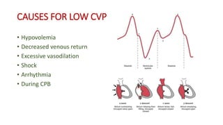
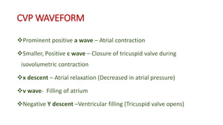
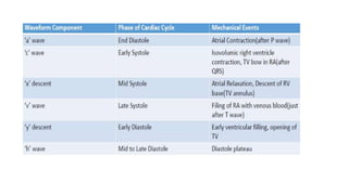
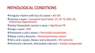
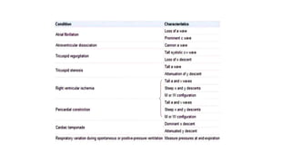
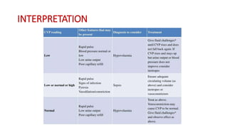
CENTRAL VENOUS
PRESSURE
By,
D. E. Nirman Kanna, BS. PT(AHS),
Perfusionist,
Cardio Thoracic and Vascular Surgery.
Central venous pressure is defined as the measure of pressure in the vena cava,
can be used as an estimation of preload and right atrial pressure.
 CVP is also defined as pressure of blood returning to or filling in the Right
atrium (Filling pressure).
Central venous pressure is often used as an assessment of hemodynamic
status, particularly in the intensive care unit.
 Pressure in IVC and SVC = Pressure in Right Atrium
The central venous pressure can be measured using a central venous
catheter advanced via the internal jugular vein and placed in the
superior vena cava near the right atrium.
A normal central venous pressure is between 8 to 12 mmHg.
SITES OF CENTRAL VENOUS CATHETER
PLACEMENT
• Superior vena cava
• Inferior vena cava
• Brachiocephalic veins
• Internal jugular veins
• Subclavian veins
• Iliac veins
• Common femoral veins
FACTORS AFFECTING CVP
Cardiac Function
Blood Volume
Capacitance of Blood vessel
Intrathoracic Pressure
Intraperitoneal Pressure
CAUSES FOR HIGH CVP
• Volume over load
• Right sided heart failure
• Cardiac Tamponade
• Constrictive Pericarditis
• Pulmonary Hypertension
• Tricuspid Stenosis and Regurgitation
• High stoke volume
INTRAOPERATIVE CVP
CAUSES FOR LOW CVP
• Hypovolemia
• Decreased venous return
• Excessive vasodilation
• Shock
• Arrhythmia
• During CPB
CVP WAVEFORM
Prominent positive a wave – Atrial contraction
Smaller, Positive c wave – Closure of tricuspid valve during
isovolumetric contraction
x descent – Atrial relaxation (Decreased in atrial pressure)
v wave- Filling of atrium
Negative Y descent –Ventricular filling (Tricuspid valve opens)
PATHOLOGICAL CONDITIONS
Irregular rhythm with loss of a wave = AF/ Afl
Cannon a wave = Complete heart block, VT, VF, TS, RVH ,PS,
Pulmonary Hypertension
Early/ Holosystolic cannon v wave = Significant TR
Large v wave = RVF
Prominent a and v waves = Pericardial constriction
Steep x and y descents = Decreased venous return
Tall a and v waves, Steep x and y descents = RV ischemia
Dominant x descent, attenuated y descent = Cardiac tamponade
INTERPRETATION
EFFECT OF CPB IN CVP
• CVP should be maintained 8 to 12 mmHg (minimum 6mmHg should be
maintained)before Cardiopulmonary bypass.
• During bypass, CVP will be equal to zero or in negative value
• During Steep Trendelenburg position, CVP will be decreased and during reverse
Trendelenburg position CVP will be increased.
• After weaning from CPB, CVP should be maintained in normal value,
• if CVP is high, Blood volume should be reduced and vasodilator drugs can be
administered.
• If CVP is low, Volume should be added and vasoconstrictive drugs can be administered.
CENTRAL VENOUS PRESSURE.pptx

CENTRAL VENOUS PRESSURE.pptx

  • 1.
    CENTRAL VENOUS PRESSURE By, D. E.Nirman Kanna, BS. PT(AHS), Perfusionist, Cardio Thoracic and Vascular Surgery.
  • 2.
    Central venous pressureis defined as the measure of pressure in the vena cava, can be used as an estimation of preload and right atrial pressure.  CVP is also defined as pressure of blood returning to or filling in the Right atrium (Filling pressure). Central venous pressure is often used as an assessment of hemodynamic status, particularly in the intensive care unit.  Pressure in IVC and SVC = Pressure in Right Atrium
  • 3.
    The central venouspressure can be measured using a central venous catheter advanced via the internal jugular vein and placed in the superior vena cava near the right atrium. A normal central venous pressure is between 8 to 12 mmHg.
  • 4.
    SITES OF CENTRALVENOUS CATHETER PLACEMENT • Superior vena cava • Inferior vena cava • Brachiocephalic veins • Internal jugular veins • Subclavian veins • Iliac veins • Common femoral veins
  • 5.
    FACTORS AFFECTING CVP CardiacFunction Blood Volume Capacitance of Blood vessel Intrathoracic Pressure Intraperitoneal Pressure
  • 6.
    CAUSES FOR HIGHCVP • Volume over load • Right sided heart failure • Cardiac Tamponade • Constrictive Pericarditis • Pulmonary Hypertension • Tricuspid Stenosis and Regurgitation • High stoke volume
  • 7.
  • 8.
    CAUSES FOR LOWCVP • Hypovolemia • Decreased venous return • Excessive vasodilation • Shock • Arrhythmia • During CPB
  • 9.
    CVP WAVEFORM Prominent positivea wave – Atrial contraction Smaller, Positive c wave – Closure of tricuspid valve during isovolumetric contraction x descent – Atrial relaxation (Decreased in atrial pressure) v wave- Filling of atrium Negative Y descent –Ventricular filling (Tricuspid valve opens)
  • 11.
    PATHOLOGICAL CONDITIONS Irregular rhythmwith loss of a wave = AF/ Afl Cannon a wave = Complete heart block, VT, VF, TS, RVH ,PS, Pulmonary Hypertension Early/ Holosystolic cannon v wave = Significant TR Large v wave = RVF Prominent a and v waves = Pericardial constriction Steep x and y descents = Decreased venous return Tall a and v waves, Steep x and y descents = RV ischemia Dominant x descent, attenuated y descent = Cardiac tamponade
  • 13.
  • 15.
    EFFECT OF CPBIN CVP • CVP should be maintained 8 to 12 mmHg (minimum 6mmHg should be maintained)before Cardiopulmonary bypass. • During bypass, CVP will be equal to zero or in negative value • During Steep Trendelenburg position, CVP will be decreased and during reverse Trendelenburg position CVP will be increased. • After weaning from CPB, CVP should be maintained in normal value, • if CVP is high, Blood volume should be reduced and vasodilator drugs can be administered. • If CVP is low, Volume should be added and vasoconstrictive drugs can be administered.