
Recommended
More Related Content
What's hot
What's hot (20)
Similar to Hydatid Disease: Causative Organism, Life Cycle, Pathology, Diagnosis and Management
Similar to Hydatid Disease: Causative Organism, Life Cycle, Pathology, Diagnosis and Management (20)
More from Ankita Singh
More from Ankita Singh (20)
Recently uploaded
Recently uploaded (20)
Hydatid Disease: Causative Organism, Life Cycle, Pathology, Diagnosis and Management
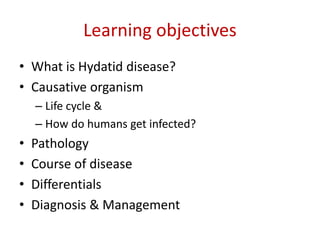
- 1. Learning objectives • What is Hydatid disease? • Causative organism – Life cycle & – How do humans get infected? • Pathology • Course of disease • Differentials • Diagnosis & Management
- 2. What is Hydatid disease? • Word hydatid means 'dew drop' (Latin). • In Greek it means 'watery vesicle‘
- 3. Cause • Echinococcus granulosus (EG)
- 4. Life cycle of EG
- 5. Pathology • It takes few years to evolve into a complete hydatid cyst. • Most commonly involved organ – Liver • Segment Vll • Commonly right lobe-66%; both lobes in 16% and only left lobe is involved in 17%. • Other organs involved- – Lung, muscles, bones – Rarely: kidneys, brain, spleen, heart
- 7. Characteristics • Hydatid fluid: – Clear – High specific gravity – Show hooklets & scolices • Pressure in the cyst is very high 70 mm of H20 – so leak and anaphylaxis becomes rapid when punctured. • Separated laminated membrane fall in the cyst causing "water lily sign" which is more often observed in lung hydatid but can be seen in liver also
- 9. Course of disease • The parasite may die and cyst eventually may get calcified. • Commonly cyst enlarges and is palpable per abdomen. • It may cause complications like jaundice due to pressure over biliary tree. • Rupture into the peritoneal cavity causes anaphylactic reaction – which may cause life-threatening shock • Rupture into biliary channels is commonest • Rupture into bowel, pleural cavity can occur.
- 10. Course of disease.. • Secondary infection causing suppuration and septicaemia. • Secondary cysts in the lung, spleen, mesentery, retroperitoneum and other organs can occur. • Hepatic dysfunction. • Disseminated abdominal hydatidosis can occur after silent rupture.
- 11. Clinical features • Asymptomatic palpable liver with classical thrill- m.c. • Jaundice and Pain • Features of anaphylaxis • Discomfort in right upper quadrant area; dyspepsia; hydatid cachexia in children; weight loss; fatigue; vomiting. • Occasionally splenomegaly, pleural effusion, cholangitis, allergic asthma, fever.
- 12. Confused with ? • Hepatoma • Amoebic liver abscess • Cystic disease of liver
- 13. Complications • Rupture – Anaphylactic reaction • Obstructive jaundice • Infection • Calcification • Liver failure • Sepsis • Death
- 14. Diagnosis • Ultrasound is diagnostic – rosettes of daughter cysts, – double contoured membrane of the cyst due to detachment of the cyst membranes, and – calcification of cyst wall
- 18. Diagnosis.. • CECT abdomen: – more accurate in identifying cyst characteristics- • cart wheel like- multivesicular rosette like
- 19. Diagnosis.. • X-ray often shows calcification • Primary serological tests- – ELISA; indirect haemagglutination test; latex agglutination test; immunofluorescence antibody test; immunoelectrophoresis. – 80-95% sensitivity for liver hydatid.
- 20. Diagnosis.. • Secondary lab tests- – Detection of precipitation line arc 5 in immunoelectrophoresis is most specific and virtually diagnostic; – immunoblotting; – Polymerase chain reaction (PCR) • Liver function tests-altered in 20% cases. • Casoni's test (intradermal test-75% sensitive); • Complement fixation test
- 21. Diagnosis.. • MRI when there is jaundice – to visualise biliary tree and its relation to hydatid cyst; – to find out cystobiliary communication; – biliary hydatids in bile duct and hepatic ducts. • ERCP can also be done to find out the communications. • Other method to find out the cystobiliary communications is intraoperative cholangiogram.
- 22. Treatment.. Medical therapy • Indications: – 10 days prior to intervention and to continue it for 1 month to 3 months after the intervention – Inoperable cysts; – Multiple or multiorgan cysts – Recurrent hydatids; – Surgically unfit patients – Cysts in lungs
- 23. Treatment.. Medical therapy • Contraindications: – Large cysts; – Honeycomb cysts (with septae) – Infected cysts; – Calcified cysts – Pregnancy
- 24. Treatment.. Medical therapy • Drugs used: – Albendazole – Praziquintal – Mebendazole • Side effects??
- 25. Treatment.. • Surgical therapy – PAIR – Open /lap surgery
- 27. Treatment