
Recommended
More Related Content
What's hot
What's hot (20)
Similar to AICHE Regional Conference Poster
Similar to AICHE Regional Conference Poster (20)
Recently uploaded
Recently uploaded (20)
AICHE Regional Conference Poster
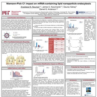
- 1. 1Department of Chemical Engineering, Massachusetts Institute of Technology, Cambridge MA, 2Koch Institute for Integrative Cancer Research, Cambridge MA, 3Department Of Pharmaceutical Sciences, College of Pharmacy, Oregon State Univ. Portland OR Niemann-Pick C1 impact on mRNA-containing lipid nanoparticle endocytosis Anastasia N. Neuman1,2, James C. Kaczmarek1,2, Gaurav Sahay3 Daniel G. Anderson1,2 Lipid Nucleic Acid Delivery Numerous diseases, including cancer, heart disease, diabetes, and the common cold, are brought on by aberrant protein expression. Delivery of nucleic acids to control such aberrant protein expression represents a promising therapeutic strategy to treat disease at its source, but nucleic acids typically require the use of delivery vehicles to be useful in a therapeutic context. Lipid nanoparticles (LNPs), in particular, have been shown to effectively deliver nucleic acids such as siRNA and mRNA both in vivo and in vitro1. Highly potent materials have been developed through the use of high throughput screening techniques; however the cellular processes that determine their effectiveness remain unclear2. mRNA Delivery with LNPs REFERENCES Approach This work was supported by Shire Pharmaceuticals, the MIT Koch Institute for Integrative Cancer Research, and the C. Michael Mohr Scholarship for Undergraduate Research. siRNA Delivery with LNPs3 mRNA Modification Impact on Efficacy Conclusions Potential for Future Studies ACKNOWLEDGMENTS Fig. 1: Nucleic acids encapsulated by a stable lipid nanoparticle (LNP) formulated with a cationic lipid, phospholipid, PEG, and cholesterol1. NPC1 Facilitated Endocytic Recycling When delivered with an LNP optimized for mRNA delivery, a lower efficacy was observed in NPC1-deficient cells than normal cells. Upon delivery with the original LNP used in the study involving siRNA, the original pattern was observed, with higher efficacy in NPC1-deficient cells than normal cells. This pattern suggests that the formulation changes impact the subcellular trafficking of the LNPs, with the original particle likely endocytosed through macropinocytosis and the optimized particle using an NPC1-dependent pathway. Formulation Modification Impact on Efficacy Changing the PEG percentage from 2.5% to 1.5% results in the particles being most effective in NPC1-/- cells, as observed with the original formulation. PEG reduces LNP aggregation and prevents nonspecific endocytosis by immune cells1. Reducing the PEG percentage may allow the particle to use multiple endocytotic pathways, and reduce reliance on NPC1 for cell entry. Changing the phospholipid from DOPE to DSPC also reversed the pattern. DOPE has a primary amine headgroup and and a tail with one degree of unsaturation while DSPC has a quaternary amine headgroup and fully saturated tail2. Conical phospholipids like DOPE adopt a hexagonal phase with lower stability while cylindrical phospholipids like DSPC adopt a more stable lamellar phase2. The unstable hexagonal phase of DOPE may promote endosomal escape. The more stable lamellar phase of DSPC inhibits endosomal escape and would depend on NPC1 deficiency to allow more LNPs to accumulate in the endosomes. Unlike the luciferase mRNA provided by Shire Pharmaceuticals used in the prior experiments, unmodified luciferase mRNA, luciferase mRNA 5-methylcytosine (5mc) and luciferase mRNA purchased from Tri-Link with both and 5- methylcytosine and psuedouridine (5mc psu) show higher efficacy in NPC1- deficient cells than normal cells in both the original and the optimized LNP. This could be due to differences in mRNA folding driven by these modifications. 1. K. J. Kauffman, M. J. Webber, D. G. Anderson, Journal of Controlled Release, 2016, 240, 227-234. 2. K. J. Kauffman, J. R. Dorkin, J. H. Yang, M. W. Heartlein, F. DeRosa, F. F. Mir, O. S. Fenton, D. G. Anderson, Nano Lett. 2015, 15, 7300–7306. 3. G. Sahay, W. Querbes, C. Alabi, A. Eltoukhy, S. Sarkar, C. Zurenko, E. Karagiannis, K. Love, D. Chen, R. Zoncu, Y. Buganim, A. Schroeder, R. Langer, D. G. Anderson, Nature Biotechnology, 2013, 31, 653-658. 4. E. Lloyd-Evans, A. J. Morgan, X. He, D. A. Smith, E. Elliot-Smith, D. J. Sillence, G. C. Churchill, E. H. Schuchman, A. Galione, F. M. Platt, Nature Medicine, 2008, 14, 1247- 1255. In conclusion, our work has demonstrated the following: • The PEG percentage and phospholipid of mRNA-loaded LNPs may impact the trafficking method. • Modifications in mRNA may impact trafficking of mRNA-loaded particles. • Use imaging to observe how changes in LNP formulation and mRNA modifications affect the trafficking of particles. • Optimize an mRNA-loaded LNP for treatment of Neimann Pick Type I Disease, the disease after which NPC1 is named. We hypothesize that differences in subcellular trafficking may account for some of the observed differences in efficacy between different nanoparticle formulations and nucleic acid cargos. As such, the two main goals of this project are as follows: 1) Observe the effect of LNP formulation differences on mRNA trafficking and delivery 2) Observe the effect of mRNA modifications on trafficking and delivery These experiments were conducted using luciferase mRNA. 24 hours following in vitro transfection of wild-type and NPC1 knockout mouse embryonic fibroblast (MEF) cells (100 ng mRNA/well), the cells were lysed and luciferase expression was quantified using luminescence. Fig. 2: LNP trafficking in i) NPC1+/+ cells and ii) NPC1-/- cells3. One cellular process known to impact the effectiveness of siRNA-loaded LNPs is endocytic recycling. siRNA-loaded LNPs often enter cells through macropinocytosis and the majority are directed to late endosomes. The siRNA must escape these endosomes for gene silencing to occur. In normal cells, most particles are recycled through either transport to the ER-Golgi route or direct fusion by the endosome to the plasma membrane3. In NPC1-deficient cells the LNPs are not recycled and accumulate in the late endosomes, allowing siRNA to continuously escape3. Thus, NPC1-deficient cells show increased gene silencing of the target gene3. Original formulation Optimized formulation C12:200:mRNA weight ratio 5:1 10:1 phospholipid DSPC DOPE C12-200 molar composition 50% 35% phospholipid molar composition 10% 16% cholesterol molar composition 38.5% 46.5% C14 PEG 2000 molar composition 1.5% 2.5% Table 1: Original and Optimized LNP Formulations2 Previous work has shown that siRNA-loaded lipid nanoparticles are recycled back out of a cell following endocytosis, rendering a large proportion of the delivered dose ineffective3. This recycling is dependent on the presence of Niemann-Pick type C1 (NPC1), a protein involved in cellular cholesterol trafficking4. Without NPC1, the recycling pathways of the late endosomes are largely inhibited. Here, we see that AF647 siRNA delivery and gene silencing in NPC1-deficient cells is increased over that in wild-type cells3. These siRNA experiments were done using a C12-200 based lipid formulation, which is here referred to as the “original particle”. Experimental design has resulted in a particle optimized for mRNA delivery, here referred to as the “optimized particle” (Table 1). The optimized particle was found to perform no better for delivery of siRNA2, and the physical basis for the difference in efficacy is unknown. cationic lipid phospholipid polyethylene glycol (PEG) cholesterol Fig. 3: Gene silencing in wild-type and NPC1 deficient cells3. Uridine Pseudouridine e Cytidine 5-methylcytidine Fig. 7: mRNA base modifications. i) ii) Fig. 4: luciferase mRNA delivery by LNPs in wild-type and NPC1 deficient cells. Fig. 5: Impact of formulation changes on luciferase mRNA delivery in wild-type and NPC1-deficient cells. The formulation of the LNPs was modified by one factor at a time from the optimized formulation to the original formulation. The optimized and original formulations can be found in Table 12. Fig. 6: mRNA modification impact on luciferase mRNA delivery in wild-type and NPC1-deficient cells.