
Recommended
More Related Content
What's hot
What's hot (20)
Similar to amgen poster
Similar to amgen poster (20)
Recently uploaded
Recently uploaded (20)
amgen poster
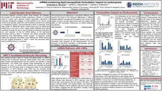
- 1. Conclusions 1Department of Chemical Engineering, Massachusetts Institute of Technology, Cambridge MA, 2Koch Institute for Integrative Cancer Research, Cambridge MA mRNA-containing lipid nanoparticle formulation impact on endocytosis Anastasia N. Neuman1,2, James C. Kaczmarek1,2, Daniel G. Anderson1,2 Lipid Nucleic Acid Delivery Numerous diseases, including cancer, heart disease, and diabetes, are brought on by aberrant protein expression. Delivery of nucleic acids to control such aberrant protein expression represents a promising therapeutic strategy to treat disease at its source, but nucleic acids typically require the use of delivery vehicles to be useful in a therapeutic context. Lipid nanoparticles (LNPs), in particular, have been shown to effectively deliver nucleic acids such as siRNA and mRNA both in vivo and in vitro1. Highly potent materials have been developed through the use of high throughput screening techniques; one such example of this for mRNA delivery is the optimized formulation found in Table 12. However, the cellular processes that determine their effectiveness remain unclear2. In particular, what makes the optimized formulation more effective for mRNA delivery than the original formulation in Table 1, which has previously been used for siRNA delivery3, is unknown. mRNA Delivery with LNPs Approach This work was supported by RaNA Therapeutics, the MIT Koch Institute for Integrative Cancer Research, and the Amgen Foundation. Formulation Impact on Endocytosis Fig. 1: Nucleic acids encapsulated by a stable lipid nanoparticle (LNP) formulated with a cationic lipid, phospholipid, PEG, and cholesterol1. NPC1 Facilitated Endocytic Recycling When delivered with an LNP optimized for mRNA delivery, a lower efficacy was observed in NPC1-deficient cells than normal cells. Upon delivery with the original LNP used in the study involving siRNA3, the original pattern was observed, with higher efficacy in NPC1-deficient cells than normal cells. This pattern suggests that the formulation changes impact the subcellular trafficking of the LNPs. To further investigate the impact LNP formulation has on endocytosis, LNPs were delivered to cells treated with an inhibitors of endocytotic pathways and the efficacy was observed. U18666A, a drug known to inhibit cholesterol trafficking5, is shown to decrease efficacy only in the optimized particle. Efficacy significantly increases in both the original and original PEG % formulations, suggesting that the change in PEG % may cause the optimized particle’s reliance on the cholesterol trafficking pathway. 1. K. J. Kauffman, M. J. Webber, D. G. Anderson, Journal of Controlled Release, 2016, 240, 227-234. 2. K. J. Kauffman, J. R. Dorkin, J. H. Yang, M. W. Heartlein, F. DeRosa, F. F. Mir, O. S. Fenton, D. G. Anderson, Nano Lett. 2015, 15, 7300–7306. 3. G. Sahay, W. Querbes, C. Alabi, A. Eltoukhy, S. Sarkar, C. Zurenko, E. Karagiannis, K. Love, D. Chen, R. Zoncu, Y. Buganim, A. Schroeder, R. Langer, D. G. Anderson, Nature Biotechnology, 2013, 31, 653-658. 4. E. Lloyd-Evans, A. J. Morgan, X. He, D. A. Smith, E. Elliot-Smith, D. J. Sillence, G. C. Churchill, E. H. Schuchman, A. Galione, F. M. Platt, Nature Medicine, 2008, 14, 1247-1255. 5. A. Eltoukhy, G. Sahay, J. M. Cunningham, D. G. Anderson, ACS Nano, 2014, 8, 7905-7913. The change in PEG % and change in phospholipid may be the cause of the efficacy differences between the original and optimized LNPs. In particular, the change in PEG % may be involved in the optimized LNPs apparent dependency on NPC1. Future studies will involve FACS analysis to determine the effect of these inhibitors on LNP uptake into cells and investigation into the cause of efficacy differences seen with inhibitors other than U18666A. We hypothesize that differences in subcellular trafficking may account for some of the observed differences in efficacy between different nanoparticle formulations and nucleic acid cargos. As such, the main goal of this work is to observe the effect of LNP formulation differences on mRNA trafficking and delivery. For experiments involving endocytosis inhibitors cells were pretreated with inhibitors (5 𝜇M chlorpromazine, 50 𝜇M dynasore, 10 𝜇M EIPA, 5 𝜇M filipin, 10 𝜇M genistein, and 5 𝜇 M U18666A) 1 hour prior to transfection. Fig. 2: LNP trafficking in i) NPC1+/+ cells and ii) NPC1-/- cells3. One cellular process known to impact the effectiveness of siRNA- loaded LNPs is endocytic recycling. siRNA-loaded LNPs often enter cells through macropinocytosis and the majority are directed to late endosomes. The siRNA must escape these endosomes for gene silencing to occur. In normal cells, particles are recycled through transport to the ER-Golgi route or endosomal fusion to the plasma membrane3. In NPC1-deficient cells LNPs are not recycled and accumulate in the late endosomes, allowing siRNA to continuously escape3. Thus, NPC1-deficient cells show increased gene silencing of the target gene3. Original formulation Optimized formulation C12:200:mRNA weight ratio 5:1 10:1 phospholipid DSPC DOPE C12-200 molar composition 50% 35% phospholipid molar composition 10% 16% cholesterol molar composition 38.5% 46.5% C14 PEG 2000 molar composition 1.5% 2.5% Table 1: Original and Optimized LNP Formulations2 cationic lipid phospholipid polyethylene glycol (PEG) cholesterol i) ii) Fig. 3: luciferase mRNA delivery by LNPs with two different formulations in wild-type and NPC1 deficient cells (mean ± SD, n=4); *** indicates p < .001. Fig. 4: Impact of formulation changes on luciferase mRNA delivery in wild- type and NPC1-deficient cells (mean ± SD, n=4); * indicates p < 0.05, ** indicates p < .01, *** indicates p < .001. Fig. 5:Endocytosis inhibitor impact on transfection efficiency of four different LNP formulations (mean ± SD, n=4); * indicates p < 0.05, ** indicates p < .01, *** indicates p < .001, compared to no inhibitor control. The formulation of the LNPs was modified by one factor at a time from the optimized formulation to the original formulation. The optimized and original formulations can be found in Table 12. Changing the PEG percentage from 2.5% to 1.5% and changing the phospholipid from DOPE to DSPC results in the particles being most effective in NPC1-/- cells, as observed with the original formulation. ACKNOWLEDGEMENTS REFERENCES In vitro transfectionLNP formulation Luciferase assay