2. Introduction
• Hepatocellular carcinoma (HCC) is the sixth most common
type of cancer and the fourth leading cause of cancer-related
deaths worldwide.
• The median survival time of patients with intermediate and
advanced HCC is less than 6 months, with a poor prognosis.
• Treatment regimen mainly includes surgery and systemic
therapy, but, patients with advanced HCC are often not
candidates for surgery.
• Many patients discontinue treatment due to significant side
effects, including hypertension, decreased weight, palmar-
plantar erythrodysesthesia, drug resistance, and poor
tolerance.
3. • Curcumin (CUR) is derived from turmeric and has been
used for centuries for its medicinal properties.
• It has various biological activities, such as antioxidant,
apoptotic and anti-inflammatory effects.
• Studies have indicated the role of CUR in cancer
suppression in gastric cancer, cervical cancer, HCC, and
so forth. (Najafi et al, 2020; Li et al 2020)
• It has been demonstrated that CUR can cause:
- DNA damage
- Cell cycle arrest at G2/M phases in HeLa cells.
- Apoptosis by enhancing reactive oxygen species (ROS)
in HepG2 cell.
- Regulation of the TGF-β1/Smad3 signalling pathway.
4. • Resveratrol (RSV) is a natural antioxidant polyphenolic
compound found in grapes, peanuts, and berries.
• Recent studies have suggested that RSV can prevent or
inhibit breast, colorectal, pancreatic, prostate, and liver
cancers. (Amini et al, 2021;Xue et al, 2014)
• RSV has been known for the following:
- Inhibits breast cancer cell proliferation by regulating the
STAT3 signaling cascade.
- Induce malignant hepatic tissue apoptosis by activating
caspase-3.
- Upregulating the ratio of Bax/Bcl-2
- Increasing p53 expression
5. • Since the cardio-, hepato-, nephro- and neuro-protective
properties of CUR and RSV offer promising results for
anti-cancer therapy for HCC.
• But these crude drugs have low water solubility, poor
chemical stability, and poor bioavailability, thus
limiting their clinical application.
• The above-mentioned problems can be solved by
nanotechnology-driven drug delivery systems (DDSs).
• 1,2-Distearoyl-sn-glycero-3-phosphoethanolamineN-
[maleimide (polyethylene glycol)-2000] (DSPEPEG2000-
Mal), (FDA approved), is a conjugated polymer widely
used in DDSs that contains hydrophilic PEG and
hydrophobic DSPE.
6. • It can be used as auxiliary material to functionalize various
DDSs,
• PEG prolongs the blood circulation time of DDSs and
DSPE is used as the hydrophobic structure of NPs for
efficient drug loading.
• Hence, encapsulating these therapeutic drugs in NPs for
cancer treatment could improve drug accumulation in
tumors via the enhanced permeability and retention (EPR)
effect.
• To further enhance the tumor targeting ability, the NP
surface was decorated with SP94 via a maleimide-thiol
reaction. This kind of NP possesses the characteristics of
higher drug loading, sustained drug release, and colloidal
stability.
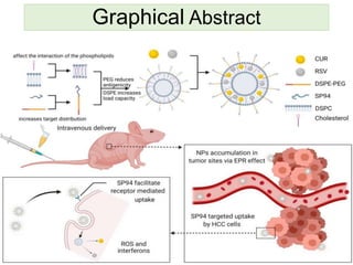
7. 1. The rational designing of, a systemically injectable
HCC-targeted NPs loaded with CUR and RSV and
DSPE-PEG2000 decorated with SP94.
2. To further enhance the tumor targeting ability, and
characteristics of higher drug loading, sustained drug
release and colloidal stability.
3. To check the potential synergistic effect of drugs as
combination therapy on HCC cells and in animal models.
Aims and Objectives
8. Fig. 1 Schematic figure of depicting major materials and methods in current
study.
9. • Cell culture: The human hepatoma cell line HepG2 was
used for cell culture experiments.
• Animals: Male 4–5-week-old BALB/c mice were used by
for all the animal experiments after approval from
institutional ethics committee.
• Synthesis of the SP94-modifed lipid DSPE-PEG2000
(DSPE-PEG2000-SP94): DSPE-PEG2000-SP94 was
prepared via a coupling reaction between a maleimide
group on DSPE-PEG2000- Mal and a cysteine residue in
SP94. Analytical high-performance liquid chromatography
(HPLC) was adopted to monitor the completion of the
reaction.
Methods
10. • Preparation of NPs loaded with CUR+RSV: The
nanoprecipitation method was adopted to prepare
CUR+RSV-loaded polymeric NPs. The NPs were
prepared with RSV, CUR, linker lipid (DSPE-PEG2000 or
DSPE-PEG2000-SP94), DSPC and cholesterol.
• Particle morphology study TEM: Morphological
examination of the NPs was performed by TEM.
• Particle size and zeta potential: The particle sizes and
zeta potentials of the NPs were determined using a
Malvern Nano-ZS90 instrument at 25 °C. The changes in
particle size over seven days were also measured by the
same method.
11. • Determination of encapsulation efficiency (EE): EE
was measured by UV spectrophotometry, concentration
curves were generated with measurements from a UV–
VIS spectrometer:
EE (%) = W(total drug in NP) − W(free drug in NP) / W
(total drug in NP) × 100%
• In vitro release kinetics study and solubility
changes: An in vitro release kinetics study was
conducted utilizing the dialysis method in phosphate
buffered-saline (PBS; pH 7.4) to quantify the drug
release behavior.
• The amounts of CUR and RSV released were measured
using a UV–Vis spectrometer.
12. • A small animal imaging instrument was used to detect the
radiant efficiency of equal masses of CUR and RSV in
PBS. The excitation wavelength was 425 nm, and the
emission wavelength was 530 nm.
• Cell-specific uptake of NPs: The cells were
counterstained with SYTO 63 (5 μL/mL) for 30 min. The
cells were observed and photographed using inverted-
type laser scanning confocal microscopy (LSCM).
• In vitro cell apoptosis studies: HepG2 cells were
seeded at 10,000 cells/well. All cells were treated with
RSV, CUR, CUR+RSV (5:1 molar ratio), NP-CUR+RSV
and SP94-NP-CUR+RSV. The concentrations of CUR and
RSV varied from 1 to 500 μM. MTT assay was performed.
13. • For combination treatments, combination index (CI)
analysis was conducted using CompuSyn software
version.
• Detection of reactive oxygen species (ROS):
Dichlorofuorescein diacetate (DCFH-DA) staining was
used to detect intracellular ROS production. Fluorescence
was quantified with a confocal microscope at 488ex
nm/525em nm.
• For ROS inhibition, all cells were pre-treated with N-
acetylcysteine (NAC; ROS scavenger) (2.5 mmol for
2 h), followed by drug treatment.
14. • Detection of caspase-3 activity: caspase-3 activity was
determined by measuring the absorbance of specific
chromogenic substrates according to the manufacturer’s
instructions. Moreover, in another group, the cell-
permeable pan-caspase inhibitor Z-Val-Ala-Asp-CH2F (Z-
VAD-FMK) (20 μmol for 30 min) was used to inhibit the
production of caspase-3 before drug treatment.
• Antitumor efficacy evaluation in vivo: Four-week-old
male BALB/c nude mice were subcutaneously implanted
with HepG2 cells.
• After the tumor sizes grew to be visible, all mice were
randomly divided into four groups: PBS, CUR+RSV, NP
and SP94-NP. The mice were gavaged with 10 mg/kg of
each compound. The treatment lasted for 21 days.
15. • The mice were sacrificed on Day 21 post-treatment,
tumour tissue sections were frozen and TUNEL assay
(terminal deoxynucleotidyl transferase-mediated dUTP
nick end labelling) was performed.
• Additionally, Ki-67 and haematoxylin and eosin H&E
immunohistochemistry was also carried out.
• Toxicity and biosafety of the NPs in vivo: Blood
samples were obtained on Day 21 to detect the ALP,
BUN, LDH and AFP levels were measured based on the
manufacturer’s instructions.
16. • In vivo tumor targeting and NPs distribution: 12, 24
and 48 h after drug administration the mice were
sacrificed. The hearts, livers, spleens, lungs, kidneys and
tumors of the xenograft model mice were collected, and
time-dependent drug distribution in the tumor tissues and
other major organs was detected using Calliper IVIS
Lumina II in vivo imaging system.
17. 1. Synthesis and characterization of DSPE-PEG2000-
SP94 loaded with CUR and RSV:
• CUR+RSV+NP and CUR+RSV+NP+DSPE-PEG2000-
SP94 constructed the targeted NPs by nanoprecipitation.
Referred to as NP and SP94-NP in Figure 1a.
Results
1 (a.)
18. • Through Electron microscopy, morphology, particle sizes
and zeta potentials of NPs were characterized, Fig. 1b–d.
EPR effect significantly impacts the penetration of NPs into
solid tumors, especially NPs below 200 nm. Therefore, the
NPs had a suitable size to aggregate at tumor sites.
1 (b.)
1 (c.)
1
19. • The long-term stability of the NPs was evaluated by storing
the NP and SP94-NP solutions at room temperature for one
week, and both NPs displayed little change in particle size
Fig. 1 e.
1
20. 2. Kinetics of CUR and RSV release
from the drug-loaded NPs:
• Next, the encapsulation
efficiency (EE), drug release
kinetics, and solubility changes
were evaluated.
• The amounts of CUR and RSV
released from the NPs by
dialysis are shown in Fig. 2f
(NPs), g (SP94 NP).
2
2
21. • A small animal imaging system was used to evaluate the
change in drug solubility in water after nanomaterial
encapsulation, and it was found that the nano-system
significantly enhanced the solubility of the drugs Fig. 2i, j.
• Although the EEs of CUR and RSV were both greater than
80%, the EE for CUR was slightly larger than that of RSV
Fig 2h.
23. 3. Intracellular assessment of the cellular uptake
of SP94-NP:
• The uptake of the drug-loaded NPs by HepG2 cells was
assessed using laser scanning confocal microscopy
(LSCM).
• After treatment with PBS, CUR+RSV, NP and SP94-NP and
SYTO 63, strong red fluorescence from SYTO 63 was
observed.
• However, in NP-treated cells, the fluorescence signal was
punctate in the cytoplasm and negligible in the nucleus
Fig. 3a, b.
25. 4. Synergistic effects of CUR and RSV and cell viability
in vitro:
• The IC50 values of RSV, CUR, CUR+RSV, NP and SP94-
NP were identified. The combination indices (CIs) of the two
combined RSV and CUR treatments are listed in Fig. 3d.
• The results showed a sequential decrease Fig. 3c, which
illustrated that CUR and RSV had synergistic effects with a
lower dosage.
• Figure 3e shows that the blank NPs had almost no toxicity
while the drug-loaded NPs had clear inhibitory effects on
cell viability after 1.5 hrs.
27. 5. Evaluation of the therapeutic mechanism
of the cytotoxicity against HCC cells in vitro:
• Cancer cell apoptosis via NPs inducing the production of
ROS and activating a cascade of caspase-3 in HepG2
cells was checked.
• The cell-generated ROS were examined by DCFH-DA
staining. As shown in Fig. 4a, b, HepG2 cells produced
significant ROS after NPs treatment, as demonstrated by
the observed bright green fluorescence.
• The apoptosis of cells pretreated with N-acetylcysteine
(NAC) was further analyzed to elucidate the effects of ROS
on apoptosis, and the green fluorescence was greatly
reduced.
28. • Apoptosis carried out by activation of cascade of caspases 3
(cysteine aspartate-specific proteinases) was also checked.
• NPs treatment significantly activated caspase-3 in HepG2
cells.
• Furthermore, the caspase-3 inhibitor Z-Val-Ala-Asp-CH2F (Z-
VADFMK) completely inhibited NP-induced apoptosis of
HepG2 cells Fig. 4c.
• In addition, NP-induced caspase-3 activity decreased under
NAC pretreatment Fig. 4c.
• Moreover, the cell viability of each group after treatment was
determined, and the related pathway by which NPs kill HepG2
cells was verified Fig. 4d.
29.
30. 6. In vivo tumor targeting and NPs distribution:
• Preferential drug aggregation in the tumors of BALB/c xenograft-
bearing nude model mice was checked.
• Distribution was observed by the fluorescence signals with an
imaging system at 12 h, 24 h, and 48 h. Fig. 5a–d
31. 7. In vivo evaluation of antitumor activity:
• In particular, targeted SP94-NP therapy resulted in more
significant tumor suppression than nontargeted NP therapy
Fig. 7e.
• Conversely, the weights of the mice treated with NPs were
remarkably higher Fig 7f.
• Examinations of blood samples from each group Fig.7g
showed that there were no significant changes in ALP, BUN,
or LDH.
• AFP is the most commonly used marker for liver cancer,
and its level was also relatively low in the NP treatment.
32.
33. • Hematoxylin and eosin (H&E) staining analysis
demonstrated that NPs treatment led to intratumoral necrosis
Fig. 8 a.
• H&E staining of the heart, liver, spleen, lung, and kidney
tissues displayed no obvious abnormalities or organ
damage, which further illustrated their efficacy and safety.
34. • Ki-67 and terminal deoxynucleotidyl transferase-mediated
dUTP nick end labelling (TUNEL) immunofluorescence
assays further confirmed the therapeutic effects of the NPs
8b–d.
35. • A new formulation was created to alleviate the
disadvantages of low water solubility, weak chemical
stability, and poor bioavailability of CUR and RSV while
enhancing their therapeutic potential against HCC with a
good safety profile.
• CUR and RSV were rationally designed based on their
shortcomings and potential anticancer activities and
assembled with the biocompatible matrix DSPE-PEG2000.
• The particle surfaces were decorated with HCC-specific
ligands (peptide SP94).
• The hardening of the liposome membrane caused by CUR
and RSV promoted the prolonged residence time of the
drugs in the NPs reservoir, thereby hindering their release
into systemic circulation.
Discussion
36. • The targeting peptide has high selectivity for cancer cells
without affecting normal cells and tissues.
• Therefore, by properly exploiting the EPR effect and the
accumulation of active NPs, the concentration of the
therapeutic drugs at tumor target site increased.
• Therefore, it has been speculated that because many
traditional chemotherapeutic drugs have substantial side
effects, this nanoplatform can be used to minimize these
side effects while improving their therapeutic effects.
Conclusion
37. • The in vitro experiments have been performed using
single cell line only.
• The NP drug treatment studies on animals have been
performed over a period of 21 days. The toxicity profile
should have been evaluated for longer period of time.
Limitations
38. • Measurement of in vivo pharmacokinetic and
pharmacodynamics parameters can be checked like:
- Compound absorption/elimination dynamics following
different routes of administration; Metabolism/
biotransformation, compound excretion dynamics
(urine/faeces, bile) etc.
- This will help to predict clinical response in humans, and
to facilitate a better understanding about the potential
clinical relevance of the designed drug.
Future Directions
39.
40. • Najaf M, Mortezaee K, Rahimifard M, Farhood B, Haghi-Aminjan
H. The role of curcumin/curcuminoids during gastric cancer
chemotherapy: a systematic review of non-clinical study. Life Sci.
2020;257: 118051.
• Li J, Wei H, Liu Y, Li Q, Guo H, Guo Y, et al. Curcumin inhibits
hepatocellular carcinoma via regulating miR-21/TIMP3 axis. Evid
Based Complement Altern Med. 2020;2020:2892917.
• Xue YQ, Di JM, Luo Y, Cheng KJ, Wei X, Shi Z. Resveratrol
oligomers for the prevention and treatment of cancers. Oxid Med
Cell Longev. 2014;2014: 765832. 15.
• Amini P, Nodooshan SJ, Ashrafzadeh M, Eftekhari SM, Aryafar T,
Khalaf L, et al. Resveratrol induces apoptosis and attenuates
proliferation of MCF-7 cells in combination with radiation and
hyperthermia. Curr Mol Med. 2021;21(2):142–50
Cross-References