1. Brief Communication
Ascending Aortic Slide for Interrupted
Aortic Arch Repair
Miguel Urencio, MD1
, Ali Dodge-Khatami, MD, PhD2
,
Chris E. Greenleaf, MD1
, Giorgio Aru, MD1
, and Jorge D. Salazar, MD2
Abstract
For repair of interrupted aortic arch, unfavorable anatomy challenges a tension-free anastomosis. We describe a useful alternative
surgical technique used in five neonates/infants, involving splitting the ascending aorta from the sinotubular junction to the arch
origin, leftward and posterior ‘‘sliding’’ of the flap with anastomosis to the distal arch creating a native tissue bridge, and
reconstruction with a patch. With wide interruption gaps between proximal and distal aortic portions, the ascending aortic slide is
a safe and reproducible technique, providing a tension-free native tissue bridge with potential for growth, and a scaffold for patch
augmentation in biventricular hearts, or for Norwood stage I in univentricular palliation.
Keywords
interrupted aortic arch, aortic arch, CHD (congenital heart disease)
Submitted February 02, 2016; Accepted May 14, 2016.
Background
Surgical repair is the definite treatment for interrupted aortic
arch (IAA). Standard techniques include direct aorta-to-aorta
anastomosis, requiring extensive mobilization of the descend-
ing aorta to reach the ascending aorta. The use of an anterior
patch at the anastomosis to augment the aortic arch serves to
reduce anastomotic tension and may reduce the incidence of
restenosis1
and tracheobronchial compression. When residual
or recurrent aortic arch obstruction occurs,2
angioplasty or reo-
peration may be required if angioplasty is unsuccessful.3
In difficult situations, when the gap between the proximal
and the distal portions of the arch is too far apart and a direct
native tissue anastomosis is not deemed possible, the use of an
interposition graft to restore continuity between the ascending
and the descending aorta may be necessary. The major draw-
back is the inability of the graft to grow with the patient, uni-
versally requiring a reoperation.3
Left bronchial compression is
also not a uncommon and troublesome complication seen after
direct anastomosis or prosthetic graft interposition.4
Native tissue-to-tissue reconstruction is always preferred
when possible to preserve the potential for growth of the repair.
Using a technique where the ascending and descending aortic
segments are incised longitudinally and anastomosed in a side-
to-side fashion, Jacobs et al incorporated the aortic branches in
the anastomosis with good results.5
The subclavian rotational
flap is another alternative, where an end-to-end anastomosis
between the subclavian artery and ascending or descending
aorta (Blalock-Park procedure) is performed. Although this
option uses autologous tissue, it has the great disadvantage of
sacrificing a major artery to the upper extremity.6
Dr William I. Norwood long ago proposed repairing IAA
using entirely autologous tissue to constitute the greater curve
of the reconstructed arch, together with a vascular homograft
patch to augment the lesser curve. During the first case
described in the present series, one of our coauthors who had
seen Dr Norwood perform arch repair decades ago suggested it
as an alternative. We have selectively needed the technique in
five neonates/infants with IAA and a wide interaortic interrup-
tion gap (Figure 1), suggest calling it an ascending aortic slide,
and review our initial experience.
Materials and Methods
This is an observational and retrospective study. After approval
by the institutional review board, we reviewed the Pediatric and
Congenital Heart Surgery database and reviewed patients who
1
Division of Cardiothoracic Surgery, University of Mississippi Medical Center,
Jackson, MS, USA
2
Children’s Heart Center, University of Mississippi Medical Center, Jackson,
MS, USA
Corresponding Author:
Ali Dodge-Khatami, Children’s Heart Center, University of Mississippi Medical
Center, 2500 North State Street, Room S345, Jackson, MS 39216, USA.
Email: adodgekhatami@umc.edu
World Journal for Pediatric and
Congenital Heart Surgery
2016, Vol. 7(5) 645-648
ª The Author(s) 2016
Reprints and permission:
sagepub.com/journalsPermissions.nav
DOI: 10.1177/2150135116655124
pch.sagepub.com
by guest on September 2, 2016pch.sagepub.comDownloaded from
2. underwent IAA with the ascending aorta slide technique from
April 2010 to September 2015.
The medical records of each patient were reviewed for
demographic data, diagnosis, pediatric intensive care unit
stay, hospital stay, cardiopulmonary bypass time, antegrade
cerebral perfusion (ACP) time, early and late complications,
echocardiographic and cardiac catheterization assessments,
recurrent or residual aortic stenosis, and the need of reinter-
vention or reoperation. Follow-up data were obtained from
the last clinic visit.
Surgical Technique
Through standard median sternotomy, either the distal ascend-
ing aorta just proximal to the innominate artery takeoff or a side
graft to the innominate artery is cannulated for proximal arter-
ial inflow, together with a Y-connector with ductal cannulation
for lower body perfusion, according to surgeon preference.
Standard bicaval cannula is performed, the head vessels pre-
pared for ACP, and bilateral cerebral and somatic near-infrared
spectroscopy monitoring used. At moderate hypothermia
(25
C-26
C), the aorta is cross clamped, cardioplegia given,
antegrade cerebral perfusion commenced with flows of 50 to
60 ml/kg/min, the ductus ligated and transected, and all ductal
tissue excised. The pulmonary artery bifurcation at the site of
ductal transection is closed. The medial ascending aorta is split
into half longitudinally from the sinotubular junction up to the
arch origin (Figure 1). The resultant flap of ascending aorta is
rotated leftward and posteriorly toward the distal aortic portion
and used as a posterior bridge of native tissue to connect the
proximal and distal parts of the interruption, thereby becoming
a neotransverse arch. Depending on the angle or rotation of the
proximal flap to its distal aortic attachment, slight kinking may
be apparent on the external appearance of this greater curvature
reconstruction (Figure 2). However, after a patch is used to
augment and reconstruct the lesser curvature of the aortic arch,
it more than makes up for the acute angle (Figure 2 insert). In
univentricular palliation, after performing a Damus-Kaye-
Stansel anastomosis, this scaffold with the ascending aortic
slide flap is used as part of the Norwood stage I operation.
Results
During the study period, eight patients underwent IAA repair,
five of whom selectively underwent this technique as a better
alternative to other standard approaches (3 males and 2
females). The average age at repair was 26.4 days (range:
7-81 days), and the average weight was 3.07 kg (range:
2.9-3.3 kg). Two (40%) were IAA type B, two (40%) were
IAA type C, and one (20%) was type A. Two patients (40%)
had an aberrant right subclavian artery which was left intact for
the repair. The sizes of the ascending aorta as measured by
preoperative transthoracic echocardiography were 3.8 to
9 mm (mean: 5.6 mm). One patient underwent hybrid palliation
due to sepsis, thrombocytopenia, and pulmonary overcircula-
tion with a ductal stent placed at 22 days followed by bilateral
pulmonary artery banding at 30 days and subsequent IAA
repair at 2.7 months. For the anterior patch of the repair, we
used homograft in three patients and photofixed xenopericar-
dium in two patients.
There was noperioperativemortality. In-hospital, 30-day post-
operative complications included one patient requiring
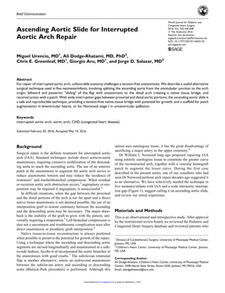
Figure 1. Insert showing an unfavorable wide gap between proximal and distal portions of the interrupted aorta and proposed incision lines.
The ascending aorta is split in half from the sinotubular junction to the arch origin. The patent ductus arteriosus (PDA) has been ligated and all
PDA tissue excised.
646 World Journal for Pediatric and Congenital Heart Surgery 7(5)
by guest on September 2, 2016pch.sagepub.comDownloaded from
3. extracorporeal membrane oxygenation on the day after surgery
due tohemodynamicinstabilitysecondarytorefractoryjunctional
ectopic tachycardia. After 24 hours of support, he was success-
fully decannulated. One patient developed subarachnoid hemor-
rhage on postoperative day 16 secondary to a ruptured left middle
cerebralarteryaneurysmrequiringsurgicalclippingonpostopera-
tive day 17; the child is asymptomatic today. Two patients had
recurrent laryngeal nerve injury causing left vocal cord paresis
diagnosed by direct laryngoscopy, one of them temporary.
The mean follow-up time was 20 months (range: 2.1-49
months). One patient had balloon angioplasty of the aortic
isthmus due to mild hypoplasia seen on the pre-Glenn cathe-
terization four months after IAA repair. One patient with type
C interruption, subaortic stenosis, and an aberrant right subcla-
vian artery had surgical reintervention due to sub- and supra-
valvar aortic stenosis requiring redo subaortic surgical
myectomy and ascending aortic patch augmentation four
months after IAA repair. Two patients underwent single-
ventricle palliation strategy. Both had successful Glenn opera-
tions and one of them eventually a Fontan completion. One late
death was recorded in the second patient awaiting Fontan com-
pletion, 2 years and 3 months after IAA repair, being 1.9 years
after Glenn palliation, secondary to influenza A infection.
Conclusion
The ascending aortic ‘‘slide’’ is a safe, effective, and reprodu-
cible technique for IAA repair, in difficult anatomic conditions
where more conventional techniques are judged suboptimal or
will not allow for a tension-free repair. It has the advantage of
using a native tissue-to-tissue anastomosis with the potential to
grow as well as providing a good posterior scaffold to facilitate
anterior patch reconstruction. This can be used for biventricular
hearts or as part of the Norwood stage I for univentricular
palliation. It is not technically more demanding, nor does it
increase antegrade cerebral perfusion, cross clamp or bypass
times, does not require sacrificing any major vessels, and does
not tether or compress the airway. Small ascending aortas may
be a contraindication or limiting factor to the ‘‘slide’’ opera-
tion; however, no cutoff value can be suggested from our lim-
ited experience. We have used the technique in neonates with
ascending aortas as small as 3.8 mm, although we recognize
that splitting an already small ascending aorta, especially in the
setting of a type C interruption and/or an aberrant right sub-
clavian artery, may lead to future supravalvar aortic stenosis.
Longer follow-up will be needed to confirm the initial satisfac-
tory results in what is proposed as a useful alternative technique
in selected cases. Limitations of the study include the retro-
spective nature of analysis, the small number of patients, and
lack of comparison to more standard techniques for the repair
of IAA in an already rare diagnosis.
Declaration of Conflicting Interests
The author(s) declared no potential conflicts of interest with respect to
the research, authorship, and/or publication of this article.
Funding
The author(s) received no financial support for the research, author-
ship, and/or publication of this article.
Figure 2. The flap has been rotated leftward and posteriorly toward the descending aorta and used as a posterior bridge of native tissue forming
the neo-greater curvature. The completed lesser curvature arch reconstruction with a patch is in the insert.
Urencio et al 647
by guest on September 2, 2016pch.sagepub.comDownloaded from
4. References
1. Mery CM, Guzman-Pruneda FA, Carberry KE, et al. Aortic arch
advancement for aortic coarctation and hypoplastic aortic arch in
neonates and Infants. Ann Thorac Surg. 2014;98(2): 625-633.
2. Oosterhof T, Azakie A, Freedom RM, Williams WG, McCrindle
BW. Associated factors and trends in outcomes of interrupted aor-
tic arch. Ann Thorac Surg. 2004;78(5): 1696-1702.
3. McCrindle BW, Tchervenkov CI, Konstantinov IE, et al; Conge-
nital Heart Surgeons Society. Risk factors associated with mortal-
ity and interventions in 472 neonates with interrupted aortic arch:
a Congenital Heart Surgeons Society study. J Thorac Cardiovasc
Surg. 2005;129(2): 343-350.
4. Schreiber C, Eicken A, Vogt M, et al. Repair of interrupted aortic
arch: results after more than 20 years. Ann Thorac Surg. 2000;
70(6): 1896-1900.
5. Jacobs ML, Rychik J, Murphy JD, Nicolson SC, Steven JM, Nor-
wood WI. Results of Norwood’s operation for lesions other than
hypoplastic left heart syndrome. J Thorac Cardiovasc Surg. 1995;
110(5): 1555-1561.
6. Brown JW, Ruzmetov M, Okada Y, Vijay P, Rodefeld MD, Tur-
rentine MW. Outcomes in patients with interrupted aortic arch and
associated anomalies: a 20-year experience. Eur J Cardiothorac
Surg. 2006;2(5): 666-673.
648 World Journal for Pediatric and Congenital Heart Surgery 7(5)
by guest on September 2, 2016pch.sagepub.comDownloaded from