2. Biology Direct 2006, 1:29 http://www.biology-direct.com/content/1/1/29
Page 2 of 27
(page number not for citation purposes)
Open peer review
This article was reviewed by W. Ford Doolittle, J. Peter
Gogarten, and Arcady Mushegian.
For the full reviews, please go to the Reviewers' comments
section.
Background
The extraordinary diversity of viruses
Viruses are ubiquitous companions of cellular life forms:
it appears that every cellular organism studied has its own
viruses or, at least, virus-like selfish genetic elements [1].
Recent environmental studies have shown that viruses,
primarily, bacteriophages, are "most abundant biological
entities on the planet" [2], with the total number of virus
particles exceeding the number of cells by at least an order
of magnitude [3,4]. Viruses actively move between
biomes and are thought to be major agents of evolution
by virtue of their capacity to operate as vehicles of hori-
zontal gene transfer (HGT) [5].
A remarkable feature of viruses is the diversity of their
genetic cycles, in a sharp contrast to the uniformity of the
cellular genetic cycle [6-9] (Fig. 1). Viruses with different
genome strategies span a vast range of genome sizes (the
genomes of the largest known virus, the mimivirus, and
the smallest viruses, e.g., circoviruses, differ by three
orders of magnitude) and show a non-uniform and non-
trivial distribution among the host taxa (Fig. 1). For exam-
ple, the extraordinary diversity of double-stranded (ds)
DNA bacteriophages is in a stark contrast to the absence
of bona fide dsDNA viruses in plants. Conversely, RNA
viruses are extremely abundant and diverse in plants and
animals but are currently represented by only two com-
pact families in bacteria, and so far have not been detected
in archaea (Fig. 1).
Given the variety of genetic strategies, genome complex-
ity, and global ecology of viruses, the problem of virus
evolution inevitably digresses into a web of interlocking
questions. What qualifies as a virus? Are viruses as a whole
monophyletic, i.e., ultimately descend from a single pri-
mordial virus or polyphyletic, i.e., have multiple origins?
If viruses are polyphyletic, how many independent line-
ages are there? What is the age distribution of different
groups of viruses – are they ancient or have they been
emerging continuously throughout life's evolution? And,
perhaps, the most fundamental questions of all: what is
the ultimate origin of viruses and what are the relation-
ships between evolution of viruses and cellular life forms?
The recent advances of comparative genomics create the
unprecedented opportunity to start tackling these issues
by inferring some of the likely answers from sequence and
structure data analysis. Here, we address several of these
questions but, primarily, the last two, most general ones,
in an attempt to outline a coherent scenario of virus origin
and evolution and delineate the connections between the
evolution of viruses and cellular life forms.
Results and discussion
Polyphyly versus monophyly in virus evolution
Comparative genomics provides no evidence of a mono-
phyletic origin of all viruses. Many virus groups simply
share no common genes, effectively, ruling out any con-
ventional notion of common origin. When applied to
viruses, the notion of "common genes" is not a simple
one because commonality is not necessarily limited to
clear-cut orthologous relationships between genes that
translate into highly significant sequence similarity.
Instead, as discussed in the next sections, distant homolo-
gous relationships among viral proteins and between viral
proteins and their homologs from cellular life forms
could convey more complex but important messages on
evolution of viruses. This complexity notwithstanding,
cases of major virus groups abound that either share no
homologous genes under any definition or have in com-
mon only distantly related domains with obviously dis-
tinct evolutionary trajectories. For example, most of the
viruses of hyperthermophilic crenarchaea have literally no
genes in common with any other viruses [10,11], whereas
RNA viruses share with DNA viruses and plasmids that
replicate via the rolling circle mechanism only extremely
distant domains in their respective replication proteins.
By contrast, monophyly of several large classes of viruses,
including vast assemblages of RNA viruses and complex
DNA viruses, can be demonstrated with confidence (Table
1). Some of these monophyletic classes of viruses cross the
boundaries set by genome strategies: thus, the retroid class
includes both RNA viruses and viruses, mobile elements,
and plasmids with DNA genomes, and the rolling circle
replication (RCR) class combines ssDNA and dsDNA
viruses and plasmids. Furthermore, based on similarities
in the structure of RNA replication complexes, along with
the presence of homologous, even if distant, replication
enzymes, it has been suggested that positive-strand RNA
viruses, double-stranded RNA viruses, and retroid ele-
ments all have a common origin [9]. On the whole, how-
ever, the conclusion seems inevitable that viruses
comprise several distinct lines of descent (Table 1).
A brief natural history of viral genes
Sequence analysis of viral proteins reveals several catego-
ries of virus genes that markedly differ in their prove-
nance. The optimal granularity of classification might be
subject to debate but at least 5 classes that can be assorted
into three larger categories seem to be readily distinguish-
able.
Genes with readily detectable homologs in cellular life forms
3. Biology Direct 2006, 1:29 http://www.biology-direct.com/content/1/1/29
Page 3 of 27
(page number not for citation purposes)
Viruses and other selfish elements: the replication strategies, genome size distribution, global ecology, and hallmark proteinsFigure 1
Viruses and other selfish elements: the replication strategies, genome size distribution, global ecology, and
hallmark proteins. For each class of viruses and related elements, the approximate range of genome sizes is indicated (kb,
kilobases). '+' denotes positive strand (same polarity as mRNA) and '-' denotes negative strand. Tr, transcription; T, translation;
R, replication; E, encapsidation; A, archaea; B, bacteria; F, fungi; Mz, Metazoa; P, plants; UE, unicellular eukaryotes. For each
class of viruses (elements), characteristic structures of hallmark proteins and characteristic electron-microscopic images of
viruses are shown. RdRp, RNA-dependent RNA polymerase; JRC, jelly-roll capsid protein; RT, reverse transcriptase; RCRE,
rolling-circle replication (initiating) endonuclease. The rightmost panel shows the host range, with the size of the respective
image and acronym roughly proportionate to the abundance of the given virus class in the respective taxon.
+
Positive-
strand
RNA
3-30 kb - +
R R, E
RdRp
T
T
+
R
JRC
UE Mz
Class Replication cycle Host range
P
Double-
strand
RNA
4-25 kb ±
R R, E
RdRp
T
+
JRC
±
Negative-
strand
RNA
11-20 kb
R R, E
RdRp
T
+-
Tr
-
+
+
Retroid
RNA
7-12 kb +
RT Tr, E
T
+Tr
±
Retroid DNA
viruses,
elements
2-10 kb +
Tr
T
+Tr
±
E, RT
±
RT
RT
B
UE
Mz
P
B
Mz
P
Mz
UE
Mz
P
B
A
F
F
F
ssDNA
viruses,
plasmids
2-11 kb
dsDNA
viruses,
plasmids
5-1,200 kb
+
RCR RCR, E
+Tr
± +
T
RCE
Mz
P
B A
Tr
±
+
T
UE Mz
B
A
S3H
±
R, E
JRC Pr-Pol
4. Biology Direct 2006, 1:29 http://www.biology-direct.com/content/1/1/29
Page 4 of 27
(page number not for citation purposes)
1. Genes with closely related homologs in cellular organ-
isms (typically, the host of the given virus) present in a
narrow group of viruses.
2. Genes that are conserved within a major group of
viruses or even several groups and have relatively distant
cellular homologs.
Virus-specific genes
3. ORFans, i.e., genes without detectable homologs
except, possibly, in closely related viruses.
4. Virus-specific genes that are conserved in a (relatively)
broad group of viruses but have no detectable homologs
in cellular life forms.
Viral hallmark genes
5. Genes shared by many diverse groups of viruses, with
only distant homologs in cellular organisms, and with
strong indications of monophyly of all viral members of
the respective gene families, – we would like to coin the
phrase "viral hallmark genes" to denote these genes that
can be viewed as distinguishing characters of the "virus
state".
The relative contributions of each of these classes of genes
to the gene sets of different viruses strongly depend on the
viral genome size which differs by more than three orders
of magnitude. Viruses with small genomes, such as most
of the RNA viruses, often have only a few genes, the major-
ity of which belong to the hallmark class. By contrast, in
viruses with large genomes, e.g., poxviruses, all 5 classes
are broadly represented. In order to illustrate the diversity
of viral "genomescapes" more concretely, we show in
Table 2 the decomposition of the gene sets of three viruses
with a small, an intermediate-sized, and a large genomes,
respectively, into the 5 classes. Notably, moderate-sized
and large genomes of bacteriophages and archaeal viruses
are dominated by ORFans that often comprise >80% of
the genes in these viruses. Rapidly evolving phage ORFans
are thought to supply many, if not most, of the ORFans
found in prokaryotic genomes (the lack of detectable
sequence conservation notwithstanding), hence playing
an important role in evolution of prokaryotes [12].
Table 1: The major monophyletic classes of viruses and selfish genetic elements
Class of viruses Constituent virus lineages Hosts Support for monophyly Refs
Positive-strand RNA
viruses
Superfamily I: picorna-like;
superfamily II: alpha-like;
superfamily III: flavi-like; the
exact affinity of RNA
bacteriophages within this class
of viruses remains uncertain
(possibly, a fourth lineage)
Animals, plants, protists,
bacteria (one family of
bacteriophages)
Conserved RdRp; JRC in
most superfamily 1 viruses,
and subsets of
superfamilies 2 and 3
viruses. Reconstructed
ancestor with RdRp and
JRC
[87]
Retroid viruses and
elements
Retroviruses, hepadnaviruses,
caulimoviruses, badnaviruses;
LTR- and nonLTR retroelements;
retrons; group II self-splicing
introns – the progenitors of
eukaryotic spliceosomal introns
Animals, fungi, plants,
protists, bacteria, archaea
Conserved RT [103, 104]
Small DNA viruses,
plasmids, and transposons
with rolling circle
replication
Gemini-, circo-, parvo-,
papovaviruses, phages (e.g.,
φX174), archaeal and bacterial
plasmids, eukaryotic helitron
transposons
Animals, plants, archaea,
bacteria
Conserved RCRE, JRC,
S3H (in eukaryotic viruses)
[17, 18, 20]
Tailed bacteriophages
(Caudovirales)
Families: Myoviridae (e.g., T4),
Podoviridae (e.g., T7),
Siphoviridae (e.g., λ)
Bacteria, euryarchaea Complex, overlapping
arrays of genes conserved
in subsets of tailed phages;
genes of all tailed phages
thought to comprise a
single pool
[11, 93, 94, 105, 106]
Nucleo-cytoplasmic large
DNA viruses (NCLDV)
Poxviruses, asfarviruses,
iridoviruses, phycodnaviruses,
mimiviruses
Animals, algae, protests Core set of 11 conserved
genes, including JRC, S3H,
and a FtsK-like packaging
ATPase, found in all
NCLDVs; reconstructed
ancestor with ~40 genes
[50–53, 107]
Abbreviations: JRC, Jelly-Roll Capsid protein; LTR, Long Terminal Repeat; RdRp, RNA-dependent RNA polymerase; RCRE, Rolling Circle
Replication (initiation) Endonuclease; RT, Reverse Transcriptase; S3H, Superfamily 3 Helicase.
5. Biology Direct 2006, 1:29 http://www.biology-direct.com/content/1/1/29
Page 5 of 27
(page number not for citation purposes)
The evolutionary origins of the 5 classes of viral genes are
likely to be very different. The least controversial are the
two classes of genes with readily detectable homologs
from cellular life forms that appear to represent, respec-
tively, relatively recent (class 1) and ancient (class 2)
acquisitions from the genomes of the cellular hosts.
Where do virus-specific genes come from is a much harder
question. In the absence of direct evidence, the default
hypothesis, probably, should be that these genes evolved
from cellular genes as a result of dramatic acceleration of
evolution linked to the emergence of new, virus-specific
functions, such that all traces of the ancestral relationships
are obliterated. This notion is compatible with the fact
that many, probably, most class 4 genes (virus-specific
genes conserved within a group of viruses) are virion com-
ponents (e.g., see the vaccinia virus case in Table 2), a
quintessential viral function. The hallmark genes that
cross the barriers between extremely diverse virus lineages
seem to be of the greatest interest and relevance for the
problem of the ultimate origins of viruses, at least, in the
context of the long argument we attempt to develop here.
Thus, we discuss the distribution among viruses, evolu-
tion and significance of these genes in a separate section.
Viral hallmark genes: beacons of the ancient virus world
Although there are no traceable vertical relationships
between large groups of viruses outside the major classes
listed in Table 1, a considerable number of genes that
encode proteins with key roles in genome replication,
expression, and encapsidation are shared by overlapping
arrays of seemingly unrelated groups of viruses. As already
noted above, some of these widespread viral genes are
genuine viral hallmarks that are found in a variety of
diverse viruses (although never in all viruses) but not in
cellular life forms except as easily recognizable proviruses
or mobile elements (Table 3, Fig. 1). The two proteins that
are most widely dispersed among viruses are the jelly-roll
capsid protein (JRC) [13-15] and the superfamily 3 heli-
case (S3H) [16]. Each of these proteins crosses the bound-
ary between RNA and DNA viruses and spans an
astonishing range of virus groups, from some of the small-
est positive-strand RNA viruses to the nucleo-cytoplasmic
large DNA viruses (NCLDV), the class of viruses that
includes the giant mimivirus (Table 3). Other proteins
listed in Table 3 are not as common as JRC or S3H but still
form multiple, unexpected connections between groups
of viruses that otherwise appear to be unrelated. A case in
point is the rolling circle replication (RCR) initiation
endonuclease (RCRE) which unites a great variety of small
ss and dsDNA replicons, including viruses, plasmids, and
transposable elements that reproduce in animals, plants,
bacteria, and archaea [17-20]. Notably, a more recent and
more careful analysis has shown that the DNA-binding
domain of the replication protein of polyoma and papil-
loma viruses (e.g., the T antigen of SV40) is a derived form
of the RCRE that has lost the catalytic amino acid residues
Table 2: Representation of the five classes of viral genes in three selected viruses with small, medium-size and large genomes
Genome size
(kb)/number of
annotated genes
Representation of the 5 classes of viral genes (number and brief description)
1. Recent
acquisitions
from cells
2. Ancient
acquisitions
from cells
3. ORFans 4. Conserved
virus-specific
genes
5. Hallmark
genes
Virus
Poliovirusa 7.4/10 None 2: a duplication of
a chymotrypsin-
like protease (3C,
2A)
1: uncharacterized
protein (3A)
1: genome-linked
protein (VPg)
6: 4 diverged
copies of JRC
(VP1-4), S3H (3C),
RdRp (3D)
Sulfolobus
turreted
icosahedral
virus (STIV)b
17.6/36 3: two predicted
transcription
regulators and an
uncharacterized
protein
5: four predicted
transcription
regulators and an
uncharacterized
protein
26:
uncharacterized
proteins
None 2: JRC, packaging
ATPase
Vaccinia virusc 194.7/~200 ~48: primarily,
proteins involved
in virus-host
interaction
~36: primarily,
proteins involved
in genome
replication and
expression
~24: poorly
characterized
proteins, possibly,
involved in virus-
host interactions
~84: primarily,
structural
components of the
virion and some
proteins involved
in genome
expression
5: JRC, S3H/
primase, packaging
ATPase, DNA
polymerase(?)
aThe classification is based on the analysis described in [87].
bThe classification is based on the analysis described in [11].
cThe classification is based on the analysis described in [108] and EVK, unpublished observations; the uncertainty in the number of genes is due to
the pseudogenization of varying subsets of genes in different strains of vaccinia virus.
6. Biology Direct 2006, 1:29 http://www.biology-direct.com/content/1/1/29
Page 6 of 27
(page number not for citation purposes)
[18]. Thus, through this detailed analysis of one of the
hallmark proteins, the well-known connection between a
variety of small ssDNA-replicons is extended to a group of
similar-sized dsDNA-replicons. A similar expansion of the
set of viral groups covered by a particular hallmark gene
resulted from the detailed analyses of the packaging
ATPase and the archaeo-eukaryotic primase (Table 3).
Replication of positive-strand RNA viruses, dsRNA
viruses, negative-strand RNA viruses, and retroid viruses/
elements is catalyzed by another idiosyncratic class of
viral enzymes, RNA-dependent RNA polymerases (RdRp)
and reverse transcriptases (RT). The positive-strand RNA
virus RdRp and the RT form a monophyletic cluster within
the vast class of the so-called palm-domains that are char-
acteristic of numerous polymerases [21-24]. The RdRps of
dsRNA viruses and negative-strand RNA viruses are likely
to be highly diverged derivatives of the same polymerase
domain, an old conjecture [24-26] that has been vindi-
cated by the recent determination of the structure of a
dsRNA bacteriophage RdRp [27,28].
The palm domain is likely to be the primordial protein
polymerase that emerged from the RNA world where
nucleotide polymerization was catalyzed by ribozymes
[29]. This is supported not only by the wide spread of this
domain in modern life forms but also by the structural
and, by inference, evolutionary link between the palm
domain and the RNA-recognition-motif (RRM) domain,
an ancient RNA-binding domain that might have, ini-
tially, facilitated replication of ribozymes [30]. The palm-
domain RdRps and RTs are excluded from the regular life
cycles of cellular life forms, although most eukaryotic
genomes encompass numerous copies of RT-containing
retroelements, and prokaryotes have some such elements
as well [31,32]. These elements, however, are selfish and,
from the evolutionary standpoint, virus-like. The most
notable incursion of an RT into the cellular domain is the
catalytic subunit of the eukaryotic telomerase, the essen-
tial enzyme that is involved in the replication of chromo-
some ends [33,34].
The list of viral hallmark genes given in Table 3 is a con-
servative one. There well might be other genes that merit
the hallmark status but for which clear evidence is hard to
come up with. An important case in point is the B-family
DNA polymerase that is the main replication enzyme of
numerous dsDNA viruses of bacteria and eukaryotes.
Homologs of these DNA polymerases are found in all
archaeal and eukaryotic genomes, so that monophyly of
all viral polymerases does not seem to be demonstrable in
phylogenetic analyses [35,36]. However, this potentially
could be explained by relatively (with respect to cellular
homologs) fast evolution of the polymerases in various
viral lineages, which would obscure their common origin.
Furthermore, monophyly of the polymerases of all viruses
that employ a protein-primed mechanism of dsDNA rep-
lication (animal adenoviruses and tailed phages like
PRD1 or φ29) has been claimed [37]. Thus, although this
currently cannot be shown convincingly, it seems possible
(and, as discussed below, even likely) that the DNA
polymerase is a viral hallmark gene in disguise. More gen-
erally, further sequencing of viral genomes, combined
with comprehensive comparative analysis, might reveal
additional genes that, despite relatively limited spread
among viral lineages, will qualify as hallmark genes.
The combination of features of viral hallmark proteins is
highly unusual and demands an evolutionary explana-
tion. Indeed, the hallmark genes are, without exception,
responsible for essential, central aspects of the viral life
cycles, including genome replication, virion formation,
and packaging of the genome DNA into the virion (Table
3). These genes span sets of extremely diverse classes of
viruses, often possessing different reproduction strategies
and differing by three orders of magnitude in genome
size. Finally, all viral hallmark genes have remote
homologs in cellular life forms (Table 3) but the viral ver-
sions appear to be monophyletic.
Three hypotheses on the origin of viral hallmark genes
immediately come to mind. The first possibility is that the
notion of hallmark virus proteins is based on an artifact.
The argument, that is commonly invoked in discussions
of unexpected patterns of homologous relationships and
could well be waged against the notion of viral hallmark
proteins, is that genuine orthologs of these proteins
(direct evolutionary counterparts, typically, with the same
function) actually do exist in cellular life forms but are not
detectable due to rapid sequence divergence between viral
and cellular proteins. However, this reasoning does not
seem to survive closer scrutiny. Firstly, the conservation of
the hallmark proteins in extremely diverse classes of
viruses with widely different replication/expression strate-
gies (Table 3) but not in cellular life forms is hardly com-
patible with the rapid divergence interpretation. Indeed,
for this to be the case, acceleration of evolution of the hall-
mark genes in diverse classes of viruses should occur in
such a manner that the similarity between viral proteins
survived, whereas the similarity between viral proteins
and their hypothetical cellular orthologs vanished. Paral-
lel conservation of hallmark protein sequences might be
perceived in the case of structural protein, such as JRC, but
is hardly imaginable for proteins involved in replication
of structurally very different genomes, such as S3H that is
conserved among viruses with RNA, ssDNA, and dsDNA
genomes. Furthermore, for most of the hallmark proteins,
distant and functionally distinct homologs from cellular
organisms are detectable (S3H and viral RNA-dependent
7. Biology Direct 2006, 1:29 http://www.biology-direct.com/content/1/1/29
Page 7 of 27
(page number not for citation purposes)
Table 3: Proteins encoded by hallmark viral genes
Protein Function Virus groups Homologs in
cellular life forms
Comments References
Jelly-roll capsid
protein (JRC)
Main capsid subunit of
icosahedral virions
Picornaviruses, comoviruses,
carmoviruses, dsRNA phage,
NCLDV, herpesviruses,
adenoviruses, papovaviruses,
parvoviruses, icosahedral
DNA phages and archaeal
viruses
Distinct jelly-roll
domains are seen in
eukaryotic
nucleoplasmins and in
protein-protein
interaction domains of
certain enzymes
Certain icosahedral
viruses, such as
ssRNA phages and
alphaviruses, have
unrelated capsid
proteins. In
poxviruses, the JRC is
not a virion protein
but forms
intermediate
structures during
virion morphogenesis
[13–15, 53, 54,
109–111]
Superfamily 3
helicase (S3H)
Initiation and
elongation of genome
replication
Picornaviruses, comoviruses,
eukaryotic RCR viruses,
NCLDV, baculoviruses, some
phages (e.g., P4), plasmids,
particularly, archaeal ones
S3H is a distinct,
deep-branching family
of the AAA+ ATPase
class
Characteristic fusion
with primase in DNA
viruses and plasmids
[16, 112]
Archaeo-
eukaryotic DNA
primase
Initiation of genome
replication
NCLDV, herpesviruses,
baculoviruses, some phages
All viral primases
appear to form a clade
within the archaeo-
eukaryotic primase
family
Characteristic fusion
with S3H in most
NCLDV, some
phages, and archaeal
plasmids
[18]
UL9-like
superfamily 2
helicase
Initiation and
elongation of genome
replication
Herpesviruses, some
NCLDV, some phages
Viral UL9-like
helicases form a
distinct branch in the
vast superfamily of
DNA and RNA
helicases
Fusion with primase in
asfarviruses,
mimiviruses
[53]
Rolling-circle
replication
initiation
endonuclease
(RCRE)/origin-
binding protein
Initiation of genome
replication
Small eukaryotic DNA viruses
(parvo-, gemini-, circo-,
papova), phages, plasmids, and
eukaryotic helitron
transposons
No cellular RCRE or
papovavirus-type
origin-binding protein;
however, these
proteins have a
derived form of the
palm domain that is
found in the majority
of cellular DNA
polymerases
Papovaviruses have an
inactivated form of
RCRE that functions
as origin-binding
protein
[17–20]
Packaging ATPase
of the FtsK family
DNA packaging into
the virion
NCLDV, adenoviruses,
polydnaviruses, some phages
(e.g., P9, M13), nematode
transposons
A distinct clade in the
FtsK/HerA
superfamily of P-loop
NTPases that includes
DNA-pumping
ATPases of bacteria
and archaea
[113]
ATPase subunit of
terminase
DNA packaging into
the virion
Herpesviruses, tailed phages The terminases
comprise a derived
family of P-loop
NTPases that is
distantly related to
Superfamily I/II
helicases and AAA+
ATPases
[109, 114]
8. Biology Direct 2006, 1:29 http://www.biology-direct.com/content/1/1/29
Page 8 of 27
(page number not for citation purposes)
polymerases are the primary examples) which makes the
existence of elusive orthologs extremely unlikely.
The other two hypotheses accept the hallmark viral pro-
teins as reality but offer contrasting evolutionary scenarios
to account for their existence and spread.
1. One hypothesis would posit that the hallmark genes
comprise the heritage of a "last universal common ances-
tor of viruses" (LUCAV). This scenario implies that,
despite all evidence to the contrary (see above) all extant
viruses are monophyletic, although their subsequent evo-
lution involved massive gene loss in some lineages as well
as extensive acquisition of new genes from the hosts in
others.
2. By contrast, under the hypothesis of polyphyletic origin
of viruses, the spread of the hallmark genes across the
range of virus groups could be explained by horizontal
gene transfer (HGT).
Upon closer inspection, none of these hypotheses seems
to be a viable general explanation for the existence and
distribution of the viral hallmark genes. Indeed, the rela-
tively small number and the mosaic spread of the hall-
mark genes (Table 3) do not seem to be conducive to the
LUCAV notion although it is apparent that a great number
of diverse viruses, if not all of them, share some common
history. Conversely, the extremely distant similarity
between the hallmark proteins from diverse virus groups
with dramatically different replication strategies is poorly
compatible with an HGT scenario.
Here, we outline a scenario of virus origin and evolution
that does not involve a LUCAV but integrates aspects of
the common origin and HGT hypotheses and is naturally
linked to specific models of evolution of cells. The sim-
plest explanation of the fact that the hallmark proteins
involved in viral replication and virion formation are
present in a broad variety of viruses but not in any cellular
life forms is that the latter never had these genes in the first
place. The alternative that we consider most likely is that
the hallmark genes antedate cells and descend directly
from a primordial gene pool. It is thought that, in such a
primordial pool, selection would act primarily on func-
tions directly involved in replication [38,39] which is
compatible with the properties of the majority of hall-
mark genes (Table 3). Given the spread of the hallmark
genes among numerous groups of dramatically different
viruses, a crucial corollary is that the major lineages of
viruses themselves derive from the same, precellular stage
of evolution. This corollary serves as the foundation for
the concept of an ancient Virus World, which we envisage
as an uninterrupted flow of genetic information through
an enormous variety of selfish elements, from the precel-
lular stage of evolution to this day; we discuss the Virus
World in the rest of this article.
Conflicting concepts of virus origin and evolution and the
inextricable link between evolution of viruses and cells
Before we discuss the full scope of the emerging concept
of the origin of viruses from the precellular gene pool, it is
necessary to briefly examine the existing trains of thought
on virus origin and evolution. Traditionally, these ideas
have revolved around three themes: i) origin of viruses
from primordial genetic elements, ii) degeneration of uni-
cellular organisms to the virus state, and iii) "escaped
genes" hypotheses deriving viruses from genes of cellular
organisms that have switched to the selfish mode of repro-
duction (reviewed in [40-45]) (Table 4). Historically, it is
remarkable that Felix d'Herelle, the discoverer of bacteri-
ophages and one of the founders of virology, proposed
that phages might be evolutionary precursors of cells as
early as 1922 [46]. Furthermore, in J.B.S. Haldane's 1928
classic on the origin of life [47], an early, "viral" stage of
evolution is considered as an integral part of the proposed
scenario for the emergence of the first life forms from the
primary soup (we revisit Haldane's prescient speculation
toward the end of this article). However, the "primordial"
hypothesis is habitually dismissed on the grounds that all
RNA-dependent
RNA polymerase
(RdRp)/reverse
transcriptase (RT)
Replication of RNA
genomes
Positive-strand RNA viruses,
dsRNA viruses, retroid
viruses/elements, possibly,
negative-strand RNA viruses
Another major group
of palm domains that
are distinct from
those in DNA
polymerases
The RdRps of dsRNA
viruses are homologs
of positive-strand
RNA virus
polymerases. The
provenance of
negative-strand RNA
virus RdRp remains
uncertain although
sequence motif and,
especially, structural
analysis suggests their
derivation from
positive-strand RNA
virus RdRps
[23–25, 28, 87,
115]
Abbreviations: NCLDV, Nucleo-Cytoplasmic Large DNA Viruses
Table 3: Proteins encoded by hallmark viral genes (Continued)
9. Biology Direct 2006, 1:29 http://www.biology-direct.com/content/1/1/29
Page 9 of 27
(page number not for citation purposes)
extant viruses are intracellular parasites, so viruses could
not exist before the emergence of modern-type cells
although antiquity of viruses has been propounded based
on the lack of cellular homologs for many virus genes
[43,48-51]. By contrast, the high prevalence of host-
related genes (as opposed to virus-specific genes) in many
viruses (particularly, those with large genomes) might be
construed as support for the "escaped genes" or even the
"cell degeneration" hypotheses.
The existence of the hallmark virus genes seems to effec-
tively falsify both the cell degeneration and the escaped-
genes concepts of viral evolution. With regard to the cell
degeneration hypothesis, let us consider the NCLDV, the
class of large viruses to which the cell degeneration con-
cept might most readily apply and, indeed, was, in the
wake of the discovery of the giant mimivirus [50-52].
Among the 11 signature genes that are shared by all
NCLDVs ([53] and Table 1), three crucial ones (JRC, S3H,
and the packaging ATPase) are virus hallmark genes. A
clear inference is that even the simplest, ancestral NCLDV
would not be functional without these genes. However,
cellular derivation of this ancestral NCLDV would have to
invoke decidedly non-parsimonious, ad hoc scenarios,
such as concerted loss of all hallmark genes from all
known cellular life forms or their derivation from an
extinct major lineage of cell evolution. The same line of
logic essentially refutes the escaped genes concept inas-
much as the hallmark genes had no cellular "home" to
escape from. Again, to save "escaped genes", an extinct cel-
lular domain would have to be postulated.
Two recent conceptual developments in the study of ori-
gin and evolution of viruses deserve special attention
(Table 4; see also discussion below). First, Bamford and
coworkers [13-15] and, independently, Johnson and cow-
orkers [54] capitalized on the conservation of the struc-
ture of the jelly-roll capsid protein in a wide variety of
viruses to propose the idea of an ancient virus lineage
spanning all three domains of cellular life (archaea, bacte-
ria, and eukaryotes). Second, Forterre presented an elabo-
rate scheme of virus-cell coevolution from the earliest
stages of life's evolution. Under this concept, viruses
emerged independently within three lineages of RNA-
Table 4: Major concepts in virus evolution
Concept Principal message References Brief critique/comment
Cell degeneration model of virus
origin
Viruses, at least complex ones, evolved as a
result of degeneration of cells, perhaps,
through a stage of intracellular parasites
[40, 43, 45, 50] This route of virus evolution appears to be
inconsistent with the results of viral
comparative genomic, in particular, the
prominence of genes without cellular
counterparts in the conserved cores of viral
genomes
Escaped-genes model of virus
origin
Viruses evolved from within cells, through
autonomization of the appropriate genes,
e.g., those coding for polymerases
[40, 43, 45, 55] Similarly, this model lacks support from
virus genome comparison
Origin of viruses from a primordial
gene pool
Viruses are direct descendants of primordial
genetic elements
[40, 43, 87] Generally, this appears to be the most
plausible path for the origin of viruses.
However, non-trivial conceptual
development is required, given that viruses
are intracellular parasites and, technically,
could not precede cells during evolution
An ancient lineage of viruses
spanning the three domains of
cellular life
The presence of JRC in a variety of groups of
DNA viruses is taken as evidence of the
existence of an ancient lineage of viruses
infecting all three domains of cellular life
[13–15] This concept capitalizes on a truly
remarkable observation of the near ubiquity
of JRC in viruses. However, inferring an
ancient lineage of viruses on the basis of the
conservation of a single protein smacks of
essentialism and does little to explain the
trajectories of most other virus-specific and
virus hallmark genes. Besides, this concept
does not specify the cellular context in
which the ancient virus lineage might have
emerged
Three DNA viruses to replicate
genomes of RNA cells
The hypothesis postulates that at least three
major lineages of RNA viruses emerged by
the escaped-genes route from RNA-based
progenitors of archaea, bacteria and
eukaryotes. These ancient RNA viruses are
thought to have given rise to three
independent lineages of DNA viruses that
imparted DNA replication onto their cellular
hosts
[49, 55] This concept is based on important general
notions of the ancient origin of viruses and
their major role in evolution of cells.
However, the specific model of Forterre
appears to be critically flawed as it stems
from a model of cellular evolution that
appears not to be defendable (see text)
10. Biology Direct 2006, 1:29 http://www.biology-direct.com/content/1/1/29
Page 10 of 27
(page number not for citation purposes)
based cells (the progenitors of archaea, bacteria, and
eukaryotes) and "invented" DNA replication that was sub-
sequently captured from different viruses by each type of
host cells, in three independent transitions to DNA
genomes [43,55].
A crucial, even if fairly obvious, aspect of viral evolution is
that it is inextricably linked to the evolution of the hosts
which, when traced back to the earliest stages of life's evo-
lution, attests to the necessity to consider scenarios of
virus origins in conjunction with models for the origin
and early evolution of cells. This dramatically ups the ante
for the study of virus evolution, bringing it to the center
stage of evolutionary biology [56,57]. Therein, however,
seem to lie some of the major problems encountered by
the current hypotheses of virus evolution (Table 4). The
concept of an ancient virus lineage advocated by Bamford
and coworkers, in addition to being based on the broad
spread of a single gene (JRC), which is construed as the
"self" of a virus [13], simply leaves a glaring gap as the
connection between viral and cellular evolution is not
considered such that it remains unclear in what kind of
cellular environment the early evolution of the primordial
viral lineage took place.
By contrast, Forterre's concept is embedded within a spe-
cific scenario of cellular evolution, and we believe that
this is the valid approach to the analysis of the origins and
evolution of viruses and cells – the only chance to achieve
understanding of these difficult problems is to consider
them in conjunction. However, the specific scenario of
cellular evolution favored by Forterre appears to be poorly
compatible with the results of comparative genomics,
thus compromising the model of virus evolution associ-
ated with it. Indeed, Forterre considers three lineages of
RNA-cells evolving from the Last Universal Common
Ancestor (LUCA) of the known life forms and giving rise
to bacteria, archaea, and eukaryotes, with the transition to
DNA-cells mediated by domain-specific viruses. This sce-
nario, while complete with respect to the evolution of
both cells and viruses, encounters at least three major,
probably, insurmountable problems. First, it is dubious at
best that the combination of complexity and genetic sta-
bility required of an even a minimal cell – a complement
of a few hundred genes that is accurately transmitted over
many cellular generations – is attainable with an RNA
genome. Indeed, given the inherent instability of RNA,
such a genome would have to consist of several hundred
RNA molecules. Accurate partitioning of this multipartite
genome between daughter cells would require an RNA
segregation system of unprecedented precision; thus,
faithful vertical inheritance of the genome is hardly imag-
inable. Stochastic segregation of RNA segments in a poly-
ploid cell might seem to be a straightforward solution to
this problem but a quick calculation shows that this is
unfeasible. Indeed, if the probability of a the two daughter
RNA segments being segregated into the two daughter
cells is 12, then the probability that, say, 100 RNA seg-
ments in a reasonable multipartite genome are all cor-
rectly segregated (i.e., none of the segments is missing in
either of the daughter cells) is (1/2)100 ≈ 10-30. Thus,
ploidy of ~1030 would be required for accurate genome
segregation; in other words, the RNA cell would have to
contain many tons of RNA. Thus, it is much more likely
that the first fully-fledged cells already had DNA genomes
resembling (even if quantitatively simpler than) those of
modern archaea and bacteria. Forterre's proposal is based
on the well-known observation that the DNA replication
systems of archaea and bacteria are, largely, unrelated,
making it unlikely that LUCA had a DNA genome [58-60].
We believe that a far more plausible implication of the
disparity of DNA replication machineries in archaea and
eukaryotes is a non-cellular LUCA, a hypothesis that finds
crucial support in the fact that the membrane lipids and
membrane biogenesis systems, as well as cell walls, are
also distinct and, largely, unrelated in archaea and bacte-
ria [39,61-63].
For efforts on reconstructing the non-cellular LUCA, a key
guiding principle is that, although modern-type cells, pre-
sumably, did not exist at this stage of evolution, some
form of compartmentalization was required to ensure
concentrations of substrates and genetic elements suffi-
cient for effective replication and, consequently, evolu-
tion. Furthermore, as discussed in greater detail below,
compartmentalization is a necessary condition of selec-
tion among evolving ensembles of genetic elements. Fol-
lowing this principle, a specific model has been
elaborated under which early evolution, from abiogenic
syntheses of complex organic molecules to the emergence
of archaeal and bacterial cells, unraveled within networks
of inorganic compartments that are found at hydrother-
mal vents and consists, primarily, of iron sulfide [39,63].
Here, in order to be concrete, we attempt a reconstruction
of the earliest events in the evolution of primordial virus-
like entities within the framework of this model although
our general conclusions do not seem to hinge on any par-
ticular model of organization of the ancient, precellular
life.
Second, the model by viral overtake meets a stumbling
block: if three different viruses brought the three distinct
DNA replication machineries into the three cell lineages,
why have not the hallmark genes that are essential for
viral DNA replication, in particular S3H and the viral-type
primase, entered the genomes of any of those cellular lin-
eages? One could argue that, in each case, an unusual
virus, not carrying any of these hallmark genes, was
involved, but this happening three times independently
stretches credulity.
11. Biology Direct 2006, 1:29 http://www.biology-direct.com/content/1/1/29
Page 11 of 27
(page number not for citation purposes)
The third major issue that is ignored in Forterre's hypo-
thesis and similar, three-domain scenarios of cellular evo-
lution [64,65], is the readily demonstrable relationship
between eukaryotes and the two prokaryotic domains.
This relationship follows the divide between the two prin-
cipal functional categories of genes in the eukaryotic cell,
the informational genes that are, almost invariably, most
closely related to archaeal homologs, and the operational
genes that are of bacterial provenance [66-68]. By far the
simplest, most parsimonious explanation of this dichot-
omy is that the eukaryotic cell emerged as a result of a
symbiosis between an archaeon and a bacterium. Given
the overwhelming evidence of the origin of mitochondria
and related organelles (hydrogenosomes and mitosomes)
from α-proteobacteria, the nature of the partners and the
direction of the symbiosis appear clear: an α-proteobacte-
rium invaded an archaeal cell [69-71]. In addition to the
genomic evidence, there is a clear biochemical rationale
for the symbiosis to actually comprise the onset of eukary-
ogenesis such that the invading α-proteobacterium
became an anaerobic symbiont that originally supplied
hydrogen to the methanogenic, archaeal host and subse-
quently gave rise to aerobic mitochondria [71]. The mas-
sive transfer of the symbiont's genes to the host genome
gave rise to the mosaic provenance of the eukaryotic
genomes. The argument against the symbiotic models of
eukaryogenesis and for three-domain models stems pri-
marily from the existence of numerous eukaryote-specific
proteins (ESPs) without readily detectable prokaryotic
homologs [64,72,73]. However, the abundance of ESPs
hardly can be taken as evidence of a third domain of life,
distinct from archaea and bacteria, and comprising the
pre-symbiosis eukaryotic line of descent. First, although
many of the ESPs are proteins with important biological
functions, they do not belong to the core of either the
informational or the metabolic proteins repertoires of
eukaryotes that are of archaeal and bacterial descent,
respectively. Second, more often than not, prokaryotic
homologs of an apparent ESP can be identified by using
sensitive techniques of protein motif analysis and, partic-
ularly, structural comparison [74]. Thus, it seems likely
that many, if not most, ESPs are cases of accelerated evo-
lution in eukaryotes which accompanies the emergence of
novel, eukaryote-specific functional systems, such as the
cytoskeleton and ubiquitin signaling. The prevalence and
nature of "true" eukaryotic innovations, i.e., proteins that
evolved through processes other than descent with (per-
haps, radical) modification from archaeal or bacterial pro-
teins, remain to be characterized. Clearly, however, even if
such novelties turn out to be numerically prominent, they
are, typically, involved in ancillary functions and hardly
can be construed as the heritage of a distinct primary line
of cell evolution [74].
In the rest of this article, we consider evolution of viruses
in conjunction with these two central concepts of cellular
evolution: i) the existence of an early non-cellular but
confined stage in life's evolution, that probably encom-
passed LUCA, and ii) the origin of the eukaryotic cell as a
result of fusion of an archaeon and a bacterium. We argue
that the scenario of virus origin and evolution informed
by comparative genomics forms a coherent whole with
these notions of early cell evolution and provides support-
ive feedback for them.
The primordial gene pool: the crucible of the major virus
lineages
Taken together, all these lines of evidence and reasoning
suggest that the principal classes of prokaryotic viruses,
including positive-strand RNA viruses, retroid elements,
and several groups of DNA viruses, emerged within the
primordial genetic pool where mixing and matching of
diverse genetic elements was incomparably more exten-
sive than it is in any modern biological community
[39,75] (Fig. 2).
As indicated above, we present a scenario of viral origins
under the recent model of the emergence of cells and
genomes within networks of inorganic compartments
[39,63]. These compartments are envisaged being inhab-
ited by diverse populations of genetic elements, initially,
segments of self-replicating RNA, subsequently, larger and
more complex RNA molecules encompassing one or a few
protein-coding genes, and later yet, also DNA segments of
gradually increasing size. Thus, early life forms, including
LUCA, are perceived as ensembles of genetic elements
inhabiting a system of inorganic compartments. This
model explains the lack of homology between the mem-
branes, membrane biogenesis systems, and the DNA rep-
lication machineries of archaea and bacteria by
delineating a LUCA that had neither a membrane nor
DNA replication. The model also outlines the processes
that might have enabled selection and evolutionary
change in such a system. Under this scenario, a transition
gradually took place from selection at the level of individ-
ual genetic elements to selection for ensembles of such
elements encoding both enzymes directly involved in rep-
lication and proteins responsible for accessory functions,
such as translation and nucleic acid precursor synthesis.
From selection for gene ensembles, there is a direct path
to selection for compartment contents such that compart-
ments sustaining rapid replication of genetic elements
would infect adjacent compartment and, effectively, prop-
agate their "genomes" [39].
This model implies that, at the early stages of evolution,
including the LUCA stage, the entire genetic system was,
in a sense, "virus-like". Initially, all RNA segments in the
population would be completely selfish, and there would
12. Biology Direct 2006, 1:29 http://www.biology-direct.com/content/1/1/29
Page 12 of 27
(page number not for citation purposes)
Evolution of the virus world: origin of the main lineages from the primordial gene poolFigure 2
Evolution of the virus world: origin of the main lineages from the primordial gene pool. Characteristic images of
RNA and protein structures are shown for each postulated stage of evolution, and characteristic virion images are shown for
the emerging classes of viruses. Thin arrows show the postulated movement of genetic pools between inorganic compart-
ments. Block arrows show the origin of different classes of viruses at different stages of pre-cellular evolution.
Pre-archaeal
compartment
Pre-bacterial
compartment
ocean
selfish
ribozymes
(group I
Introns)
positive-
strand,
ds
RNA
viruses
retrons,
group II
introns
crust
dsDNA
and RCR
viruses,
plasmids
Bacteria with
plasmids, retrons,
group I & II introns
Archaea with
plasmids, group
I introns
Escape of cells with
their viruses and other
parasitic elements
RNA World
RNA-DNA Retro
World
inorganic compartments
RNA-Protein World
DNA World
13. Biology Direct 2006, 1:29 http://www.biology-direct.com/content/1/1/29
Page 13 of 27
(page number not for citation purposes)
be no distinction between parasitic, "viral" elements and
those that would give rise to genomes of cellular life
forms. However, this distinction would emerge as soon as
the first "selfish cooperatives" – relatively stable ensem-
bles of co-inherited genetic elements [39] – evolve. Inevi-
tably, the selfish cooperatives would harbor parasitic
genetic elements that would divert the resources of the
ensemble for their own replication. The separation of par-
asitic elements from cooperators would be cemented by
the emergence of physical linkage of the latter: genes that
encode components of the replication machinery and are
physically linked to genes for accessory functions would
be locked into ensembles of cooperators, whereas solitary
genes (or small gene arrays) encoding replication func-
tions would occupy the parasitic niche. From a comple-
mentary standpoint, within the inorganic compartment
model, the distinction between the progenitors of viruses
and the ancestors of cellular life forms may be described
as one between primary infectious agents, that routinely
moved between compartments, and increasingly complex
and "sedentary" ensembles of selfish cooperators that
would spill from one compartment into another on much
rarer occasions.
Like other models of the early stages of evolution of bio-
logical complexity, this scenario faces the problem of
takeover by selfish elements [76-78]. If the primordial
parasites become too aggressive, they would kill off their
hosts within a compartment and could survive only by
infecting a new compartment. Devastating "epidemics"
sweeping through entire networks and eventually elimi-
nating all their content are imaginable, and indeed, this
might have been the fate of many, if not most, primordial
"organisms". The evolution of those primordial life forms
that survived would involve, firstly, emergence of temper-
ate virus-like agents that do not kill the host, and sec-
ondly, early invention of primitive defense mechanisms,
likely, based on RNA interference (RNAi). The ubiquity of
both temperate selfish elements and RNAi-based defense
systems [79-81] in all life forms is compatible with the
origin of these phenomena at a very early stage of evolu-
tion.
From the very beginning, the parasites would have differ-
ent levels of genome complexity, from the minimal repli-
cable unit, like in viroids, to more complex entities that
might encode one or more proteins facilitating their own
replication. Capsid proteins, in particular, JRC were the
crucial innovation that might have led from virus-like
genetic elements to entities resembling true viruses (as
long as viruses are defined as intracellular parasites, we are
not entitled to speak of viruses evolving prior to the
appearance of membrane-bounded cells; however, this is
a semantic problem only). Under this model, the advan-
tage conferred on a selfish genetic element by encapsida-
tion is obvious: stabilization of the genome and increased
likelihood of transfer between compartments and even
between different networks of compartments. Given this
advantage of the capsid and the extensive reassortment
and recombination that is thought to have reigned in the
primordial gene pool, it is not surprising that different
genes that could contribute to virus replication, such as
polymerases, helicases, ATPases, and nucleases, combined
with the JRC gene on multiple occasions, and several such
combinations were propagated by selection to give rise to
distinct viral lineages. Alternative capsid proteins that
form helical capsids in diverse RNA and DNA viruses [82]
might have evolved already at this early stage of evolution.
However, the capsid is by no means a pre-requisite of evo-
lutionary success for selfish genetic elements. Indeed,
virus-like entities, such as the prokaryotic retroelements
(retrons and group II introns) and various small plasmids,
have maintained the capsid-less, selfish life style from
their inferred emergence within the primordial gene pool
to this day.
Thus, under this scenario, virus-like entities are older than
typical, modern-type cells: agents resembling modern
viruses, some of them even with capsids, existed before
the emergence of membrane-bounded cells with large
dsDNA genomes. Moreover, these agents would, effec-
tively, have a virus-like life style because the primordial
life forms are perceived as non-cellular but compartmen-
talized and would support ancient virus-like agents mov-
ing between compartments much like modern cells
support viruses. The origin of ancient virus-like agents, the
ancestors of the major classes of modern viruses, would
follow the stages of evolution of genetic systems: RNA-
only selfish agents resembling group I introns or viroids
might have emerged in the primitive RNA world. The
RNA-protein world begot RNA viruses, the postulated
stage of an RT-based, mixed RNA-DNA system spawned
retroid elements, and the DNA stage yielded several line-
ages of DNA viruses (Fig. 2). It appears likely that the pri-
mordial gene pool harbored an extraordinary variety of
virus-like entities. When archaeal and bacterial cells
escaped the compartment networks, they would carry
with them only a fraction of these viruses that subse-
quently evolved in the cellular context to produce the
modern diversity of the virus world.
General considerations on the course of evolution from
the RNA world to modern-type systems, together with the
spread of different virus classes among modern life forms
(Table 1 and Fig. 1), suggest that the following classes of
virus-like selfish elements emerged from the primordial
gene pool and infected the first bacteria and/or archaea
(Fig. 2): i) RNA-only elements, such as group I introns, ii)
positive-strand RNA viruses (and, possibly, dsRNA viruses
as well), iii) retroid elements similar to prokaryotic ret-
14. Biology Direct 2006, 1:29 http://www.biology-direct.com/content/1/1/29
Page 14 of 27
(page number not for citation purposes)
rons and group II introns, iv) small RCR replicons, v) at
least one, and probably, more groups of larger, dsDNA
viruses ancestral to the order Caudovirales (tailed
phages). Incidentally, the logic of this inference suggests
that the dsDNA viruses emerging from the primordial
gene pool already possessed DNA polymerases and,
accordingly, that the DNA polymerase is a true viral hall-
mark gene, notwithstanding the difficulty in obtaining
strong evidence of this (see above).
To conclude the discussion of viral origin from the pri-
mordial genetic pool, we would like to emphasize, once
again, the tight coupling between the earliest stages of
viral and cellular evolution. Indeed, the presence of viral
hallmark genes in extremely diverse groups of viruses,
combined with a variety of other genes, constitutes strong
evidence of extensive reassortment/recombination associ-
ated with the origin of viruses, which is best compatible
with a primordial gene pool with rampant gene mixing
and matching. Thus, viral comparative genomics seems to
provide substantial support for the non-cellular model of
early evolution. However, it should be noticed that the
virus world concept does not necessarily require a non-
cellular LUCA; the concept would survive even if LUCA
was, actually, a cell. What is germane to our model is the
existence of an advanced pre-cellular stage of evolution at
which substantial genetic diversity was already attained;
whether LUCA existed at that or at a later stage, while an
extremely important and intriguing issue in itself, is not
central to our argument.
Origin of eukaryotic viruses: the second melting pot of viral
evolution
Origin of the eukaryotic viruses is a distinct and fascinat-
ing problem. Two features of eukaryotic viruses are most
relevant for this discussion:
i) with the sole exception of large dsDNA viruses, all
major classes of viruses display greater diversity in eukary-
otes than in prokaryotes;
ii) although eukaryotic viruses share a substantial number
of genes with bacteriophages and other selfish genetic ele-
ments of prokaryotes, the relationships between prokary-
otic and eukaryotic viral genomes are always complex, to
the extent that direct, vertical links between specific
groups of eukaryotic and prokaryotic viruses often are not
traceable (Table 5).
This implicates the emerging eukaryotic cell as a second,
after the primordial gene pool, melting pot of virus evolu-
tion, in which extensive mixing and matching of viral and
cellular genes molded a new domain of the virus world
(Fig. 3).
Table 5: Evolutionary connections between prokaryotic and eukaryotic viruses and related selfish genetic elements
Lineages of eukaryotic
viruses
Lineages of prokaryotic
viruses
Shared genes Type of relationships References
Positive-strand RNA
viruses
Positive-strand RNA
bacteriophages (MS2, etc)
RdRp Possible direct vertical link (monophyly)
although capsid proteins of RNA phages
are unrelated to those of eukaryotic
viruses
[87]
Retroid viruses and
elements
Retrons, group II introns RT Possible direct vertical relationship
although eukaryotic viruses/elements
have many additional genes including
proteases and virion components; none
of the prokaryotic elements have capsids.
[32, 103, 104]
Parvoviruses,
papovaviruses,
circoviruses,
geminiviruses, helitron
transposons
Small DNA bacteriophages
(e.g., φX174) and plasmids
RCRE Generic evolutionary relationship linked
to the common mode of replication
[17–19]
Adenoviruses Tailed bacteriophages with
genome-linked terminal
proteins (e.g., PRD1)
JRC, DNA polymerase,
terminal protein, packaging
ATPase
Possible direct relationship suggested by
the coherent set of conserved proteins
[116]
Herpesviruses Tailed bacteriophages JRC, large terminase
subunit, UL9 helicase,
DNA polymerase,
assemblin (virion
morphogenetic protease)
Possible direct relationship suggested by
the coherent set of conserved proteins.
However, a more complex relationship
with different phages might be more
likely
[109, 117]
Nucleo-Cytoplasmic
Large DNA viruses
(NCLDV)
Tailed bacteriophages,
plasmids
JRC, S3H, primase,
packaging ATPase, Holliday
junction resolvase,
helicases
Complex relationships with different
groups of phages and plasmids; in
particular, the fusion primase-S3H
protein most closely resembles a
homolog from archaeal plasmids.
[53, 107]
15. Biology Direct 2006, 1:29 http://www.biology-direct.com/content/1/1/29
Page 15 of 27
(page number not for citation purposes)
The second melting pot of virus evolution: origin of eukaryotic virusesFigure 3
The second melting pot of virus evolution: origin of eukaryotic viruses. Characteristic images of archaeal, bacterial,
and eukaryotic viruses are shown.
A host: Archaeon with
plasmids, group I
introns, and viruses
mitochondrion
(from bacterium)
plasma
membrane
endoplasmic
reticulum
chromatin
forming
nucleus
nuclear
pore
cytosol
(from archaeon)
phages
Eukaryotic cell
A symbiont: -Proteobacterium
with plasmids, group I & II
introns, retrons, and phages
group II introns
archaeal
viruses
16. Biology Direct 2006, 1:29 http://www.biology-direct.com/content/1/1/29
Page 16 of 27
(page number not for citation purposes)
As discussed above, it seems most likely that the eukaryo-
tic cell emerged via an archaeo-bacterial fusion. Specifi-
cally, the most parsimonious scenario seems to be that
engulfment of an α-proteobacterium by a methanogenic
archaeon, leading to the origin of the first, initially, anaer-
obic mitochondriate cell, had been the starting point of
eukaryogenesis [70,71]. Conceivably, mitochondrial sym-
biogenesis set off a dramatic series of events that led to the
emergence of the eukaryotic cell in a relatively short time.
The mitochondria, probably, have spawned the invasion
of group II introns into the host genes; this onslaught of
introns could have been the driving force behind the ori-
gin of the nucleus [70]. The mitochondria also donated
numerous genes that integrated into the host genome,
including genes coding for components of the essential
organelles of the eukaryotic cells, such as the endoplasmic
reticulum, the nucleus, and the bacterial-type plasma
membrane that displaced the original archaeal membrane
[66,83]. Furthermore, several bacteriophage genes, in all
likelihood, from the mitochondrial endosymbiont's
phages, have been recruited for replication and expression
of the mitochondrial genome [84,85]. Even more notably,
the catalytic subunit of the telomerase, the pan-eukaryotic
enzyme that is essential for the replication of linear chro-
mosomes appears to have evolved from a prokaryotic ret-
roid element [33,34]. Thus, prokaryotic selfish elements
apparently supplied at least one key component of the
eukaryotic cell replication machinery.
The relationships between bacteriophage genes and those
of eukaryotic viruses (Table 5) imply that the mitochon-
dria and, possibly, other, transient endosymbionts also
contributed many genes from their phage gene pool to the
emerging eukaryotic viruses [53,85]. Based on the current
knowledge of bacterial and archaeal virus genomics, bac-
teriophages of the endosymbiont(s) played a much
greater role in the origin of eukaryotic viruses than
archaeal selfish elements (Table 5). In particular, eukary-
otic RNA viruses apparently could have been derived only
from the respective phages inasmuch as no archaeal RNA
viruses have been so far discovered. Retroid elements are
also most characteristic of bacteria, α-proteobacteria in
particular (those few retroids that have been discovered in
isolated archaeal species are thought to be the results of
relatively recent HGT from bacteria), such that the
remarkable proliferation of these elements in eukaryotes,
most likely, was spawned by mitochondrial endosymbio-
sis [86]. The case of DNA viruses is more complicated
because both bacteriophages and archaeal viruses and
plasmids share genes with eukaryotic DNA viruses (Table
5). Thus, different groups of eukaryotic DNA viruses
might have inherited genes from either bacteriophages or
archaeal viruses (and other selfish elements), or a mixture
thereof.
The pivotal contribution of prokaryotic viruses to the ori-
gin of eukaryotic viral genomes appears to be as strongly
supported by the conservation of the hallmark genes
across most of the virus world as the original emergence
of viruses from the primordial gene pool. Indeed, since
the viral hallmark genes have never become integral parts
of the cellular gene pool, the only source from which
eukaryotic viruses could inherit these essential genes was
the gene pool of prokaryotic viruses, plasmids, and other
selfish elements. Obviously, while the hallmark genes
comprise a large fraction of the gene complement in small
viruses, such as most of the RNA viruses and retroid
viruses/elements, in large DNA viruses, these genes form
only a small genomic kernel (see Table 2 and discussion
above). Accordingly, as far as small viruses are concerned,
the notion of direct evolutionary derivation of eukaryotic
viruses/elements from prokaryotic counterparts might be
justified. In particular, although positive-strand RNA
viruses of eukaryotes share only one gene, the RdRp, with
RNA bacteriophages, this gene occupies more than half of
the genome coding capacity in the phages and the small-
est eukaryotic viruses, and its product is either the sole
virus-specific component of the replication machinery of
the respective viruses or, at least, constitutes its catalytic
core. This prominence of the conserved RdRp gene sup-
ports the view that bacterial and eukaryotic RNA viruses
are linked by a direct, vertical relationship. The same logic
could apply to retroid viruses/elements, where the com-
mon denominator is the RT, and to RCR elements (viruses
and plasmids) that are unified by the presence of the
RCRE. However, even small viruses of eukaryotes carry
signs of recombination bringing together genes from dif-
ferent sources, including hallmark genes that are not
found next to each other in prokaryotic viruses. An obvi-
ous case in point is the juxtaposition of the RdRp gene
with the genes for JRC and/or S3H in eukaryotic positive-
strand RNA viruses [87]. The latter two genes are not seen
in RNA bacteriophages, suggesting, however counter-intu-
itively, that DNA phages (and plasmids, in the case of
S3H), many of which possess genes for these proteins,
made major contributions to the evolution of eukaryotic
RNA viruses. Even more dramatically, eukaryotic retroid
viruses possess, in addition to the RT, a diverse array of
genes of apparently different provenance, including
RNAse H, a ubiquitous cellular enzyme whose exact origin
in retroviruses is hard to infer [88], the integrase, probably
derived from bacterial transposons [89,90], the protease
of likely mitochondrial origin [91], and the capsid/enve-
lope proteins whose ancestry remains uncertain. Thus, the
conclusion is inevitable that eukaryotic retroviruses and
retrotransposons evolved through a complex series of
recombination events between selfish elements of diverse
origins [88] and, possibly, genes of cellular origin as well.
17. Biology Direct 2006, 1:29 http://www.biology-direct.com/content/1/1/29
Page 17 of 27
(page number not for citation purposes)
The large dsDNA viruses of eukaryotes are a different lot
altogether, with several classes of genes of different origins
and ages (see discussion above and Table 2). The con-
served core in each lineage of large eukaryotic dsDNA
viruses consists of a small set of hallmark genes along with
some less common genes shared with bacteriophages,
other bacterial selfish elements, and/or bacteria, and a few
genes that appear to be unique to eukaryotes and have
been acquired by the viruses at an early stage of eukaryo-
genesis. In addition to the core genes, each group of large
viruses acquired numerous genes, mostly, from eukaryo-
tes but, in some cases, also from bacteria; often, these
genes play roles in virus-host interactions, and some of
them seem to have been scavenged by viruses relatively
recently (Table 2; [53,92]). Large viruses also have many
"unique", lineage-specific genes the provenance of which
remains uncertain. This "multilayer" genome organiza-
tion of large viruses, along with the observations that even
the hallmark genes often connect the same group of
eukaryotic viruses to different prokaryotic selfish elements
(Table 5), does not seem to support direct derivation of
these viruses from individual phages.
The above data and considerations clearly point to a great
amount of genomic perturbation during the emergence of
eukaryotic viruses. However, from a more general perspec-
tive, one must remember the Darwinian principle that
evolution can only proceed via a succession of managea-
ble steps each of which is associated with an increase in
fitness. Hence the emergence of eukaryotic viruses
involves an apparent paradox: an uninterrupted succes-
sion of viruses must connect the prokaryotic and eukaryo-
tic domains of the virus world, even if, in most cases, this
succession cannot be unequivocally identified in terms of
shared genes. From this standpoint, the notion that even
a single shared protein, such as the JRC, represents a line-
age of virus evolution [13-15] appears relevant. However,
other viral hallmark genes, as well as less widespread
genes shared by phages and eukaryotic viruses (Table 5),
comprise additional lines of descent connecting the
prokaryotic domain of the virus world with the eukaryotic
domain. These lines of viral gene evolution form multiple
intersections in the melting pot of eukaryogenesis: the
path between the two domains was not smooth but rather
involved drastic remolding of the viral genomes. Cer-
tainly, this conclusion must be taken with the caveat that
the available bacteriophage genomes account for but a
tiny slice of the enormous phage diversity, so it is impos-
sible to rule out that direct progenitors of the main groups
of eukaryotic viruses are lurking in the unexplored parts of
the phage domain. Still, given the numerous connections
at the level of individual genes between prokaryotic and
eukaryotic viruses discovered so far and the lack of whole-
sale genomic links, we venture to extrapolate that the
future, massive sequencing of phage genomes brings more
of the same: we will be able to identify phage roots of
additional eukaryotic viral genes but (in most cases) not
genome-wide vertical relationships.
As pointed out above, the representation of different
genome strategies are dramatically different between
prokaryotic and eukaryotic viruses, the foremost distinc-
tion being the higher prevalence of DNA viruses in
prokaryotes contrasted by the preponderance of RNA
viruses in eukaryotes (Fig. 1). The cause(s) of this disparity
is a major puzzle in virology but consideration of the dif-
ferences in the cellular organization of prokaryotes and
eukaryotes might offer a clue. Conceivably, the emergence
of the nucleus substantially changed the distribution of
niches available for viruses by sequestering the machiner-
ies for host cell DNA replication, repair, and transcription
within the nucleus and hence hampering the exploitation
of these systems by DNA viruses, a mode of reproduction
that is readily accessible to bacteriophages. Hence the rel-
ative decline in DNA virus diversity in eukaryotes and the
massive spread of RNA viruses into the freed niches.
Thus, the birth of the eukaryotic virus realm can be envis-
aged as a turbulent process of mixing and matching of
genes from several different sources, the most important
one being, probably, the gene pool of prokaryotic viruses,
in the melting pot of the emergent eukaryotic cell. At this
juncture, we must, once again, stress the importance of
the links between viral and cellular evolution and the
coherence of the emerging scenarios. Indeed, the cataclys-
mic events of eukaryogenesis appear to have precipitated
the birth of a new domain of the virus world. Of course,
our models of the virus world evolution depend on the
adopted scenario for evolution of cells; to wit, a "eukary-
otes first" scenario [64,65] would call for a very different
perspective on viral evolution as well. As explicated above,
comparative-genomic data do not seem to be compatible
with this scheme at all but, instead, strongly favor an
archaeo-bacterial fusion scenario which dictates the polar-
ity of the virus world evolution discussed in this section.
Moreover, the apparent mixed contribution of bacteri-
ophages and archaeal selfish elements to the formation of
the genomes of some eukaryotic viruses, such as the
NCLDV (Table 5), provides supportive feedback for the
fusion models of eukaryogenesis.
Conclusion
The ancient Virus World and its connections to the
evolution of cells
Our principal conclusions are encapsulated in the concept
of the ancient Virus World. We envisage this world as a
distinct flow of genes that emerged from within the pri-
mordial gene pool and retained its identity ever since, the
numerous gene transfers between viral and cellular
genomes notwithstanding. The notion of the virus world,
18. Biology Direct 2006, 1:29 http://www.biology-direct.com/content/1/1/29
Page 18 of 27
(page number not for citation purposes)
one of its central tenets being intense gene mixing at the
precellular stage of life's evolution, explains the spread of
several essential genes (which we dubbed viral hallmarks)
among apparently unrelated groups of viruses, observa-
tions that otherwise might seem completely baffling. The
virus world concept blurs the distinction between mono-
phyletic and polyphyletic origins of virus classes and
implies a "third path". The major classes of viruses do not
have a common origin in the traditional sense but neither
are they unrelated, being connected through overlapping
arrays of common genes, some of which are hallmarks of
the Virus World.
The Virus World, apparently, remained continuous in
time, in terms of uninterrupted inheritance of substantial
sets of virus-specific genes, since its emergence from the
primordial gene pool at the precellular stage of evolution
to this day. The temporal continuity of the Virus World is
complemented by its current interconnectedness as dem-
onstrated by numerous observations of intervirus HGT,
especially, the rampant gene exchange among phages [93-
95]. The routes of HGT within the virus world can be most
unexpected as spectacularly illustrated by the recent dis-
covery of the homology of the surface glycoproteins of
herpesviruses (large dsDNA viruses) and rhabdoviruses
(negative-strand RNA viruses) [96,97]. In yet another
dimension, the discovery of extensive movement of
viruses between environments [2] attests to the spatial
continuity of the virus community on earth. Hence the
Virus World appears to be a spatial-temporal continuum
that transcends the entire history of life on this planet.
The continuity of the Virus World notwithstanding, dur-
ing eukaryogenesis, one of the most remarkable, turbulent
transitions in life's evolution, it has gone through a sec-
ond melting pot that gave rise to the realm of eukaryotic
viruses. In this new domain of the Virus World, a few pri-
meval lineages (primarily, viruses with small genomes)
retained their identity, whereas most have been remolded
into novel ones, albeit linked to the prokaryotic domain
of the virus world through a variety of genes.
Despite the upheaval associated with eukaryogenesis, we
submit that, in the Virus World, all viruses evolve from
other viruses, even if via circuitous routes: Omnis virus e
virus. Granted, this requires an important caveat: viruses
do exist that seem to be disconnected form the rest of the
virus world. Examples include several viruses of hyper-
thermophilic Crenarchaeota [11] and some of the insect
polydnaviruses [98]. In terms of the classes of virus genes
delineated here (see Table 2), the genomes of these viruses
consist, mainly, of class 1 genes (relatively recent acquisi-
tions from the host) and class 3 genes (ORFans). These
viruses, although they are exceptions rather than the rule,
might be viewed as a challenge to the Virus World con-
cept, and their genome composition seems to lend some
support to the escaped-genes hypothesis. However, given
the strong evidence of the continuity of the virus world
provided by the results of comparative-genomic analyses
of other viruses, we are inclined to believe that these
agents evolved from other, "normal" viruses via step-by-
step gene substitution and loss of original viral genes. The
escaped-genes scenario for the origin of these viruses
could be falsified by delineation of the path leading from
"normal" to "unique" viruses via gradual loss of hallmark
genes in related viral genomes. Hints at such a solution
might be coming from the discovery of the JRC and the
hallmark packaging ATPase in some of the crenarchaeal
viruses [11,54] and of the latter gene in one of the polyd-
navirus genomes (EVK, unpublished observations).
Having formulated the concept of the ancient Virus
World, it makes sense to define the denizens of this world
in more biologically relevant terms. The most meaningful
definition appears to be that the Virus World consists of
selfish genetic elements whose reproduction depends on
certain basic capacities of cellular life forms, such as trans-
lation and energy transformation. This is an inclusive def-
inition that accommodates not only traditional viruses
but also a plethora of other selfish genetic elements, in
particular, viroids that do not even have genes, and vari-
ous kinds of mobile elements that integrate into the host
genomes but propagate in the selfish mode. Once such
elements lose their ability to reproduce autonomously,
they emigrate from the virus world to the cellular world.
Most often, having no function, they perish upon immi-
gration but, on some occasions, the deteriorating selfish
elements are functionally integrated into the host genome
(especially, regulatory regions) which could be an impor-
tant factor of the host's evolution [99,100]. Under this
concept, the eukaryotic spliceosomal introns are a major
incursion of the virus world into the cellular world.
Indeed, the selfish nature of self-splicing group I and
group II introns and their legitimate place in the virus
world are evident, and the myriad spliceosomal introns of
eukaryotes are believed to have evolved from group II
introns, probably, coming from the mitochondrial endo-
symbiont, at the dawn of eukaryotic evolution
[70,101,102]. In complex eukaryotes, such as mammals,
introns comprise ~30% of the genome; another ~60% of
the genome consists of intergenic regions that are chock-
full of mobile elements in various stages of decay. Thus,
paradoxically, the bulk of our genome seems to be derived
from the ancient Virus World.
A crucial aspect of the Virus World concept is its inextrica-
ble connection with certain classes of models for the evo-
lution of cells. In particular, the results of viral
comparative genomics appear to demand a complex, pre-
cellular stage of life's evolution, characterized by extensive
19. Biology Direct 2006, 1:29 http://www.biology-direct.com/content/1/1/29
Page 19 of 27
(page number not for citation purposes)
mixing and matching of genetic elements, and an
archaeo-bacterial fusion model of eukaryogenesis. Under
this view, the various replication and expression strategies
employed by viruses (Fig. 1) emerge as relics of an
ancient, "experimental" stage of evolution. Two distinct
versions of one of these experimental designs, the inven-
tion of dsDNA replication, were particularly successful –
so much so that they gave rise to the two cell types that
survived throughout the rest of life's history. It is interest-
ing to note that this concept of pre-cellular evolution
reverberates with Haldane's early vision: "...life may have
remained in the virus stage for many millions of years
before a suitable assemblage of elementary units was
brought together in the first cell" [47]. The concept of the
virus world and its connections with the evolution of cells
has an important corollary: further mapping of the viral
world will not only reveal the vagaries of virus evolution
but should substantially inform the study of the origin of
different types of cells.
Reviewers' comments
Reviewer's report 1
W. Ford Doolittle, Dalhousie University
Fond as I am of many of Dr. Koonin's imaginative ideas,
I'm not so keen on this one. My reasons are three-fold.
First, Koonin et al. acknowledge that HGT among and
between viruses and hosts is one of the major forces in the
evolution of their genomes. Their viral hallmark genes
have had plenty of opportunity in the last three-four bil-
lion years to make their way into one or another host
genome. That they have not done so must mean that the
proteins they encode are of no use to hosts. And if they are
of no use to hosts, then that in itself is sufficient explana-
tion for their exclusive presence in viruses. It is irrelevant
to the question of how long these genes have been associ-
ated with viruses.
Author response: Certainly, this is an ingenious and perti-
nent argument. However, matters are not quite so simple.
Firstly, it is not absolutely true that cellular life forms have not
recruited viral hallmark genes for their own purposes. A clear
counterexample (it is discussed in the present paper but I will
rehash the issue in brief) is the telomerase, a reverse tran-
scriptase that apparently has been acquired by an ancestral
eukaryote from a mobile retroelement and employed in an
essential eukaryotic function, the replication of chromosomal
ends. Let us note the pattern here: reverse transcriptase is a hall-
mark of the virus world that is common in both prokaryotic and
eukaryotic selfish elements but performs an essential cellular
function in eukaryotes only. It is hard to deny that this pattern
is strongly suggestive of the origin of the gene in question within
the virus world, with subsequent recruitment for the cellular
function (telomerase) in eukaryotes. Granted, this is an excep-
tion. Most of the viral hallmark genes, indeed, are amiss from
cellular life forms, which does seem to suggests that they are of
no use to cells or might even be deleterious. In some cases, e.g.,
the JRC, this is understandable (cells do not need capsids), in
others, the reasons remain mysterious, e.g., S3H. However, is
exclusive presence of these genes in a broad variety of viruses
(and this is how the hallmark genes are defined), indeed, irrel-
evant for the antiquity of their association with viruses? Cer-
tainly, the hallmark genes must have entered the virus world by
one route or another, whenever that might have happened,
time-wise. If the source of these genes was not the primordial
gene pool, they must have come from genomes of cellular organ-
isms. That means that these genes, at some time, have been of
some use to cellular organisms. It seems extremely far fetched to
suppose that, after being acquired by a virus, these genes all of
a sudden became useless for cells. A different, probably, more
viable version of a late, cellular origin of the hallmark genes
might be considered (as, indeed, pointed out by Doolittle in the
second part of his comment). This alternative involves acquisi-
tion of the progenitor of a hallmark gene from a cellular genome
by a particular virus accompanied by a dramatic acceleration of
evolution caused by the functional alteration. The gene, then,
would sweep the viral world. The assumption here is that there
is a set of functional constraints that is common to a variety of
viruses. This seems to be plausible for some of the hallmark
genes but not others, e.g., it does stand to reason that the cap-
sids have common design even in very different viruses but, in
the cases of S3H or the primase, a viral common denominator
hardly can be gleaned, which does not bode well for the HGT
scenario. We continue with this line of reasoning in our
response to Doolittle's second point.
Second, and again, Koonin et al. acknowledge the role of
HGT between viral "lineages" in the evolution of their
genomes. The presence of genes in diverse viruses, though
consistent with their presence in some LUCAV or primor-
dial gene pool, is not proof of it. These genes could have
arisen in one lineage and been transferred to others. By
"arisen" I mean evolved so dramatically away from what-
ever viral or host function they used to perform that we
can no longer detect the relationship. This headlong eras-
ing-all-traces-of-the-past phase would have eased up
before the born-again genes began their inter-viral odys-
sey, explaining the "conservation of the hallmark proteins
in extremely diverse classes of viruses with widely differ-
ent replication/expression strategies." Koonin et al. dis-
miss the HGT scenario because of "the extremely distant
similarity between the hallmark proteins from diverse
virus groups". But they are not so extremely distant that
their homology is undetectable by sequence, and no one
says the HGTs have to have been recent. Koonin would
not, I think, argue that the extreme dissimilarity (hom-
ology not detectable by sequence) of many eukaryotic
proteins to any in prokaryotes argues for their primordial
pre-cellular origin.
20. Biology Direct 2006, 1:29 http://www.biology-direct.com/content/1/1/29
Page 20 of 27
(page number not for citation purposes)
Author response: This point is very closely related to the first
one and their separation might be somewhat arbitrary. Never-
theless, we follow the structure of Doolittle's argument and
respond separately. First of all, let us notice that, beyond any
doubt, HGT is an extremely important aspect of viral evolution.
Not only do not we deny the role of HGT but, on the contrary,
we emphasize that HGT is the glue that holds the virus world
together. Furthermore, it is likely that the currently observed
distribution of the hallmark genes among viruses has been
affected by HGT. However, we still maintain that HGT is not
the preferred and, indeed, not a good explanation at all for the
general pattern of that distribution. We attempted to present
the arguments in this article but let us consolidate them here.
1. The distant similarity between the versions of the hallmark
genes in diverse viral lineages suggests the ancient spread of
these genes in the virus world. However, we must agree with
Doolittle that ancient here does not necessarily equal primor-
dial. 2. The essentiality of the functions of the hallmark genes
is not well compatible with HGT being the principal mode of
their dissemination over the virus world. Indeed, a question
seems inevitable: HGT of a hallmark to what? Without the
given hallmark gene, e.g., the JRC or the RdRp, there would be
no competent recipient virus. Thus, the HGT scenario necessar-
ily would involve displacement of ancestral genes with the same
function, e. g., of a rod-shaped capsid protein gene by the JRC
gene or of a gene for a DNA polymerase with the RCRE gene.
Such displacements are not impossible in principle but some
will turn out to be awkward when the pre-existing viral replica-
tion machinery is poorly suited to accommodate the newcomer.
More importantly, what kind of virus world does this translate
into? Seemingly, one with perpetual displacement of essential
genes as a result of rampant HGT. Where would these essential
genes come from in the first place? They would be there as long
as viruses exist – suspiciously similar to the model of the virus
world discussed in this paper. 3. The conservation of the hall-
mark genes between prokaryotic and eukaryotic viruses seems to
be another telltale sign of primordial origin of these genes. We
are unaware of any extensive gene movements between prokary-
otic and eukaryotic viruses in modern times. However, at some
point(s) in the past, such exchanges must have happened, and
as discussed in this paper, the epoch of eukaryogenesis seems to
be the most likely period for these events to occur (the second
"melting pot" of virus evolution). Should that be the case, the
spread of hallmark genes across the virus world is at least as old
as the eukaryotes. However, that is a completely unrealistic
upper bound because the hallmark genes must have been
already contained in bacteriophage genomes in order to contrib-
ute to the emergence of the eukaryotic viruses. Taking the rea-
sonable uniformitarian approach to bacterial evolution, we are
justified to deduce that the prevalence of the hallmark genes in
the virus world is as old as bacteria. From there, it is but a small
– though not necessarily easy, given that uniformitarianism is
hardly applicable here, – step back to the primordial gene pool.
The final point on this second comment of Doolittle is about
eukaryotes and whether or not we would take the existence of
eukaryotic proteins without detectable prokaryotic homologs (or
with extremely distant homologs) as evidence of a primordial,
pre-cellular emergence of eukaryotes. Surely, we won't although
it is notable that others make this argument quite earnestly
(perhaps, not exactly pre-cellular origin of eukaryotes but, def-
initely, a distinct, primordial eukaryotic lineage – see [60]and
references therein). The difference from the situation with
viruses is clear and straightforward: it is demonstrable in many
cases and seems highly plausible in others that emergence of
novel eukaryotic functions entails major acceleration of the evo-
lution of the genes that were inherited from prokaryotes but
were exapted for these novel functions (the cytoskeleton and the
ubiquitin system are obvious cases in point). Once emerged, the
novel, eukaryote-specific cellular structures were rapidly fixed
and then changed minimally throughout the evolution of
eukaryotes. Thus the acceleration of evolution was dramatic but
very brief, explaining the chasm between highly conserved, pan-
eukaryotic proteins and their (sometimes, barely recognizable)
prokaryotic progenitors. In our view, this is, by far, a simpler,
better explanation of the observed pattern than any claim of a
"new entity", let it be cellular or pre-cellular. The case of viruses
is in a stark contrast: no pan-viral genes, no perceptible set of
common functional constraints across diverse viral lineages
(with some possible exceptions like JRC), hence no basis for
rapid acceleration upon the entrance of a hallmark gene into
the viral world followed by fixation in the new functional niche
and the accompanying, equally dramatic deceleration of evolu-
tion. In a sense, this is the crux of our argument: viral hallmark
genes are altogether a different lot from the sets of conserved
pan-eukaryotic genes (or pan-archaeal, or pan-bacterial ones).
Hence a qualitatively different evolutionary scenario is called
for, and we try to step up to the plate in this paper.
Third, I totally agree with Koonin et al. that the habitual
dismissal of an early viral origin "on the grounds that all
extant viruses are intracellular parasites" is jejune by any
standards, and that virus-like entities surely predated the
appearance of modern cells. But today's viruses do not
have to descend directly – in the sense that any of their
genes descend directly – from these entities. Adam's sins
are not my sins, even though I'm pretty sure the lineage of
sinning is unbroken.
Author response: It is important that we agree on the antiq-
uity of virus-like entities and (presumably) their importance in
the evolution of life from the get go. We also do not disagree on
the possibility that today's viruses have nothing to do with those
of old. What we do disagree about is the conception that this
possibility is as realistic as the alternative outlined in this paper,
namely, that the major lineages of modern viruses derive
directly from the primordial, pre-cellular gene pool. Not only is
there no shred of evidence in support of the presumed sweep of
new genes over the virus world but there are (we believe) sub-

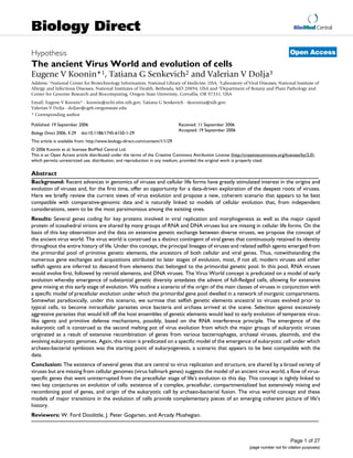
![Biology Direct 2006, 1:29 http://www.biology-direct.com/content/1/1/29
Page 2 of 27
(page number not for citation purposes)
Open peer review
This article was reviewed by W. Ford Doolittle, J. Peter
Gogarten, and Arcady Mushegian.
For the full reviews, please go to the Reviewers' comments
section.
Background
The extraordinary diversity of viruses
Viruses are ubiquitous companions of cellular life forms:
it appears that every cellular organism studied has its own
viruses or, at least, virus-like selfish genetic elements [1].
Recent environmental studies have shown that viruses,
primarily, bacteriophages, are "most abundant biological
entities on the planet" [2], with the total number of virus
particles exceeding the number of cells by at least an order
of magnitude [3,4]. Viruses actively move between
biomes and are thought to be major agents of evolution
by virtue of their capacity to operate as vehicles of hori-
zontal gene transfer (HGT) [5].
A remarkable feature of viruses is the diversity of their
genetic cycles, in a sharp contrast to the uniformity of the
cellular genetic cycle [6-9] (Fig. 1). Viruses with different
genome strategies span a vast range of genome sizes (the
genomes of the largest known virus, the mimivirus, and
the smallest viruses, e.g., circoviruses, differ by three
orders of magnitude) and show a non-uniform and non-
trivial distribution among the host taxa (Fig. 1). For exam-
ple, the extraordinary diversity of double-stranded (ds)
DNA bacteriophages is in a stark contrast to the absence
of bona fide dsDNA viruses in plants. Conversely, RNA
viruses are extremely abundant and diverse in plants and
animals but are currently represented by only two com-
pact families in bacteria, and so far have not been detected
in archaea (Fig. 1).
Given the variety of genetic strategies, genome complex-
ity, and global ecology of viruses, the problem of virus
evolution inevitably digresses into a web of interlocking
questions. What qualifies as a virus? Are viruses as a whole
monophyletic, i.e., ultimately descend from a single pri-
mordial virus or polyphyletic, i.e., have multiple origins?
If viruses are polyphyletic, how many independent line-
ages are there? What is the age distribution of different
groups of viruses – are they ancient or have they been
emerging continuously throughout life's evolution? And,
perhaps, the most fundamental questions of all: what is
the ultimate origin of viruses and what are the relation-
ships between evolution of viruses and cellular life forms?
The recent advances of comparative genomics create the
unprecedented opportunity to start tackling these issues
by inferring some of the likely answers from sequence and
structure data analysis. Here, we address several of these
questions but, primarily, the last two, most general ones,
in an attempt to outline a coherent scenario of virus origin
and evolution and delineate the connections between the
evolution of viruses and cellular life forms.
Results and discussion
Polyphyly versus monophyly in virus evolution
Comparative genomics provides no evidence of a mono-
phyletic origin of all viruses. Many virus groups simply
share no common genes, effectively, ruling out any con-
ventional notion of common origin. When applied to
viruses, the notion of "common genes" is not a simple
one because commonality is not necessarily limited to
clear-cut orthologous relationships between genes that
translate into highly significant sequence similarity.
Instead, as discussed in the next sections, distant homolo-
gous relationships among viral proteins and between viral
proteins and their homologs from cellular life forms
could convey more complex but important messages on
evolution of viruses. This complexity notwithstanding,
cases of major virus groups abound that either share no
homologous genes under any definition or have in com-
mon only distantly related domains with obviously dis-
tinct evolutionary trajectories. For example, most of the
viruses of hyperthermophilic crenarchaea have literally no
genes in common with any other viruses [10,11], whereas
RNA viruses share with DNA viruses and plasmids that
replicate via the rolling circle mechanism only extremely
distant domains in their respective replication proteins.
By contrast, monophyly of several large classes of viruses,
including vast assemblages of RNA viruses and complex
DNA viruses, can be demonstrated with confidence (Table
1). Some of these monophyletic classes of viruses cross the
boundaries set by genome strategies: thus, the retroid class
includes both RNA viruses and viruses, mobile elements,
and plasmids with DNA genomes, and the rolling circle
replication (RCR) class combines ssDNA and dsDNA
viruses and plasmids. Furthermore, based on similarities
in the structure of RNA replication complexes, along with
the presence of homologous, even if distant, replication
enzymes, it has been suggested that positive-strand RNA
viruses, double-stranded RNA viruses, and retroid ele-
ments all have a common origin [9]. On the whole, how-
ever, the conclusion seems inevitable that viruses
comprise several distinct lines of descent (Table 1).
A brief natural history of viral genes
Sequence analysis of viral proteins reveals several catego-
ries of virus genes that markedly differ in their prove-
nance. The optimal granularity of classification might be
subject to debate but at least 5 classes that can be assorted
into three larger categories seem to be readily distinguish-
able.
Genes with readily detectable homologs in cellular life forms](data:image/gif;base64,R0lGODlhAQABAIAAAAAAAP///yH5BAEAAAAALAAAAAABAAEAAAIBRAA7)